LUMBO-PERITONEAL SHUNT
A common indication for a LP shunt is in an adult with a communicating hydrocephalus, as in "normal pressure" hydrocephalus. The technique is suitable in patients with small ventricles as well as in cases with benign intracranial hypertension. In the paediatric age, an LP shunt is applied rarely, because communicating hydrocephalus is rarely found in this age group.
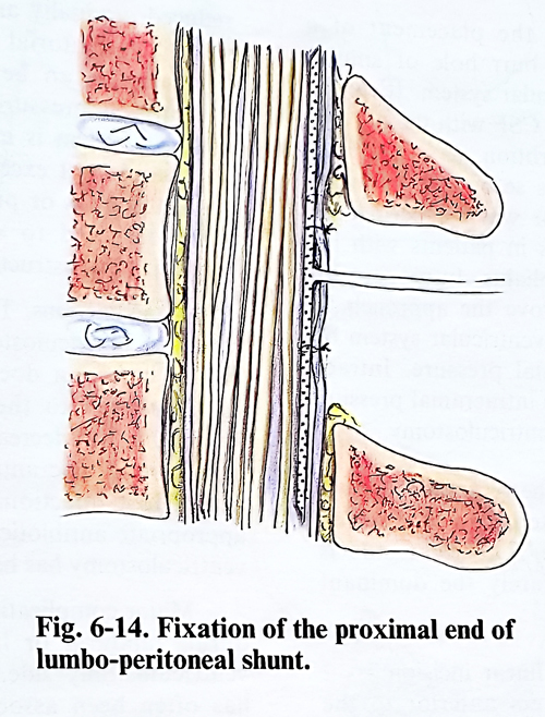
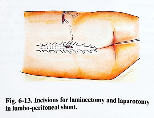
Surgical technique. The patient is placed in a lateral position on the operative table, to make the lumbar spine and lateral abdomen accessible for exploration. A standard laminectomy is performed removing the lamina of L3. The second incision of 5 - 6 cm is made below and parallel to the twelfth rib starting from the lateral border of the paravertebral muscles (Fig. 6-13). Penetrating between muscle fibres, the peritoneum is reached. One end of the shunting tube is introduced about 5 - 10 cm into the peritoneal cavity. The peritoneum is stitched around the tube. The other end of the shunting tube is introduced through the tunnel, in the subcutaneous tissue until it reaches the laminectomy. The dura mater and the arachnoid are opened. The end of the catheter is introduced into the subarachnoid space between the roots of the cauda equina and the tube is fixed to the dura (Fig. 6-14). There are many side holes along the shunting tube, facilitating the drainage.
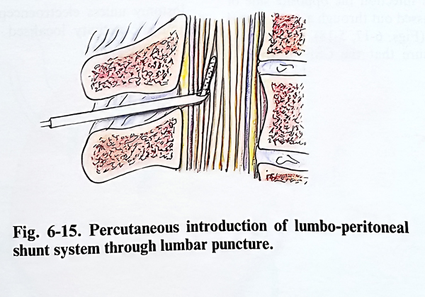
The shunting system can also be inserted more easily and less invasively using a percutaneous technique. This technique employs a 14G needle through which the shunt tubing is passed. The ability of the tubing to pass through the needle should be checked before the shunt placement. A small incision is made in the low midline lumbar area through which the needle is advanced with its tip bevel parallel to the longitudinal fibres of the dura mater to allow easier penetration. When CSF fluid is obtained, the tip is advanced slightly, rotated 90 degrees so that its opening is facing cephalic, and a predetermined length of tubing is then passed through the needle. The position of the catheter tip should be above the conus into the low thoracic region, thereby avoiding later traction of the lumbar roots (Fig. 6-15). The needle is withdrawn over the tubing, with care taken not to pull the catheter back.
When CSF flow is uninterruptedly running from the catheter, it is tunnelled around the flank to the peritoneum, and another opening is made to enter the peritoneal cavity. Occasionally, if the patient is obese or the subarachnoid space cannot be entered with certainty, a small laminotomy is necessary to open the dura under direct vision. Available LP shunt systems have either distal slit valves or a variety of in-line systems, some of which require careful horizontal-vertical orientation when they are positioned. The shunt tubing should be anchored at the lumbar incision with a small silastic collar or other similar device to prevent its subsequent migration.
Complications. Shunt complications can be divided into those that are common to all types of shunts and those that are unique to a particular type.
All shunts carry the risk of obstruction, disconnection and infection. The frequency and location of obstruction and disconnection depend to some degree on the type of shunt hardware used. The most common obstruction place is within the ventricular catheter lumen or around its tip. Paediatric patients most frequently suffer an obstruction of the ventricular catheter by choroid plexus, ependymal, or glial tissue, which can grow into or surround the catheter. A distal slit-valve catheter is the next most frequent site of obstruction.
Another source of malfunction is disconnection, which can occur at any point in the system but is most often found where the different shunt components join. In the paediatric age group, increased protein levels in the CSF appear to be associated with more frequent ventricular catheter obstruction. As noted previously, the use of radio-opaque materials for the entire length of the shunt system is recommendable, making it possible to determine the continuity of the shunt system on plain X-ray films. A reservoir in the system allowing access to CSF sampling is also helpful. By tapping the reservoir, pressure measurements can be made, the system's function can be ascertained, and CSF can be obtained for examination. Each type of shunting system has its own peculiarities which dictate how best to establish its functional adequacy. Most systems contain a pumping mechanism, either in the valve or the reservoir, which should test proper functioning, but the response to pumping and proper functioning of the shunt alone may not correlate absolutely with the overall pressure regulating function.
The management of shunt infection is subject to considerable debate. In principle it requires complete removal of the infected shunt system, with drainage intermittently through a reservoir or continuously by the establishment of an external ventricular drainage system.
A chronic low-grade infection shunt complication specific to VA shunts is nephritis.
Complications specific to VP shunts are ascites; pseudocyst; perforation of viscus; spread of infection or very rarely seeding of a neoplasm from the ventricles to the abdominal cavity. The use of coiled spring tubing inside the peritoneal cavity prevents its kinking but increases the possibility of perforation of abdominal organs.
Complications specific to a VA shunt involve the heart, lungs, and vascular system. They include such major problems as subclavian and vena cava obstruction, mural thrombosis, bacterial endocarditis, cardiac tamponade (secondary to perforation of the heart wall), embolization by the distal catheter into the pulmonary artery and chronic pulmonary thromboembolism.
The development of significant subdural fluid accumulations after ventricular shunting relates to the patient's age and the size of the ventricles; the older the patient, the bigger the risk for this complication, both during and after shunt insertion. Even a minor head injury may produce a significant subdural fluid accumulation if the shunted ventricles are large. The use of a high-pressure valve helps to reduce but does not eliminate this problem. If the subdural collection is asymptomatic and relatively small, treatment is not necessarily indicated, but if a progressive increase in volume is documented with clinical symptoms, it is necessary to drain the subdural fluid. This can be treated with a subdural peritoneal shunt without a valve to provide a pressure gradient across the cortical mantle between the two shunting systems.