Legacy: "Atlast of Neurosurgery" / L.Karaguiosov, A. Ramadan, K.Karaguiosov / Kiwait/ 1998
NEXT > 8, 9, 10 chapter - 3D Atlas of Neurological Surgery
7. SURGICAL TREATMENT OF INTRACRANIAL INFECTIONS
In this chapter we consider bacterial infections and some parasitic lesions as cysticercosis and hydatid cysts that may require neurosurgical treatment for the control of infection or to relieve pressure on the intracranial content. These infections are mostly metastatic, but they can occur by direct spread from a nearby infected focus, or as a result of a surgical procedure.
OSTEOMYELITIS OF SKULL BONES
An osteomyelitis of the cranial bones is usually the result of direct penetration of the infection. It can be seen affecting the bone flap after craniotomy, or after skeletal traction with tongs in cervical fractures or adjacent to purulent sinusitis or otitis. A metastatic haematogeneous osteomyelitis is extremely rare. The most common organism causing osteomyelitis is staphylococcus, but other organisms or a combination of them can be identified. Local pain and oedema are the main clinical manifestations.
Fever and the other usual signs of infection can also be present. X-rays films reveal destruction of the bone tissue.
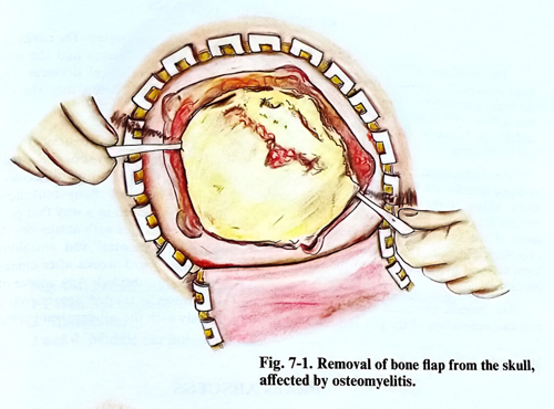
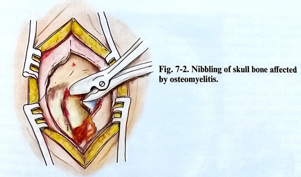
Osteomyelitis of the skull bones needs surgical treatment. The incision of the scalp over the osteomyelitic focus should be sufficiently large, permitting exploration of the entire affected bone surface. When it is a case of postcraniotomy osteomyelitis, it often requires total removal of the bone flap, using the previous surgical incision (Fig. 7-1). When the osteomyelitis doesn't show the integrity of the calvaria to be broadly affected, a burr hole is drilled at the border of osteomyelitic focus and the affected bone is removed completely by nibbling (Fig. 7-2). The affected bone differs in colour and pus comes out of the diploic tissue instead of blood. The nibbling continues until a normal appearing bone tissue has been reached and the epidural space appears unaffected. Sinogenic osteomyelitis must be treated simultaneously with the source of infection. Intraoperative antibiotics are used i.v., and for wound irrigation.
Closure is as usual with a drainage left epidurally for a few days. Antibiotic therapy depends upon the results of the bacteriological investigations and continues for up to 3 - 4 weeks after the surgical treatment.
The subsequently required cranioplasty is postponed for at least 6 months after complete wound healing.
Some forms of cranial osteomyelitis (with very small and multiple foci) can be treated initially with antibiotics followed up frequent clinical and X-ray follow-up of the lesion.
EPIDURAL ABSCESS
The epidural abscess is a result of direct penetration of the infection in the epidural space from sinusitis, inflammation of the middle ear and osteomyelitis of the cranial bones. The clinical manifestations begin with headache, vomiting and generalised signs and symptoms of bacterial infection. The growth of the abscess provokes increased intracranial pressure and deterioration of the level of consciousness. In some patients, penetration of the infection into the subdural space leads to a sudden deterioration because of additional meningitis, brain abscess formation or thrombosis of the dural sinuses. CT and MRI studies are essential to detect the location and the size of the epidural abscess.
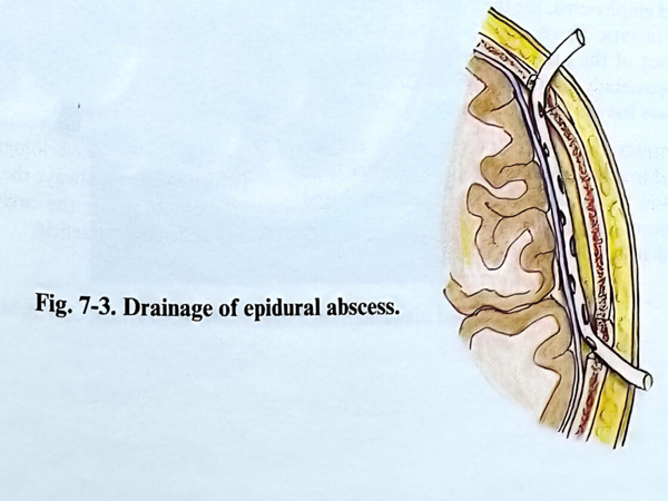
The surgical treatment consists of craniotomy and removal of the pus with the produced granulation tissue. The cavity is washed with saline and antibiotics and the wound is closed with placing an epidural drainage (Fig. 7-3). Antibiotic treatment continues for several weeks after complete healing of the wound, and the wound must be treated with local instillation of antibiotics occasionally.
SUBDURAL ABSCESS
The subdural abscesses (mentioned also as “empyema”) usually occupy large areas on the convexity and basal surface of the brain hemispheres. The origin of the infection is frequently in the paranasal sinuses or the middle ear. It can be also a complication after any craniotomy.
Headache, fever, neck rigidity, and focal epileptic attacks are the early signs and symptoms. Many of the patients with a subdural abscess deteriorate suddenly with rapid impairment of the level of consciousness and focal neurological deficit. The resulting mortality is high. CT and MRI are essential for early and precise diagnosis.
The surgical treatment consists of a craniotomy, evacuation of the pus and removal of the granulation tissue. The cavity is washed with saline and antibiotics and the wound is closed with a fine subdural drainage line left inside. Antibiotic instillation into the cavity may be required.
Urgent surgical treatment in these cases is the rule. The removal of the extensive granulation tissue and multiple abscesses needs wider craniotomies and many burr holes. Drainage tubes are inserted in a way that permits washing of all the cavities with antibiotics. CT follow-up control is essential and antibiotic therapy is continued for 3 weeks after clinical signs have completely subsided. Any source of infection in the sinuses is treated aggressively and simultaneously with the subdural empyema.
BRAIN ABSCESS
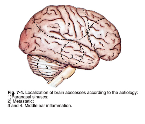
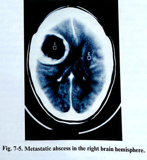
Irrespective of the fact that an abscess may develop in any part of the cranial cavity, it has a predominant localisation depending on the aetology (Fig. 7-4). Statistical data show nearly 60% of the intracranial abscesses to have an otological origin and 93% out of these are situated in the temporal lobe or in the cerebellum. About 10% of abscesses originate from infection of the air sinuses (frontal and ethmoidal and are located in the frontal lobes. Chronic pleuropulmonary sepsis, mainly in bronchiectasia and emphysema, are the most frequent causes of metastatic abscesses. They are also about 10% out of the total and are primarily located frontoparietally (Fig. 7-5). About 20% of brain abscesses have no obvious source.
Intracranial abscesses very often require surgical treatment as soon as possible after their diagnosis, as patients with abscesses, compared with those with intracranial tumours, have a bigger risk of sudden deterioration. When the general condition of the patient is good and without clinical data suggesting increased intracranial pressure, the operation is performed after complete and detailed investigations.
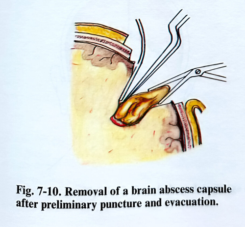
There have always been controversies in the evolution of the surgical treatment of brain abscesses regarding the methods to be used. The main surgical methods considered in current neurosurgical practice are puncturing and washing with antibiotics, opening and draining the abscess cavity, and excision of the capsule after puncture and aspiration.
Abscesses in an early stage (local cerebritis) can be followed up and treated conservatively. It should be a rule to evacuate any abscess larger than 2 - 2.5 cm in diameter, when a specimen for bacteriological study is required (which is almost always the case), when there are air bubbles inside the cavity or the patient has any neurological deficit.
Multiple abscesses are always treated surgically for early sampling of the causative organism when it is unknown or because of severe brain affection threatening the patient's life. All abscess cavities larger than 2 - 2.5 cm are then evacuated.
ABSCESSES OF BRAIN HEMISPHERES
The choice of surgical technique depends on location, volume of the abscess, the patient's condition, is it a recurrence, and the possibility of fungal infection.
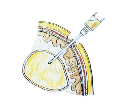
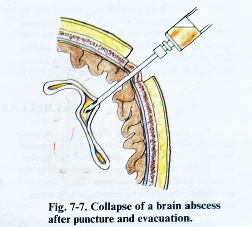
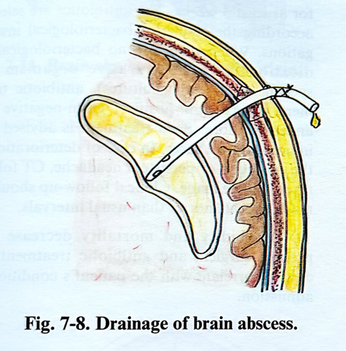
PUNCTURE AND EVACUATION. A single burr hole is made. Its position is determined from the location of the abscess. The shortest distance to the lesion and the avoidance of eloquent cortex are the principles to follow in choosing the burr hole position. A ventricular cannula is slowly inserted into the brain tissue, not touching the edge of the burr hole to be able to feel very clearly the resistance when the abscess wall penetration. In abscesses with a long evolution the resistance on reaching the capsule is rather strong. The depth is measured and the abscess cavity is penetrated. If the pus is thick and does not drain out spontaneously, it should be aspirated with a syringe. After the evacuation of the abscess the cavity is washed with antibiotics, the ventricular cannula is removed, and the operative wound is closed leaving a fine drainage tubing in the cavity (Figs. 7-6; 7-7; 7-8).
"Free hand" finding of the abscess with the cannula can be difficult at times. More and more, CT guided and neuronavigation techniques are being applied for these lesions, especially when the lesion is not in contact with the cortex. For lesions deeper than 4 - 5cm, a stereotactic technique can also be applied.
The drainage is kept in for several days and antibiotic washes of the cavity are performed daily.
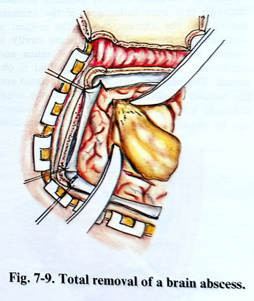
COMPLETE REMOVAL. The total removal of the brain abscess with its capsule is the most radical method. It can be applied to abscesses located superficially with a thick capsule when the patient's condition is good. The abscess should be located in non-eloquent brain.This method is very efficient against recurrences (Figs. 7-9; 7-10).
However, the brain damage induced by dissection can be significant. Currently few cases have their abscesses excised, and the technique is reserved for recurrent cases, or when the existence of a foreign body undetectable by investigations is suspected. Fungal abscesses, where the microorganisms are inside the capsule, require excision for their radical treatment.
Surgery is similar to that for the removal of a subcortical or cortical space occupying lesion. The craniotomy overlays the abscess location, the lesion is approached by a small cortical incision and the capsule is dissected. This technique also has been much improved with the introduction of the CT guided methods, neuro-navigation and intraoperative ultrasound.
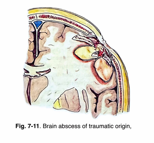
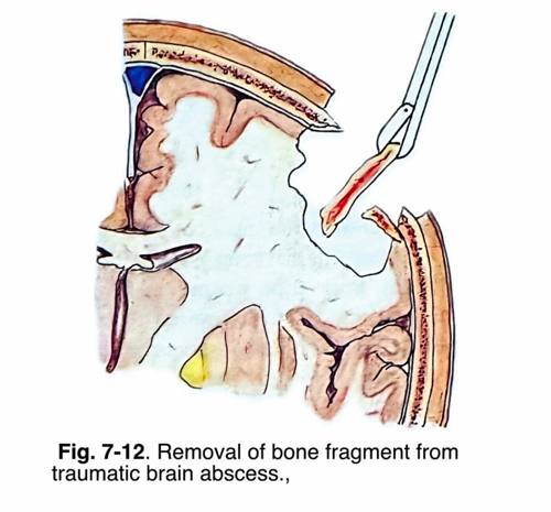
BRAIN ABSCESSES AFTER OPEN HEAD INJURIES. The brain abscesses developing after open head injuries have specific irregular shapes and their location is related to the scar tissue provoked by the injury. They are more often multiple, with their irregular shapes resulting from extension and adhesions to the bone edges. Frequently more then one abscess cavity is found. Some of the cavities have a thick capsule, while the others - a very thin one. The abscesses of traumatic origin contain foreign bodies, as bone fragments, hairs, and at times even metallic pieces. In some cases there can be fistulae through the skin. The presence of a foreign body makes abscess gliosis and cure impossible without its surgical removal.
The shape of the scalp incision depends on the size and location of the residual scar tissue, provoked by the injury. If a primary wound repair has not been done, the skin edges are excised. The existing bone defect is widened until normal dura around it is exposed. Bone parts affected by osteomyelitis should be removed. The presence of a fistula indicates the way to penetrate toward the abscess cavity. The content of the abscess is evacuated and the capsule is excised only in cases of massive penetration of contaminated material. In the remaining cases the surgeon proceeds as described above. The majority of abscesses of traumatic origin are situated near the cranial vault, with adherences of the dura and the brain. As much as possible they should not be torn on penetrating so as to avoid massive contamination between the abscess and the CSF spaces. During the excision the foreign bodies, such as bone fragments, hairs, metallic pieces, etc., should be completely removed (Figs. 7 - 11; 7-12). If the X-ray data indicate that there are foreign bodies far from the abscess cavity, they are left intact and followed up. At the end of the operation the wound is closed, leaving drainage tubes in the ordinary way.
CEREBELLAR ABSCESSES
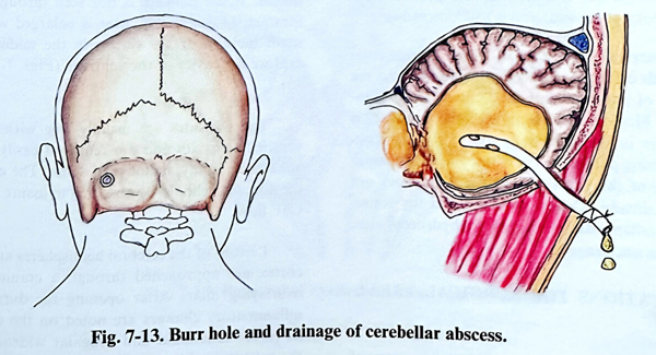
The abscesses of the cerebellum are most often located in the hemispheres and are caused by suppurative otitis or mastoiditis. The cerebellar surface is usually found adherent to the pyramid of the temporal bone. These abscesses are treated surgically by punctures and drainage (Fig. 7-13).
Puncture and evacuation of the abscess are carried out in the following way: under local anaesthesia a burr hole is made 3 cm laterally and below the external occipital protuberance that is enlarged 2 - 3 cm in diameter. The dura is opened in the usual way. A puncture is made through this burr hole, and, if the cannula enters into the abscess cavity, the content is completely evacuated. The abscess cavity is washed with copious amounts of saline and antibiotics, until a clear washing fluid is obtained. The place of puncture is coagulated and the wound is closed in layers with the usual drainage left. Excision of cerebellar abscesses can be required in recurrences. Complete control of the otological infection should be obtained in all cases.
POSTOPERATIVE CARE AND RESULTS
After an operation for an intracranial abscess, antibiotic treatment must be administered for at least 3 weeks. The antibiotics are selected according the results of bacteriological investigations. When there are no bacteriological indications for the causative organism (no specimens or sterile cultures), antibiotic treatment covers gram-positive, gram-negative and anaerobic flora. Such treatment is advised also in non-surgical cases. In case of deterioration of the condition or persistent headache, CT follow-up should be made. Clinical follow-up should be made also at shorter than usual intervals.
Morbidity and mortality decrease with prompt surgical and antibiotic treatment and clearly correlate with the patient's condition on admission.
PARASITIC LESIONS OF THE BRAIN
Among the different types of parasites, invading the cranial cavity that require surgical treatment the most common conditions are cysticercosis and hydatidosis,
CYSTICERCOSIS
Cysticercosis is the most common parasitic disease affecting the nervous system. The larval forms of the pork tape worm (taenia solium) cause it. The infection is acquired through the gastrointestinal tract. The organisms reach the nervous system via haematogeneous route. Upon reaching the nervous system, the parasite may localise in the brain parenchyma, the ventricular system (through the choroid plexus), the subarachnoid space or in a combination of all these. The presenting clinical picture depends on the predominant localisation of the larvae. In case of parenchymal localisation, when the parasite dies inducing an inflammatory reaction, it may cause more harm than the larva itself. These lesions in the parenchyma may be single or multiple, and are usually rounded soft tissue masses or cystic lesions.
With time, lesions may calcify and usually become spherical. In case of a meningobasal (basal cisterns) or intraventricular localisation, the predominant pathology of presentation is hydrocephalus.
Surgical treatment of brain cysticercosis depends on the localisation, the number and the shape of parasites and their clinical manifestations. Medical treatment with praziquantel is effective in parenchymally and cortically localised living parasites. This drug affects the metabolism of the parasite, and therefore does not affect already calcified parasites with the consequent inflammatory reaction or hydrocephalus remaining unchanged.
INDICATIONS FOR SURGICAL TREATMENT
Surgical treatment is applied in a case of cysticercosis with progressive deterioration of neurological signs and symptoms, most frequently hemiparesis and epilepsy, refractory to medical treatment. In such cases if the CT and MRI reveal parasites corresponding with the clinical data, and they are not calcified, medical treatment is tried first. If this is ineffective, patients are operated.
The second group of patients indicated for surgical treatment are those with increased intracranial pressure. When the intracranial hypertension is due to hydrocephalus, provoked by intraventricular localisation of parasites or the racemous form found inside the basal cisterns, the initial surgical method is a shunt operation. After stabilising the patient's condition, removal of parasites and medical treatment can follow.
The parasites located in the chiasmatic cistern and provoking visual disturbances also require selective removal.
SURGICAL TECHNIQUE
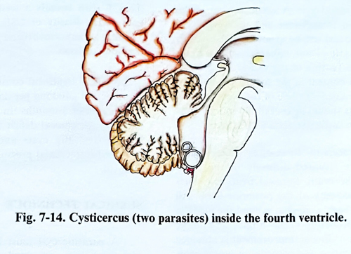
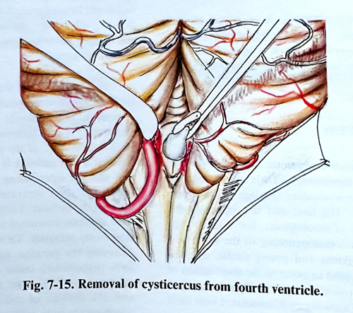
In a case of cysticercus inside the fourth ventricle, a standard craniectomy is made in the posterior fossa with midline incision of the soft tissues. If the parasite is not seen through the foramen Magendi, the latter is enlarged with a small incision of the vermis in the midline to explore the cavity of the ventricle (Figs. 7-14; 7-15).
The parasites are usually free within the ventricular cavity and are removed easily. The adherence, if any, is not very dense. The exit of the aqueduct should be observed to assure a free CSF outflow.
Lesions of the cerebral hemispheres and the cortex are approached through a craniotomy overlaying them. After opening the dura, the inflammatory changes are noted on the cortex - the turbid arachnoid and irregular widening of the subarachnoid space. The parasite vesicles in the subarachnoid space are easily seen as small cysts surrounded by fine arachnoid adhesions. Opening of the arachnoid should be sufficient to mobilise the cyst and to remove it. The parasites located under the pia are detected as small bulges of cerebral gyri. A small incision of the pia-arachnoid is sufficient and the vesicle becomes visible and will be exposed by the brain's pulsation, as it is not adherent to the brain parenchyma.
Rupture of the vesicle and spreading its liquid content in the subarachnoid space should be avoided. If some of the cysts ruptures, its contents should be aspirated out immediately and the brain surface washed with saline.
In the racemous form of cysticercosis, the parasites are situated diffusely in the basal cisterns of the brain. Surgical treatment starts with a craniectomy of the posterior fossa. All cysts in the cisterna magna, around the medulla and both cerebellopontine angles should be removed. If no clinical improvement is obtained after this operation and parasites in supratentorial basal cisterns are present, a frontotemporal craniotomy (unilateral or bilateral) follows for the removal of as many of the lesions inside and around the chiasmatic and sylvian cisterns as is possible.
COMPLICATIONS
Meningial irritation is frequently observed in the postoperative period with neck rigidity, Kernig's sign, fever, and a high WBC count in the CSF. Usually th reaction is aseptic.
HYDATID CYST
The causative organism is the larval stage of the dog tapeworm (Taenia echinococcus).
The infection is acquired through the gastrointestinal tract. The liver and the lungs are the primary sites of involvement, but some organisms spread haematogenously to the brain and bones. In children and young adults, cerebral hydatid cysts tend to occur in the distribution of the middle cerebral artery. Usually it is a case of a single, slowly growing cyst, associated with little or no inflammatory reaction or oedema.
Children usually present with signs and symptoms of increased intracranial pressure whereas young adults often have hemispheric signs and symptoms. Radiography may show thinning of the bone overlying the lesion, and the CT scan reveals a well-delineated cystic lesion with density of CSF, with little if any capsular enhancement and without any surrounding oedema.
Medical treatment consists of mebendazol (Vermox) 50 - 100 mg per day per kilogram for a minimum of 3 months. In case of small cysts and a minor clinical deficit this treatment may be effective. Big cysts and the presence of increased intracranial pressure indicate surgical removal.
SURGICAL TECHNIQUE
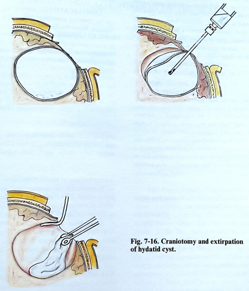
A parasitic cyst must be removed totally without being perforated, so that its liquid content is not spread inside the cranial cavity. In a case of a single, not very large cyst, its extirpation can be easily performed (Fig. 7-16).
A craniotomy is made in the region over the cyst, usually located near the cortical surface and it will be seen immediately after opening the dura. In other cases the cortex is atrophic, and the thinned cortical layer determines the place of the lesion. The separation of the cyst from the brain tissue is easily accomplished, as the germinate capsule is not adherent to the brain tissue. However, it is very thin and may be easily torn. Removal 'in toto' of the parasitic cyst is easier, if a soft catheter is inserted between the cyst and the surrounding gliotic brain and the capsule is then separated by flushing saline through the catheter. Large cysts are punctured, their contents evacuated and the collapsed wall then easily removed. To avoid recurrence after removing the parasite, its bed is washed with 20% (hypertonic) saline solution. Alternative solutions are 0.5% silver nitrate, 5% cetrimide, and 5% tincture of chlorhexidine. The cavity which results from extirpation of the parasite is washed with the above-mentioned antiseptic solutions if there is no communication with the ventricular system.
In case of multiple hydatidosis a broad craniotomy is performed over the area where the cysts are situated and they must then be removed if possible, without been opened. In such cases parasites occupy a large part of the intracranial space and very often they penetrate into the lateral ventricles. Intraoperative ultrasound in these cases is of great value. Their removal is very difficult, because they are of various sizes, some of them being so small so they cannot be seen even under a surgical micro-scope.
COMPLICATIONS
Aseptic meningitis can be observed postoperatively and it quickly subsides. When the hydatid fluid has penetrated inside the ventricular cavity, the manifestation of aseptic meningitis may be more pronounced: fever, vomiting, clear signs of menigioradicular irritation, increased white cell count in the CSF.