HYDATID CYST
The causative organism is the larval stage of the dog tapeworm (Taenia echinococcus).
The infection is acquired through the gastrointestinal tract. The liver and the lungs are the primary sites of involvement, but some organisms spread haematogenously to the brain and bones. In children and young adults, cerebral hydatid cysts tend to occur in the distribution of the middle cerebral artery. Usually it is a case of a single, slowly growing cyst, associated with little or no inflammatory reaction or oedema.
Children usually present with signs and symptoms of increased intracranial pressure whereas young adults often have hemispheric signs and symptoms. Radiography may show thinning of the bone overlying the lesion, and the CT scan reveals a well-delineated cystic lesion with density of CSF, with little if any capsular enhancement and without any surrounding oedema.
Medical treatment consists of mebendazol (Vermox) 50 - 100 mg per day per kilogram for a minimum of 3 months. In case of small cysts and a minor clinical deficit this treatment may be effective. Big cysts and the presence of increased intracranial pressure indicate surgical removal.
SURGICAL TECHNIQUE
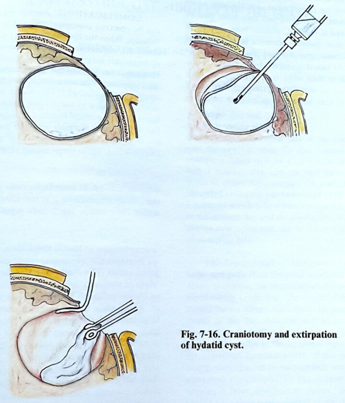
A parasitic cyst must be removed totally without being perforated, so that its liquid content is not spread inside the cranial cavity. In a case of a single, not very large cyst, its extirpation can be easily performed (Fig. 7-16).
A craniotomy is made in the region over the cyst, usually located near the cortical surface and it will be seen immediately after opening the dura. In other cases the cortex is atrophic, and the thinned cortical layer determines the place of the lesion. The separation of the cyst from the brain tissue is easily accomplished, as the germinate capsule is not adherent to the brain tissue. However, it is very thin and may be easily torn. Removal 'in toto' of the parasitic cyst is easier, if a soft catheter is inserted between the cyst and the surrounding gliotic brain and the capsule is then separated by flushing saline through the catheter. Large cysts are punctured, their contents evacuated and the collapsed wall then easily removed. To avoid recurrence after removing the parasite, its bed is washed with 20% (hypertonic) saline solution. Alternative solutions are 0.5% silver nitrate, 5% cetrimide, and 5% tincture of chlorhexidine. The cavity which results from extirpation of the parasite is washed with the above-mentioned antiseptic solutions if there is no communication with the ventricular system.
In case of multiple hydatidosis a broad craniotomy is performed over the area where the cysts are situated and they must then be removed if possible, without been opened. In such cases parasites occupy a large part of the intracranial space and very often they penetrate into the lateral ventricles. Intraoperative ultrasound in these cases is of great value. Their removal is very difficult, because they are of various sizes, some of them being so small so they cannot be seen even under a surgical micro-scope.
COMPLICATIONS
Aseptic meningitis can be observed postoperatively and it quickly subsides. When the hydatid fluid has penetrated inside the ventricular cavity, the manifestation of aseptic meningitis may be more pronounced: fever, vomiting, clear signs of menigioradicular irritation, increased white cell count in the CSF.