PARASITIC LESIONS OF THE BRAIN
Among the different types of parasites, invading the cranial cavity that require surgical treatment the most common conditions are cysticercosis and hydatidosis,
CYSTICERCOSIS
Cysticercosis is the most common parasitic disease affecting the nervous system. The larval forms of the pork tape worm (taenia solium) cause it. The infection is acquired through the gastrointestinal tract. The organisms reach the nervous system via haematogeneous route. Upon reaching the nervous system, the parasite may localise in the brain parenchyma, the ventricular system (through the choroid plexus), the subarachnoid space or in a combination of all these. The presenting clinical picture depends on the predominant localisation of the larvae. In case of parenchymal localisation, when the parasite dies inducing an inflammatory reaction, it may cause more harm than the larva itself. These lesions in the parenchyma may be single or multiple, and are usually rounded soft tissue masses or cystic lesions.
With time, lesions may calcify and usually become spherical. In case of a meningobasal (basal cisterns) or intraventricular localisation, the predominant pathology of presentation is hydrocephalus.
Surgical treatment of brain cysticercosis depends on the localisation, the number and the shape of parasites and their clinical manifestations. Medical treatment with praziquantel is effective in parenchymally and cortically localised living parasites. This drug affects the metabolism of the parasite, and therefore does not affect already calcified parasites with the consequent inflammatory reaction or hydrocephalus remaining unchanged.
INDICATIONS FOR SURGICAL TREATMENT
Surgical treatment is applied in a case of cysticercosis with progressive deterioration of neurological signs and symptoms, most frequently hemiparesis and epilepsy, refractory to medical treatment. In such cases if the CT and MRI reveal parasites corresponding with the clinical data, and they are not calcified, medical treatment is tried first. If this is ineffective, patients are operated.
The second group of patients indicated for surgical treatment are those with increased intracranial pressure. When the intracranial hypertension is due to hydrocephalus, provoked by intraventricular localisation of parasites or the racemous form found inside the basal cisterns, the initial surgical method is a shunt operation. After stabilising the patient's condition, removal of parasites and medical treatment can follow.
The parasites located in the chiasmatic cistern and provoking visual disturbances also require selective removal.
SURGICAL TECHNIQUE
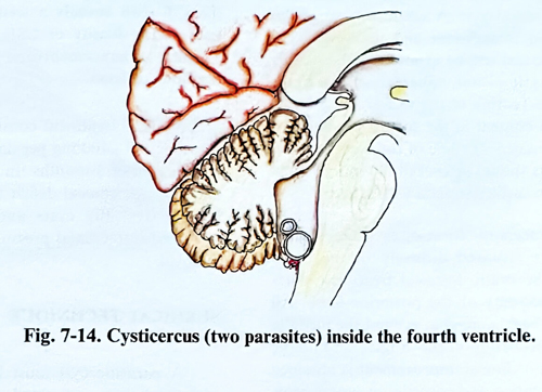
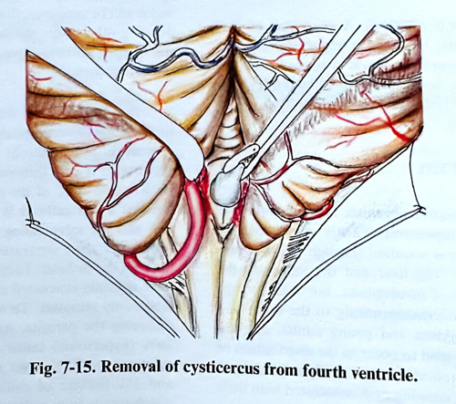
In a case of cysticercus inside the fourth ventricle, a standard craniectomy is made in the posterior fossa with midline incision of the soft tissues. If the parasite is not seen through the foramen Magendi, the latter is enlarged with a small incision of the vermis in the midline to explore the cavity of the ventricle (Figs. 7-14; 7-15).
The parasites are usually free within the ventricular cavity and are removed easily. The adherence, if any, is not very dense. The exit of the aqueduct should be observed to assure a free CSF outflow.
Lesions of the cerebral hemispheres and the cortex are approached through a craniotomy overlaying them. After opening the dura, the inflammatory changes are noted on the cortex - the turbid arachnoid and irregular widening of the subarachnoid space. The parasite vesicles in the subarachnoid space are easily seen as small cysts surrounded by fine arachnoid adhesions. Opening of the arachnoid should be sufficient to mobilise the cyst and to remove it. The parasites located under the pia are detected as small bulges of cerebral gyri. A small incision of the pia-arachnoid is sufficient and the vesicle becomes visible and will be exposed by the brain's pulsation, as it is not adherent to the brain parenchyma.
Rupture of the vesicle and spreading its liquid content in the subarachnoid space should be avoided. If some of the cysts ruptures, its contents should be aspirated out immediately and the brain surface washed with saline.
In the racemous form of cysticercosis, the parasites are situated diffusely in the basal cisterns of the brain. Surgical treatment starts with a craniectomy of the posterior fossa. All cysts in the cisterna magna, around the medulla and both cerebellopontine angles should be removed. If no clinical improvement is obtained after this operation and parasites in supratentorial basal cisterns are present, a frontotemporal craniotomy (unilateral or bilateral) follows for the removal of as many of the lesions inside and around the chiasmatic and sylvian cisterns as is possible.
COMPLICATIONS
Meningial irritation is frequently observed in the postoperative period with neck rigidity, Kernig's sign, fever, and a high WBC count in the CSF. Usually th reaction is aseptic.