ABSCESSES OF BRAIN HEMISPHERES
The choice of surgical technique depends on location, volume of the abscess, the patient's condition, is it a recurrence, and the possibility of fungal infection.
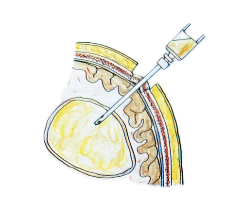
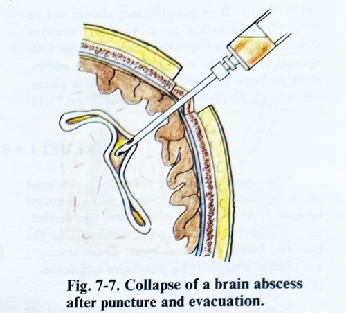
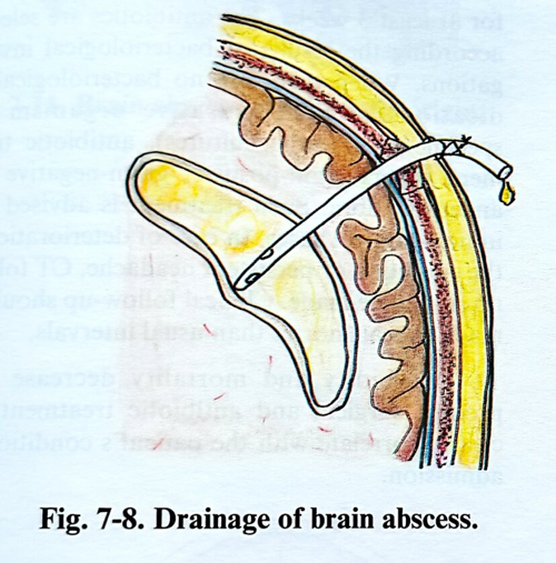
PUNCTURE AND EVACUATION. A single burr hole is made. Its position is determined from the location of the abscess. The shortest distance to the lesion and the avoidance of eloquent cortex are the principles to follow in choosing the burr hole position. A ventricular cannula is slowly inserted into the brain tissue, not touching the edge of the burr hole to be able to feel very clearly the resistance when the abscess wall penetration. In abscesses with a long evolution the resistance on reaching the capsule is rather strong. The depth is measured and the abscess cavity is penetrated. If the pus is thick and does not drain out spontaneously, it should be aspirated with a syringe. After the evacuation of the abscess the cavity is washed with antibiotics, the ventricular cannula is removed, and the operative wound is closed leaving a fine drainage tubing in the cavity (Figs. 7-6; 7-7; 7-8).
"Free hand" finding of the abscess with the cannula can be difficult at times. More and more, CT guided and neuronavigation techniques are being applied for these lesions, especially when the lesion is not in contact with the cortex. For lesions deeper than 4 - 5cm, a stereotactic technique can also be applied.
The drainage is kept in for several days and antibiotic washes of the cavity are performed daily.
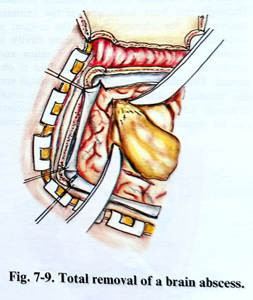
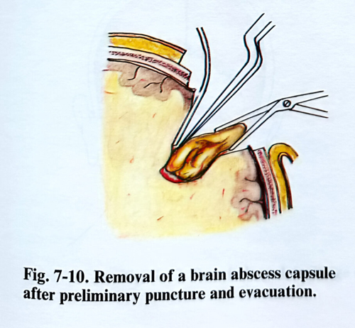
COMPLETE REMOVAL. The total removal of the brain abscess with its capsule is the most radical method. It can be applied to abscesses located superficially with a thick capsule when the patient's condition is good. The abscess should be located in non-eloquent brain.This method is very efficient against recurrences (Figs. 7-9; 7-10).
However, the brain damage induced by dissection can be significant. Currently few cases have their abscesses excised, and the technique is reserved for recurrent cases, or when the existence of a foreign body undetectable by investigations is suspected. Fungal abscesses, where the microorganisms are inside the capsule, require excision for their radical treatment.
Surgery is similar to that for the removal of a subcortical or cortical space occupying lesion. The craniotomy overlays the abscess location, the lesion is approached by a small cortical incision and the capsule is dissected. This technique also has been much improved with the introduction of the CT guided methods, neuro-navigation and intraoperative ultrasound.
BRAIN ABSCESSES AFTER OPEN HEAD INJURIES. The brain abscesses developing after open head injuries have specific irregular shapes and their location is related to the scar tissue provoked by the injury. They are more often multiple, with their irregular shapes resulting from extension and adhesions to the bone edges. Frequently more then one abscess cavity is found. Some of the cavities have a thick capsule, while the others - a very thin one. The abscesses of traumatic origin contain foreign bodies, as bone fragments, hairs, and at times even metallic pieces. In some cases there can be fistulae through the skin. The presence of a foreign body makes abscess gliosis and cure impossible without its surgical removal.
The shape of the scalp incision depends on the size and location of the residual scar tissue, provoked by the injury. If a primary wound repair has not been done, the skin edges are excised. The existing bone defect is widened until normal dura around it is exposed. Bone parts affected by osteomyelitis should be removed. The presence of a fistula indicates the way to penetrate toward the abscess cavity. The content of the abscess is evacuated and the capsule is excised only in cases of massive penetration of contaminated material. In the remaining cases the surgeon proceeds as described above. The majority of abscesses of traumatic origin are situated near the cranial vault, with adherences of the dura and the brain. As much as possible they should not be torn on penetrating so as to avoid massive contamination between the abscess and the CSF spaces. During the excision the foreign bodies, such as bone fragments, hairs, metallic pieces, etc., should be completely removed (Figs. 7 - 11; 7-12). If the X-ray data indicate that there are foreign bodies far from the abscess cavity, they are left intact and followed up. At the end of the operation the wound is closed, leaving drainage tubes in the ordinary way.