Legacy: "Atlast of Neurosurgery" / L.Karaguiosov, A. Ramadan, K.Karaguiosov / Kiwait/ 1998
PREV> 17. Infections of the Spine and Spinal Cord
NEXT< 19. General Principles of Peripheral Nerve Repair
18. FUNCTIONAL SURGERY OF THE SPINAL CORD
Some surgical procedures can be made on the spinal cord with the aim of modifying its function and by that improving the condition in some diseases. The majority of these procedures consist of the interruption of afferent pathways to relieve painful syndromes, spasticity, and other neurological manifestations.
HIGH THORACIC CORDOTOMY
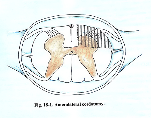
Cordotomy is based on the concept that the spinothalamic tract, located in the anterolateral quadrant of the spinal cord, transmits the pain and temperature sensations (Fig. 18-1).
Cordotomy is indicated in patients with cancer pain, when the expected survival period is not more than 3 years. This condition is mandatory because the recurrence rate of pain is very high and after this period painful dysesthesia increases. The high percentage of postoperative complications such as weakness, sphincter and sexual disturbances is another argument against applying this operation frequently. After cordotomy the level of analgesia drops caudally in the first 3 weeks postoperatively with 3 - 6 dermatomes and after 6 months another 3-6 dermatomes further down. That is why, with a cordotomy done at the level of 2-3 thoracic vertebrae, the level of permanent hypoalgesia or analgesia usually is at about the 10th thoracic dermatome. Cordotomy should be contraindicated in benign diseases such as spondylotic pain, postherpetic neuralgia, causalgia, different forms of neuropathies and neuritis, phantom pain and particularly when the cause of the pain syndrome is unknown.
The spinothalamic tract which transmits pain and temperature sensation crosses the midline, and the operation should be done on the opposite side to that of the pain.
Unilateral cordotomy is most effective in patients with cancer pain, when the internal organs are not involved and when the cancer is situated out of the midline and below the lower thoracic area. The best candidates for high thoracic cordotomy are those patients with cancer involving the lower limbs, buttocks, and the lateral retroperitoneal area inside the pelvis, abdomen, and the chest wall.
When the cancer is near to the midline, unilateral cordotomy cannot offer long prolonged relief of pain. In similar cases the cordotomy must be bilateral, but the second procedure should be done at a different level and it is preferable that one of the two cordotomies be percutaneous.
SURGICAL TECHNIQUE
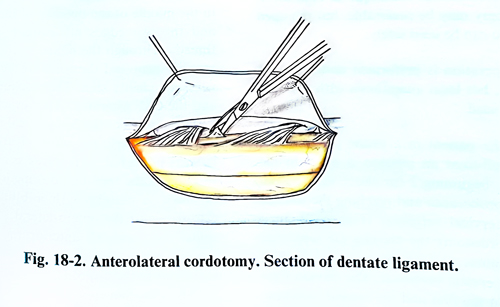
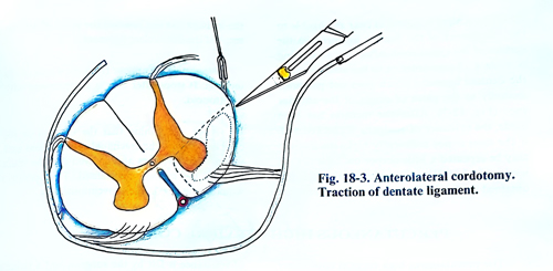
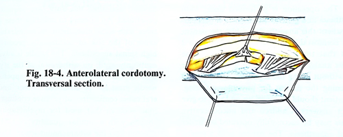
The open cordotomy is performed at the level of the 2nd d-3rd thoracic vertebrae. The patient is in a prone position on the operating table. General anaesthesia is usually administered, but local anaesthesia with sedation permits control of any provoked analgesia and prevents some complications during the operation. The laminectomy is performed in the conventional way with the opening of two laminae. The dura mater is opened with an arch-form incision with its convexity on the side of the planned cordotomy. If possible, bipolar electrodes are placed within the epidural space in the midline rostrally and caudally for recording sensory evoked potentials from bilateral peroneal stimulation. After that the operation is done under optical magnification. The width of the spinal cord is measured precisely, and using a piece of wax the scalpel is marked in such a way that the cutting part of the scalpel is only half the width of the spinal cord. The dentate ligaments are identified on both sides and their attachments to the dura, on the side of the cordotomy, are cut. The arachnoid is torn on the surface on the side where the cordotomy is to be made. One of the dentate ligaments is caught with traction thread using a clip (Figs.18-2; 18-3). The spinal cord can be rotated by 45 degrees by pulling the traction tread. If the dentate ligament is not sufficiently strong it may be torn during the rotation. In such a case, traction threads are introduced under the anterior roots to establish some rotation, which should permit the anterior spinal artery to be seen. For this purpose a small dental mirror can be used. A small perforation is first made in the pia mater immediately anterior to the place of attachment of the dentate ligament and after that the scalpel is driven into the necessary depth up to 4 - 5 mm to where the piece of wax is limiting its entry. If the pia mater is not perforated before, an incision is made so, that the dentate ligament may not be torn with a wrong movement and the incision surpasses the required limits. After the scalpel enters to the sufficient depth of 4 - 5 mm, it is taken out anteriorly, its tip having to come out 1-2 mm medially from the origin of the anterior root. A utmost care should be taken of not damaging the anterior spinal artery. The incision should be made not closer that 2 mm from the midline, where the anterior spinal artery is usually located (Fig. 18-4).
If the operation is carried out with local anaesthesia and sedation, in the beginning of the incision of the spinothalamic tract, the patient should be awakened to verify the area of analgesia and the motor function of the lower limbs. If the level of provoked analgesia is not sufficiently high to cover the area of the pain, the incision should be deepened and enlarged anteriorly. If the level of analgesia is not sufficiently caudal and the sacral area and perineum are not affected, the incision is enlarged toward the transverse diameter of the spinal cord. After obtaining sufficient analgesia in the desired dermatomes, the anaesthesia is deepened to permit closing of the operative wound.
ANTERO-LATERAL CORDOTOMY IN THE HIGH CERVICAL REGION
At the level of the superior cervical segments of the spinal cord, the spinothalamic tract is greater in volume and every region of the body has its topographic representation in the antero-lateral column of the spinal cord: fibres transmitting pain and temperature sensations from the pelvic region and the lower limbs are superficially situated and those from the thoracic region lie deeper and medially. This topographic disposition and the large volume, which the spinothalamic tract occupies, permit a selective section, depending on the painful area.
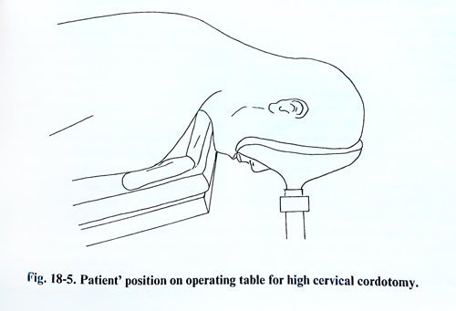
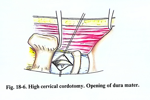
The percutaneous technique of high cervical cordotomy may be preferable, but the open method also can be used safely. The operation is performed under general anaesthesia, but local anaesthesia with sedation can also be used. With the patient in a prone position and the head well-fixed, an incision is made in the nuchal area, beginning 2 cm above the external occipital protuberance and reaching the level of the 4th §th cervical vertebrae (Fig. 18-5). In a unilateral cordotomy the muscles are separated from the laminae and the spinous process of the second cervical vertebrae. The flavum ligament between the first and the second vertebrae is removed. In this region there are usually a relatively thick extradural veins, which can be coagulated if rupture. Next, a self-retaining retractor is placed transversally to the muscles and a second one – vertically for effective exposure between the first and second cervical vertebrae. The second retractor is placed between the external occipital protuberance and the second spinous process. This makes possible the maximum widening of the space between the first and second vertebrae (Fig. 18-6).
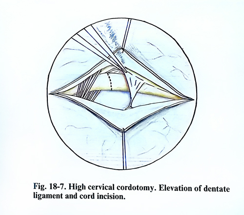
The dura is opened with a vertical incision in the middle of the opening of the spinal canal and the two edges are lifted with traction threads. Through the intact arachnoid the dentate ligament and the roots of the 2nd cervical nerve are clearly seen. The arachnoid is opened and the CSF aspirated. After evacuation of the CSF, the roots of the 2nd cervical nerve and the dentate ligament are more clearly seen. The latter is divided, as it serves as a brake, when rotation of the spinal cord is made. The posterior root is caught with a blunt hook and elevated until the antero-lateral surface of the spinal cord and the anterior root of the 2nd cervical nerve are clearly exposed. There are large variations in the disposition of the blood vessels in this area, but a small zone without blood vessels can always be found, where the incision is to be made. A slightly curved knife is used, which is 4.5 - 5 mm long and 2 mm broad. The tip of the scalpel is placed as near as possible to the attachment of the dentate ligament on the surface of the spinal cord. After this, an incision is made till 4 - 5 mm anterior to the attachment of the dentate ligament which does not penetrate medially to the place of origin of the anterior root (Fig. 18-7). After the incision has been made, the existence of analgesia is checked and, if the level is not sufficiently high, the incision may be repeated a little deeper until the desired level is reached. When complete analgesia is not obtained on the lumbar region, this iindicates that the incision has not reached the place of attachment of the denticulate ligament. Bleeding may appear from the edges of the incision. It stops after a light compression with a cottonoid.
After establishing that the incision of the cord is sufficient, the arachnoid space is filled with saline. The retractors are removed and the space between the laminae normalised. The operative wound is sutured in the conventional way.
PERCUTANEOUS HIGH CERVICAL CORDOTOMY
The percutaneous high cervical cordotomy has the same indications as does open cordotomy. The operation is performed under local anaesthesia and general sedation. In a cooperative patient this anaesthesia enables the patient to lie comfortably during the 45 minutes to 2 hours that the procedure requires. In some periods analgesia should be at a deeper level without losing the capability of repeatedly awakening the patient for physiologic testing. The patient should be conscious and able to give correct answers. Children and uncooperative patients may be successfully operated on under general anaesthesia.
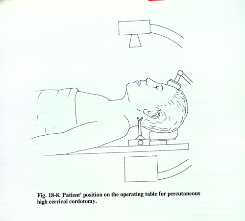
The patient is in a supine position on the operative table. The upper part of the cervical spine should be horizontal to trap contrast medium in the operative area. The patient's head and neck should be placed in a strictly anteroposterior position to permit the electrode introduction as precisely as possible. Radiological control is necessary to provide a true lateral projection of the upper cervical spine and an A-P view of the region of the odontoid process. A special stand is put on the side for the needle in the form of a small fork, which is fixed at the level of the base of the auricle (Fig. 18-8). The needle through which the electrode is to be introduced is positioned strictly horizontally to reach the skin at the centre between C1 - C2 space as seen in the lateral projection on the screen.
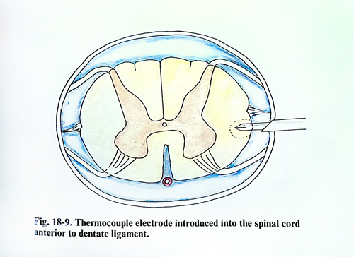
After the soft tissues have been injected with local anaesthetic, the needle penetrates horizontally, until a sense of resistance is felt by the surgeon and a twinge of pain by the patient. First the ligamentum flavum, and then the dura, are sequentially penetrated. An attempt to infiltrate these structures with local anaesthetic is undesirable because of the risk of accidentally producing a high spinal anaesthesia. The needle is advanced cautiously until, on withdrawal of the stylet, a flow of CSF occurs. Once this happens, the needle must not be advanced further, and several cubic centimetres of positive contrast medium, such as that used for myelography, are shaken with a similar quantity of CSF and injected into the subarachnoid space. The contrast outlines the subarachnoid space anterior and posterior to the spinal cord and, most importantly, allows the dentate ligament to be seen. An electrode is inserted through the needie, which through the micromanipulator of the needle guide must enter toward the anterior margin of the dentate ligament under radiological and impedance control, 2 - 3 mm behind the anterior outline of the spinal cord on the lateral projection. In the anteroposterior projection the tip of electrode must reach 4 mm laterally from the midline, passing through a level at the the middle of the odontoid process of the 2nd cervical vertebra (Fig. 18-9). Once the needle has appropriately penetrated the cord at the anterior margin of the dentate ligament, its position in the lateral spinothalamic tract must be confirmed. This can be done in different ways. Impedance monitoring can show a sharp rise from approximately. 400 to 500 ohms characteristic of CSF to over 1000 ohms when the tip of the electrode is within the spinal cord. Electrostimulation can also be applied, but the best method is clinical confirmation during progressive coagulation. A minimal lesion is produced with a 25 mA flow for at least 20 - 40 seconds during which current is delivered. It is best to start with a minimal level, assessing the results at the end of each 60 seconds of current flow until the desired level of analgesia is achieved or a current fall occurs. If analgesia is still inadequate when fall-off occurs, the electrode may be advanced minimally deeper into the cord and the lesion repeated. If the lesion is performed with the patient conscious the ipsilateral leg power is simultaneously tested.
Special postoperative care is usually not required. The patient is kept in bed rest for 24 hours against post-puncture headache. Getting out of bed is permitted the next day, but with an accompanying person, as ataxia or weakness is frequently observed in the first days of the postoperative period.
There are comparatively rare complications. In about 5% of the cases hemiparesis may be observed on the side of the operation. it disappears within 24 days. Ataxia is more frequent and is usually transitory. It may remain in not more than 3% as a lasting complication. Sphincter troubles that need catheterization of the bladder are met in 10%. Dysesthesia in the area of analgesia is encountered in 1%. Respiration disturbances are also observed when the cordotomy is made bilaterally. Their characteristic is that when the patient is awake the respiration is normal, but when falling asleep he usually lapses into apnoea. Such serious disturbances may need intubation and ventilation. This complication usually passes in some days or after up to 3 weeks.
After the cordotomy, the pain disappears and analgesics are not necessary in about 90% of the cases. However within about 3 months, the percentage of excellent results falls to 84%. At the end of the first year about 40% once again have a pain syndrome, which of course, is not the same as the previous one. At the end of the second year the percentage with good results falls to 50%.
COMMISSUROTOMY
Commissurotomy consists of interruption of fibres, transmitting the pain and temperature sensations at the place where they pass through the anterior commissure. The operation has the advantage that it provokes analgesia on both sides. The analgesia is spread in the area along the dermatomes, corresponding to the segments where the incision of the spinal cord has been made.
The indications for this operation are similar to those of antero-lateral cordotomy, but the extension of the pain syndrome should not exceed the region by more than 3 - 4 dermatomes, as it would not be possible to make an incision longer than the level of 3 - 4 spinal cord segments. It is not advisable to perform this operation in the cervical region or in the conus medullaris, owing the to risks of provoking serious neurological deficit.
SURGICAL TECHNIQUE
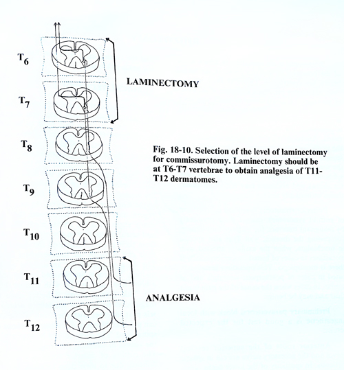
A standard laminectomy is made at the level of the segments where the commissurotomy has to be made. To determine the level of the laminectomy, it must be kept in mind that the tract transmitting the pain and temperature sensation crosses at two to three segments above the place where the posterior roots for the corresponding segment enter the spinal cord. Besides this consideration the relationship should be considered the correspondence between the spinal cord segments and the vertebrae. For instance, if by a commissurotomy analgesia has to be obtained about dermatomes T11 and T12, one must take into account that it must be made in the segments and the roots located 3 vertebrae above the respective vertebrae, i.e. at the level of the T8 - T9 vertebrae. The tracts that transmit the pain and temperature sensations cross 2 segments above and therefore the laminectomy and the incision of the spinal cord must be made at the level of the T6 - T7 vertebrae (Fig. 18-10). After the dura and the arachnoid are opened, the median fissure along the posterior surface of the spinal cord must be localised and an incision must be made along the midline 8 mm deep. It is performed with a very thin and sharp scalpel (Fig. 18-11).
After this operation, complete analgesia on the dermatomes is obtained in 60% of the patients; in 20 -30% there is diminished pain and in 10% no favour-able effect. The possible complications are some sphincter disturbances, weakness of the lower limbs and dysesthesia.
RHIZOTOMY
Rhizotomy is an operation in which some of the posterior or anterior roots of the spinal nerves are interrupted. Different from cordotomy, rhizotomy provokes analgesia of all kinds of sensation. Complete loss of touch and deep sensation of the limbs leads to serious difficulties in their function. Even though the muscle power is preserved, the movements remain uncoordinated. Anaesthesia or even hemianaesthesia of the penis leads to impotence, if the roots of the second lumbar nerve are interrupted. Cutting of the second and third sacral roots is followed by sphincter incontinence. However, wide areas of the neck, the thorax and abdomen may remain in anaesthesia without any serious complaints.
Posterior rhizotomy is applied in treatment of painful syndromes, caused most frequently by malignant tumours, for instance lung cancer, involving the thoracic wall. It is also applied in brachialgia, when the axilla is affected with metastases in cases of breast cancer, especially if there is monoplegia. This operation is not indicated in cases of postherpetic neuralgia and of course, in cases when the cause of the pain is not clear and may be benign.
Preliminary paravertebral block with local anaesthetic is a good test for the expected results.
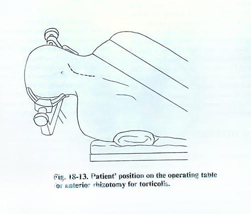
Anterior roots of the superior cervical nerves and the accessory nerve are cut in spastic torticollis. In spasticity of the lower limbs, anterior roots at the level of the laminae T12 - L1 can be interrupted. Of every 5 roots, 4 are cut and the cutting ends with the disappearance of the ankle jerk reflex.
SURGICAL TECHNIQUE
The spinal roots are exposed by a strandard laminectomy whose extension depends on the number of roots that need to be cut. The laminectomy level is determined very carefully, taking into consideration the existing relations between the spinal cord segment and the vertebrae. One must bear in mind also the disposition and length of the roots in the different regions of the spinal canal. In the cervical region they are horizontally located and form an arc with slight convexity downward. In the thoracic region their length is larger and they appear at an acute angle to the spinal cord. The lumbar and sacral roots have an almost vertical location from their place of origin on the spinal cord to their exit through the intervertebral foramina.
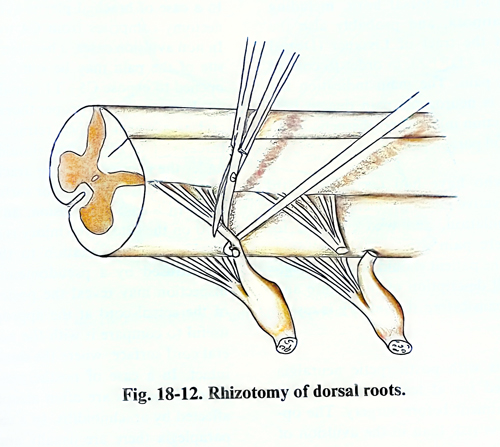
The dura is opened by an incision along the midline. The roots are elevated one by one with a blunt hook and are cut with scissors (Fig. 19-12). This manipulation is better done under magnification, because sometimes arterial vessels pass together with the roots, whose cutting can provoke bleeding or ischaemic lesions of the spinal cord. The roots should be separated from the vessels in advance; the vessels are left intact.
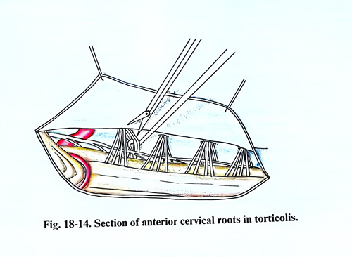
The cutting of anterior roots is a little more difficult, due to their deep location, but cutting the dentate ligaments in advance facilitates this (Figs. 18-13; 18-14).
DORSAL ROOT ENTRY ZONE LESIONS
This method is based on the lesion of the posterior part of the dorsal horn, including substantia gelatinosa, and probably also the medial part of the tract of Lissauer (Dorsal Root Entry Zone - DREZ, in order to control deafferentation pain. The main indication for this procedure is neurogenic pain due to brachial plexus traction injuries, postherpetic neuralgia, and painful paraplegia. Patients who have had treatment with different conservative methods, including transcutaneous stimulation, and who continue to experience severe pain are candidates for a DREZ lesion. The patient should be psychologically stable. Full description of the nature and severity of the pain before the surgery is essential.
The patients with postherpetic neuralgia should be treated for at least one year with conservative treatment before surgery. The operation carries less risk than in the avulsion of the brachial plexus.
Patients with a spinal cord injury may experience chronic pain at or around the site of the injury due to spinal instability or some other mechanical factor in the bones and the joints. Stabilising procedures or facet injections can treat this pain. Another kind of pain is that of central origin experienced in anaesthetic areas, consisting of burning and tingling feelings. This type of pain and the pain triggered by touch experienced in the anaesthetic areas can be tingling or lancinating in nature. In such cases a DREZ lesion at 2 or 3 segments above the spinal cord injury may be beneficial.
SURGICAL TECHNIQUE
The operation is performed with the patient in a prone position and under general anaesthesia. In patients with brachial plexus avulsion it is possible to determine the spinal level accurately by palpating the prominent C7 vertebra. X-ray control is essential in the dorsal region and at the level of the conus medullaris.
In a case of brachial plexus avulsion, the laminectomy comprises from C4 to TI inclusively. In non-avulsion cases, a hemilaminectomy at the site of the pain may be sufficient. The dura is opened to expose C5 - TI spinal cord segments, or alternatively, the proper thoracic or lumbosacral area which is then retracted with traction sutures. The next stage should be performed under the microscope. The arachnoid is opened to expose clearly the posterior surface of the spinal cord. In a case of avulsion, an atrophy of the cord on the side of the injury is seen, often with displacement or rotation to the contralateral side caused by a pseudomeningocele. Visual inspection may reveal the posterolateral sulcus of the spinal cord at the appropriate site. It is useful to compare it with the normal contralateral cord surface, where the dorsal entry zone is intact. In a case of postherpetic neuralgia the affected rootlets are often atrophic and slightly affected by arachnoiditis. In a case of traumatic paraplegia there are usually arachnoidal adhesions expressed at and above the level of the injured cord.
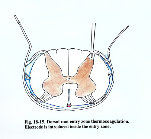
Once the sulcus between the posterior and lateral columns has been identified, it is possible to proceed with the lesion. The rootlets in the region of interest are retracted gently toward the midline to allow visualisation of the DREZ. The scalpel is inserted to a depth of approximately 2 mm at a 30 - 45 degree angle to the horizontal plane. The incision is made along the entire extent of the DREZ at the segment of interest.
An interval of 2 - 3 mm is skipped between lesions of each spinal segment to preserve the integrity of the cord. Any bleeding from the incision can be controlled by bipolar microcoagulation. The lesion may be made by the radio-frequency thermocoagulation - a method developed by Nashold et al. According to this method a thermocouple electrode with a 0.25 mm diameter is introduced 2 mm into the cord parenchyma at an angle of 25 degrees to a vertical plane through the posterolateral sulcus (Fig. 18 - 15). The current starts from 0 and gradually increases to reach a temperature of 70-75 degrees centigrade constantly for 15 seconds. Than it is switched off and the electrode is removed. Prior to making a lesion in the cord, the impedance in the system should be tested, checking that it is about 450 to 700 ohms and making sure that all the electrical connections are properly cleaned. In a case of brachial plexus avulsion, such lesions are done from C5 to Tl, and 20 - 24 of them are placed 2 mm apart. To reach the temperature of 70 - 75 degrees, 45 mA of current is usually required, whereas in the dorsal region it may be only 25 mA. In the dorsal region, for postherpetic neuralgia, or in a case of paraplegia, fewer lesions may be necessary.
If bleeding occurs after puncture of the cord, this will stop after application of oxidised cellulose. It is usually possible to avoid bleeding by insuring under the microscope that the electrode is not passed through significant vessels on the cord surface. Complete control of pain is possible in brachial plexus avulsion in approximately one third of patients, and pain improvement in another one third. Because of the pain from the laminectomy wound and postoperative sedation, only few days after surgery is possible to evaluate the exact degree of pain relief.
Temporary neurological deficit can be observed in 50% of the patients, although with the improvement in electrode technology and lesion making the percentage has been reduced. In about 5 to 10% of the patients there will be some permanent neurological complication such as weakness and ataxia in the ipsilateral lower limb accompanied by sensory loss in the upper part of the trunk on the side of the lesion.