RHIZOTOMY
Rhizotomy is an operation in which some of the posterior or anterior roots of the spinal nerves are interrupted. Different from cordotomy, rhizotomy provokes analgesia of all kinds of sensation. Complete loss of touch and deep sensation of the limbs leads to serious difficulties in their function. Even though the muscle power is preserved, the movements remain uncoordinated. Anaesthesia or even hemianaesthesia of the penis leads to impotence, if the roots of the second lumbar nerve are interrupted. Cutting of the second and third sacral roots is followed by sphincter incontinence. However, wide areas of the neck, the thorax and abdomen may remain in anaesthesia without any serious complaints.
Posterior rhizotomy is applied in treatment of painful syndromes, caused most frequently by malignant tumours, for instance lung cancer, involving the thoracic wall. It is also applied in brachialgia, when the axilla is affected with metastases in cases of breast cancer, especially if there is monoplegia. This operation is not indicated in cases of postherpetic neuralgia and of course, in cases when the cause of the pain is not clear and may be benign.
Preliminary paravertebral block with local anaesthetic is a good test for the expected results.
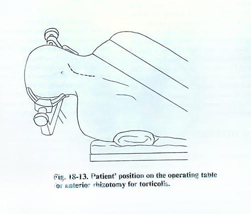
Anterior roots of the superior cervical nerves and the accessory nerve are cut in spastic torticollis. In spasticity of the lower limbs, anterior roots at the level of the laminae T12 - L1 can be interrupted. Of every 5 roots, 4 are cut and the cutting ends with the disappearance of the ankle jerk reflex.
SURGICAL TECHNIQUE
The spinal roots are exposed by a strandard laminectomy whose extension depends on the number of roots that need to be cut. The laminectomy level is determined very carefully, taking into consideration the existing relations between the spinal cord segment and the vertebrae. One must bear in mind also the disposition and length of the roots in the different regions of the spinal canal. In the cervical region they are horizontally located and form an arc with slight convexity downward. In the thoracic region their length is larger and they appear at an acute angle to the spinal cord. The lumbar and sacral roots have an almost vertical location from their place of origin on the spinal cord to their exit through the intervertebral foramina.
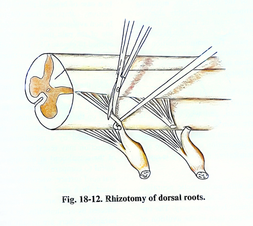
The dura is opened by an incision along the midline. The roots are elevated one by one with a blunt hook and are cut with scissors (Fig. 19-12). This manipulation is better done under magnification, because sometimes arterial vessels pass together with the roots, whose cutting can provoke bleeding or ischaemic lesions of the spinal cord. The roots should be separated from the vessels in advance; the vessels are left intact.
The cutting of anterior roots is a little more difficult, due to their deep location, but cutting the dentate ligaments in advance facilitates this (Figs. 18-13; 18-14).