DORSAL ROOT ENTRY ZONE LESIONS
This method is based on the lesion of the posterior part of the dorsal horn, including substantia gelatinosa, and probably also the medial part of the tract of Lissauer (Dorsal Root Entry Zone - DREZ, in order to control deafferentation pain. The main indication for this procedure is neurogenic pain due to brachial plexus traction injuries, postherpetic neuralgia, and painful paraplegia. Patients who have had treatment with different conservative methods, including transcutaneous stimulation, and who continue to experience severe pain are candidates for a DREZ lesion. The patient should be psychologically stable. Full description of the nature and severity of the pain before the surgery is essential.
The patients with postherpetic neuralgia should be treated for at least one year with conservative treatment before surgery. The operation carries less risk than in the avulsion of the brachial plexus.
Patients with a spinal cord injury may experience chronic pain at or around the site of the injury due to spinal instability or some other mechanical factor in the bones and the joints. Stabilising procedures or facet injections can treat this pain. Another kind of pain is that of central origin experienced in anaesthetic areas, consisting of burning and tingling feelings. This type of pain and the pain triggered by touch experienced in the anaesthetic areas can be tingling or lancinating in nature. In such cases a DREZ lesion at 2 or 3 segments above the spinal cord injury may be beneficial.
SURGICAL TECHNIQUE
The operation is performed with the patient in a prone position and under general anaesthesia. In patients with brachial plexus avulsion it is possible to determine the spinal level accurately by palpating the prominent C7 vertebra. X-ray control is essential in the dorsal region and at the level of the conus medullaris.
In a case of brachial plexus avulsion, the laminectomy comprises from C4 to TI inclusively. In non-avulsion cases, a hemilaminectomy at the site of the pain may be sufficient. The dura is opened to expose C5 - TI spinal cord segments, or alternatively, the proper thoracic or lumbosacral area which is then retracted with traction sutures. The next stage should be performed under the microscope. The arachnoid is opened to expose clearly the posterior surface of the spinal cord. In a case of avulsion, an atrophy of the cord on the side of the injury is seen, often with displacement or rotation to the contralateral side caused by a pseudomeningocele. Visual inspection may reveal the posterolateral sulcus of the spinal cord at the appropriate site. It is useful to compare it with the normal contralateral cord surface, where the dorsal entry zone is intact. In a case of postherpetic neuralgia the affected rootlets are often atrophic and slightly affected by arachnoiditis. In a case of traumatic paraplegia there are usually arachnoidal adhesions expressed at and above the level of the injured cord.
Once the sulcus between the posterior and lateral columns has been identified, it is possible to proceed with the lesion. The rootlets in the region of interest are retracted gently toward the midline to allow visualisation of the DREZ. The scalpel is inserted to a depth of approximately 2 mm at a 30 - 45 degree angle to the horizontal plane. The incision is made along the entire extent of the DREZ at the segment of interest.
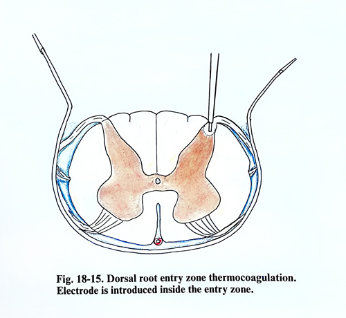
An interval of 2 - 3 mm is skipped between lesions of each spinal segment to preserve the integrity of the cord. Any bleeding from the incision can be controlled by bipolar microcoagulation. The lesion may be made by the radio-frequency thermocoagulation - a method developed by Nashold et al. According to this method a thermocouple electrode with a 0.25 mm diameter is introduced 2 mm into the cord parenchyma at an angle of 25 degrees to a vertical plane through the posterolateral sulcus (Fig. 18 - 15). The current starts from 0 and gradually increases to reach a temperature of 70-75 degrees centigrade constantly for 15 seconds. Than it is switched off and the electrode is removed. Prior to making a lesion in the cord, the impedance in the system should be tested, checking that it is about 450 to 700 ohms and making sure that all the electrical connections are properly cleaned. In a case of brachial plexus avulsion, such lesions are done from C5 to Tl, and 20 - 24 of them are placed 2 mm apart. To reach the temperature of 70 - 75 degrees, 45 mA of current is usually required, whereas in the dorsal region it may be only 25 mA. In the dorsal region, for postherpetic neuralgia, or in a case of paraplegia, fewer lesions may be necessary.
If bleeding occurs after puncture of the cord, this will stop after application of oxidised cellulose. It is usually possible to avoid bleeding by insuring under the microscope that the electrode is not passed through significant vessels on the cord surface. Complete control of pain is possible in brachial plexus avulsion in approximately one third of patients, and pain improvement in another one third. Because of the pain from the laminectomy wound and postoperative sedation, only few days after surgery is possible to evaluate the exact degree of pain relief.
Temporary neurological deficit can be observed in 50% of the patients, although with the improvement in electrode technology and lesion making the percentage has been reduced. In about 5 to 10% of the patients there will be some permanent neurological complication such as weakness and ataxia in the ipsilateral lower limb accompanied by sensory loss in the upper part of the trunk on the side of the lesion.