HIGH THORACIC CORDOTOMY
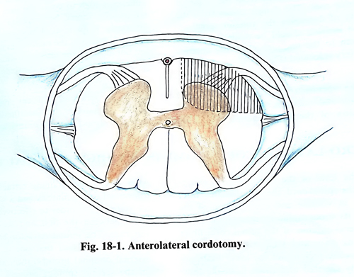
Cordotomy is based on the concept that the spinothalamic tract, located in the anterolateral quadrant of the spinal cord, transmits the pain and temperature sensations (Fig. 18-1).
Cordotomy is indicated in patients with cancer pain, when the expected survival period is not more than 3 years. This condition is mandatory because the recurrence rate of pain is very high and after this period painful dysesthesia increases. The high percentage of postoperative complications such as weakness, sphincter and sexual disturbances is another argument against applying this operation frequently. After cordotomy the level of analgesia drops caudally in the first 3 weeks postoperatively with 3 - 6 dermatomes and after 6 months another 3-6 dermatomes further down. That is why, with a cordotomy done at the level of 2-3 thoracic vertebrae, the level of permanent hypoalgesia or analgesia usually is at about the 10th thoracic dermatome. Cordotomy should be contraindicated in benign diseases such as spondylotic pain, postherpetic neuralgia, causalgia, different forms of neuropathies and neuritis, phantom pain and particularly when the cause of the pain syndrome is unknown.
The spinothalamic tract which transmits pain and temperature sensation crosses the midline, and the operation should be done on the opposite side to that of the pain.
Unilateral cordotomy is most effective in patients with cancer pain, when the internal organs are not involved and when the cancer is situated out of the midline and below the lower thoracic area. The best candidates for high thoracic cordotomy are those patients with cancer involving the lower limbs, buttocks, and the lateral retroperitoneal area inside the pelvis, abdomen, and the chest wall.
When the cancer is near to the midline, unilateral cordotomy cannot offer long prolonged relief of pain. In similar cases the cordotomy must be bilateral, but the second procedure should be done at a different level and it is preferable that one of the two cordotomies be percutaneous.
SURGICAL TECHNIQUE
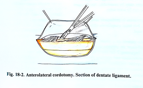
The open cordotomy is performed at the level of the 2nd d-3rd thoracic vertebrae. The patient is in a prone position on the operating table. General anaesthesia is usually administered, but local anaesthesia with sedation permits control of any provoked analgesia and prevents some complications during the operation. The laminectomy is performed in the conventional way with the opening of two laminae. The dura mater is opened with an arch-form incision with its convexity on the side of the planned cordotomy. If possible, bipolar electrodes are placed within the epidural space in the midline rostrally and caudally for recording sensory evoked potentials from bilateral peroneal stimulation. After that the operation is done under optical magnification. The width of the spinal cord is measured precisely, and using a piece of wax the scalpel is marked in such a way that the cutting part of the scalpel is only half the width of the spinal cord. The dentate ligaments are identified on both sides and their attachments to the dura, on the side of the cordotomy, are cut. The arachnoid is torn on the surface on the side where the cordotomy is to be made. One of the dentate ligaments is caught with traction thread using a clip (Figs.18-2; 18-3). The spinal cord can be rotated by 45 degrees by pulling the traction tread. If the dentate ligament is not sufficiently strong it may be torn during the rotation. In such a case, traction threads are introduced under the anterior roots to establish some rotation, which should permit the anterior spinal artery to be seen. For this purpose a small dental mirror can be used. A small perforation is first made in the pia mater immediately anterior to the place of attachment of the dentate ligament and after that the scalpel is driven into the necessary depth up to 4 - 5 mm to where the piece of wax is limiting its entry. If the pia mater is not perforated before, an incision is made so, that the dentate ligament may not be torn with a wrong movement and the incision surpasses the required limits. After the scalpel enters to the sufficient depth of 4 - 5 mm, it is taken out anteriorly, its tip having to come out 1-2 mm medially from the origin of the anterior root. A utmost care should be taken of not damaging the anterior spinal artery. The incision should be made not closer that 2 mm from the midline, where the anterior spinal artery is usually located (Fig. 18-4).
If the operation is carried out with local anaesthesia and sedation, in the beginning of the incision of the spinothalamic tract, the patient should be awakened to verify the area of analgesia and the motor function of the lower limbs. If the level of provoked analgesia is not sufficiently high to cover the area of the pain, the incision should be deepened and enlarged anteriorly. If the level of analgesia is not sufficiently caudal and the sacral area and perineum are not affected, the incision is enlarged toward the transverse diameter of the spinal cord. After obtaining sufficient analgesia in the desired dermatomes, the anaesthesia is deepened to permit closing of the operative wound.