PERCUTANEOUS HIGH CERVICAL CORDOTOMY
The percutaneous high cervical cordotomy has the same indications as does open cordotomy. The operation is performed under local anaesthesia and general sedation. In a cooperative patient this anaesthesia enables the patient to lie comfortably during the 45 minutes to 2 hours that the procedure requires. In some periods analgesia should be at a deeper level without losing the capability of repeatedly awakening the patient for physiologic testing. The patient should be conscious and able to give correct answers. Children and uncooperative patients may be successfully operated on under general anaesthesia.
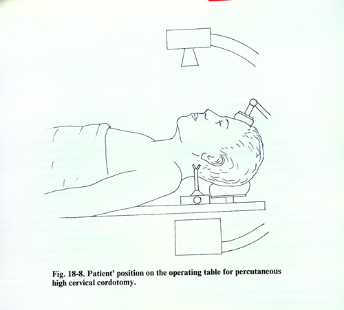
The patient is in a supine position on the operative table. The upper part of the cervical spine should be horizontal to trap contrast medium in the operative area. The patient's head and neck should be placed in a strictly anteroposterior position to permit the electrode introduction as precisely as possible. Radiological control is necessary to provide a true lateral projection of the upper cervical spine and an A-P view of the region of the odontoid process. A special stand is put on the side for the needle in the form of a small fork, which is fixed at the level of the base of the auricle (Fig. 18-8). The needle through which the electrode is to be introduced is positioned strictly horizontally to reach the skin at the centre between C1 - C2 space as seen in the lateral projection on the screen.
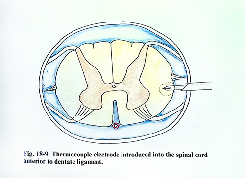
After the soft tissues have been injected with local anaesthetic, the needle penetrates horizontally, until a sense of resistance is felt by the surgeon and a twinge of pain by the patient. First the ligamentum flavum, and then the dura, are sequentially penetrated. An attempt to infiltrate these structures with local anaesthetic is undesirable because of the risk of accidentally producing a high spinal anaesthesia. The needle is advanced cautiously until, on withdrawal of the stylet, a flow of CSF occurs. Once this happens, the needle must not be advanced further, and several cubic centimetres of positive contrast medium, such as that used for myelography, are shaken with a similar quantity of CSF and injected into the subarachnoid space. The contrast outlines the subarachnoid space anterior and posterior to the spinal cord and, most importantly, allows the dentate ligament to be seen. An electrode is inserted through the needie, which through the micromanipulator of the needle guide must enter toward the anterior margin of the dentate ligament under radiological and impedance control, 2 - 3 mm behind the anterior outline of the spinal cord on the lateral projection. In the anteroposterior projection the tip of electrode must reach 4 mm laterally from the midline, passing through a level at the the middle of the odontoid process of the 2nd cervical vertebra (Fig. 18-9). Once the needle has appropriately penetrated the cord at the anterior margin of the dentate ligament, its position in the lateral spinothalamic tract must be confirmed. This can be done in different ways. Impedance monitoring can show a sharp rise from approximately. 400 to 500 ohms characteristic of CSF to over 1000 ohms when the tip of the electrode is within the spinal cord. Electrostimulation can also be applied, but the best method is clinical confirmation during progressive coagulation. A minimal lesion is produced with a 25 mA flow for at least 20 - 40 seconds during which current is delivered. It is best to start with a minimal level, assessing the results at the end of each 60 seconds of current flow until the desired level of analgesia is achieved or a current fall occurs. If analgesia is still inadequate when fall-off occurs, the electrode may be advanced minimally deeper into the cord and the lesion repeated. If the lesion is performed with the patient conscious the ipsilateral leg power is simultaneously tested.
Special postoperative care is usually not required. The patient is kept in bed rest for 24 hours against post-puncture headache. Getting out of bed is permitted the next day, but with an accompanying person, as ataxia or weakness is frequently observed in the first days of the postoperative period.
There are comparatively rare complications. In about 5% of the cases hemiparesis may be observed on the side of the operation. it disappears within 24 days. Ataxia is more frequent and is usually transitory. It may remain in not more than 3% as a lasting complication. Sphincter troubles that need catheterization of the bladder are met in 10%. Dysesthesia in the area of analgesia is encountered in 1%. Respiration disturbances are also observed when the cordotomy is made bilaterally. Their characteristic is that when the patient is awake the respiration is normal, but when falling asleep he usually lapses into apnoea. Such serious disturbances may need intubation and ventilation. This complication usually passes in some days or after up to 3 weeks.
After the cordotomy, the pain disappears and analgesics are not necessary in about 90% of the cases. However within about 3 months, the percentage of excellent results falls to 84%. At the end of the first year about 40% once again have a pain syndrome, which of course, is not the same as the previous one. At the end of the second year the percentage with good results falls to 50%.