ANTERO-LATERAL CORDOTOMY IN THE HIGH CERVICAL REGION
At the level of the superior cervical segments of the spinal cord, the spinothalamic tract is greater in volume and every region of the body has its topographic representation in the antero-lateral column of the spinal cord: fibres transmitting pain and temperature sensations from the pelvic region and the lower limbs are superficially situated and those from the thoracic region lie deeper and medially. This topographic disposition and the large volume, which the spinothalamic tract occupies, permit a selective section, depending on the painful area.
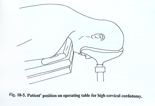
The percutaneous technique of high cervical cordotomy may be preferable, but the open method also can be used safely. The operation is performed under general anaesthesia, but local anaesthesia with sedation can also be used. With the patient in a prone position and the head well-fixed, an incision is made in the nuchal area, beginning 2 cm above the external occipital protuberance and reaching the level of the 4th §th cervical vertebrae (Fig. 18-5). In a unilateral cordotomy the muscles are separated from the laminae and the spinous process of the second cervical vertebrae. The flavum ligament between the first and the second vertebrae is removed. In this region there are usually a relatively thick extradural veins, which can be coagulated if rupture. Next, a self-retaining retractor is placed transversally to the muscles and a second one – vertically for effective exposure between the first and second cervical vertebrae. The second retractor is placed between the external occipital protuberance and the second spinous process. This makes possible the maximum widening of the space between the first and second vertebrae (Fig. 18-6).
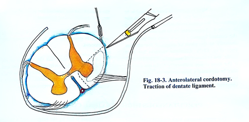
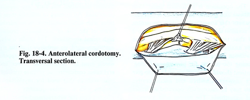
The dura is opened with a vertical incision in the middle of the opening of the spinal canal and the two edges are lifted with traction threads. Through the intact arachnoid the dentate ligament and the roots of the 2nd cervical nerve are clearly seen. The arachnoid is opened and the CSF aspirated. After evacuation of the CSF, the roots of the 2nd cervical nerve and the dentate ligament are more clearly seen. The latter is divided, as it serves as a brake, when rotation of the spinal cord is made. The posterior root is caught with a blunt hook and elevated until the antero-lateral surface of the spinal cord and the anterior root of the 2nd cervical nerve are clearly exposed. There are large variations in the disposition of the blood vessels in this area, but a small zone without blood vessels can always be found, where the incision is to be made. A slightly curved knife is used, which is 4.5 - 5 mm long and 2 mm broad. The tip of the scalpel is placed as near as possible to the attachment of the dentate ligament on the surface of the spinal cord. After this, an incision is made till 4 - 5 mm anterior to the attachment of the dentate ligament which does not penetrate medially to the place of origin of the anterior root (Fig. 18-7). After the incision has been made, the existence of analgesia is checked and, if the level is not sufficiently high, the incision may be repeated a little deeper until the desired level is reached. When complete analgesia is not obtained on the lumbar region, this iindicates that the incision has not reached the place of attachment of the denticulate ligament. Bleeding may appear from the edges of the incision. It stops after a light compression with a cottonoid.
After establishing that the incision of the cord is sufficient, the arachnoid space is filled with saline. The retractors are removed and the space between the laminae normalised. The operative wound is sutured in the conventional way.