
Legacy: "Atlast of Neurosurgery" / L.Karaguiosov, A. Ramadan, K.Karaguiosov / Kiwait/ 1998
Read PREVIOUS > 20. Cranial Nerves
BRACHIAL PLEXUS
Injuries of the brachial plexus most often occur after open or closed trauma or gunshot injuries below and above the clavicle, first rib, and scapular fractures, or dislocation of the shoulder joint. A frequent type injury of the brachial plexus is by its traction, which leads to avulsion of its roots from the spinal cord. This injury occurs as a result of strong and sudden traction of the upper limb or impacts on the shoulder region.
Injuries of the brachial plexus occur also during delivery, when the newborn has been pulled out by the upper limb.
Aneurysms of the subclavian artery and tumours in the subclavian region are considered comparatively rare causes of injury to the brachial plexus. Sometimes the brachial plexus is damaged by a cervical rib, by stenosis between the scalene muscles, through which the plexus roots pass or by compression by the subclavian muscle below the clavicle.
SURGICAL ANATOMY
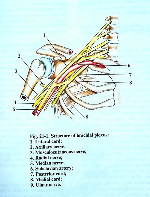
The brachial plexus is formed by the anterior branches of C5 - C8 and Tl spinal nerves which are roots of the brachial plexus. In approximately half of the cases, the C4 spinal nerve is included, while in others - it is the T2 nerve. In this way the brachial plexus is in some cases formed by inclusion of C4 - T1, in others by C5 - T2. The first is designated as of plexus superior type, while the second - as of inferior type. The anterior branch of the C5 spinal nerve joins the C6 at a sharp angle, forming the superior trunk of the plexus. The anterior branch of the C7 spinal nerve remains independent and forms the middle trunk, while the C8, and Tl merge in the inferior plexus trunk. The three trunks come out of interscalenus slit and continue downward anteriorly and laterally, reaching the superior surface of the first rib (Fig. 21-1). Every trunk is divided into an anterior and posterior branch, which pass below the middle part of the clavicle and enter the axillary cavity. The anterior and posterior branches of the three trunks form three cords in the axillary cavity: the lateral, medial and posterior cords, which envelop the axillary artery.
The lateral cord is formed by the union of the anterior branches of the superior and middle trunks and contains fibres from the CS, C6, and C7 spinal nerves. The medial cord is a continuation of the anterior branch of the inferior trunk and therefore contains fibres from the C7 and Tl spinal nerves. The posterior cord is formed from the posterior branches of the three trunks and consequently contains fibres of all nerves, which form the brachial plexus. There are variations in the formation of the plexus. Sometimes the brachial plexus is formed simply - without the formation of trunks. The roots of the plexus are divided laterally from the spinal column into anterior and posterior branches. The posterior branches of all plexus roots are united in the posterior cord. The anterior branches of the CS, Co and C7 roots are merged in the lateral cord, while those of the C8 and Tl merge in the medial cord.
The entire brachial plexus extends from the cervical spine to the distal region of the axillary cavity, i.e. to the level of the “surgical” neck of the humerus. The clavicle divides the plexus into the two parts: supraclavicular and infraclavicular. The first is situated in the supraclavicular fossa and comprises the roots, the trunks of the plexus, and their division into anterior and posterior branches. The infraclavicular part, which consists of the plexus cords, is situated in the axillary cavity.
At their point of exit from the spinal canal the plexus roots are situated in the sulci of the transverse processes of the cervical vertebrae. Anterior to the roots in a vertical direction are situated the vertebral artery and vein. The first thoracic nerve is situated immediately posterior to the stellate ganglion of the sympathetic chain. Just lateral from the spinal column, the brachial plexus is situated in the slit between the anterior and medial scalene muscles. The subclavian artery is situated in the inferior part of this slit anterior to the C8 and Tl nerves and their unification occurs in the inferior plexus trunk. If there is a well-developed scalenus minimus muscle, the artery remains separated from the plexus by it. In the supraclavicular fossa the brachial plexus is situated posterior to the sternomastoid muscle, omohyoid muscle, and the superior and middle fascias of the neck. The suprascapular artery goes anterior to the plexus and immediately above the clavicle, while the cervical transverse artery passes between the middle and superior plexus trunk.
The cords of the infraclavicular part of the brachial plexus are situated around the axillary artery in the axillary fossa. Lateral from the artery is found the lateral cord. The musculocutaneous nerve and the lateral branch of the median nerve separate from it. Medial from the artery is located the medial cord, from which separate the medial cutaneous nerve of the arm, the medial cutaneous nerve of the forearm, the ulnar nerve, and the medial branch of the median nerve. Posterior to the artery is the posterior cord of the brachial plexus, which is the thickest, and which divides at the level of the quadrangular foramen of the axilla into the axillary and radial nerves.
SURGICAL INDICATIONS
Traumatic lesions of brachial plexus do not always need surgical treatment. Open injuries, due to a sharp traumatic agent need urgent exploration and primary suturing. Lacerated wounds and gunshot injuries should be kept under observation. The surgical treatment is indicated after some weeks or 2 - 3 months, when clinically and electrophysiological investigations reveal any serious lesion. The closed injuries of brachial plexus also need surgical exploration after some period of observation. Surgical treatment is useless in a case of avulsion of brachial plexus roots from the spinal cord. When end to end suture is impossible, autotransplantation with sural nerve or neurotisation with intercostal nerves is indicated.
SURGICAL APPROACHES
Surgical approaches to the brachial plexus differ, depending on which part of the plexus must be reached. For limited injuries approaches of the plexus are used exposures to only pre-determined locations. In extensive injuries, the entire plexus must be exposed from its roots, situated lateral from the spine, to the origin of all nerves which separate from the plexus to the upper limb.
SURGICAL APPROACH TO THE BRACHIAL PLEXUS IN THE SUPRACLAVICULAR FOSSA
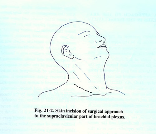
The patient is placed in a supine position on the operating table, a cushion being put under the shoulders, and the head is turned slightly to the opposite side of the operation site. The most suitable incision is the one coinciding with the skin creases in this region, i.e. the incision is oblique regarding the clavicle or almost to the half of the angle formed by the posterior border of the sternomastoid muscle and the clavicle (Fig. 21-2). Together with the skin and the subcutaneous tissue, the platysma, and the superficial cervical fascia are divided. The superficial jugular vein, which goes under the skin is also divided between ligatures or is retracted. After that the middle cervical fascia is exposed where the sleeve of the omohyoid muscle can be seen. This muscle is situated under the clavicle upon the brachial plexus; it should be freely retracted laterally and superiorly; the middle cervical fascia must be incised inferior to the muscle. If this does not ensure a sufficiently wide operative field, the muscle can be divided in the middle, at its tendon between its two bellies. The brachial plexus is revealed in the loose fatty and fibrous tissues under the fascia.
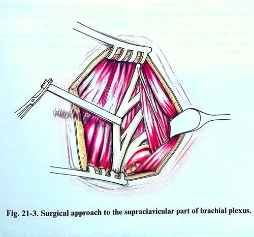
In seeking the nerve trunks it is best to reach the surface of the scalenus anterior muscle. The phrenic nerve is exposed on its anterior surface and should be preserved. Laterally from the scalenus muscle the C5, C6, C7, C8 and the T1 nerves are exposed, which are the roots of the brachial plexus. The anterior branches of the C5 and C6 spinal nerves go downward and forward and join into one common trunk (superior trunk). Immediately below them the anterior branch of C7 nerve is separated and it continues laterally and inferiorly as the middle trunk of the plexus. Deep behind the clavicle, the subclavian artery and vein are revealed, and behind them the anterior branches of C8 and TI nerves, which soon after their exit from the spinal canal join together as the inferior trunk of the plexus. Owing to their deep location their dissection is more difficult and it is necessary that the patient's shoulder be lowered as much as possible (Fig. 21-3).
In dissecting the roots and the trunks of the plexus one must have in mind that, superficially on the nerve trunks and parallel to the clavicle, pass the suprascapular artery and the superficial artery of the neck, while deep between the C6 and C7 nerves may pass the transverse artery of the neck. These arteries must be preserved, especially when the plexus injury is accompanied by injury of the subclavian or axillary arteries, as they serve the collateral blood circulation of the upper limb. Utmost attention is given to the preservation of the pleural cupola, internal jugular vein, and subclavian artery situated within the limits of the operative field, as even small damage leads to haemorrhage, which is difficult to stop due to the depth of the blood vessels. In this region the injury of veins may provoke an air embolism, if the body is too elevated. On this account the surgical manipulations should be performed under visual control and without the use of sharp instruments.
Depending on the pathological changes found, neurolysis or resection of the neuroma is made with subsequent suturing of the nerve trunks. The technique of neurolysis and suturing of the nerves does not differ from those on other peripheral nerves. For overcoming the nerve gaps when suturing, the patients head has to be turned to the injured side, and the shoulder must be elevated as high as possible.
SURGICAL APPROACH TO THE BRACHIAL PLEXUS IN THE INFRACLAVICULAR FOSSA
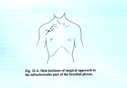
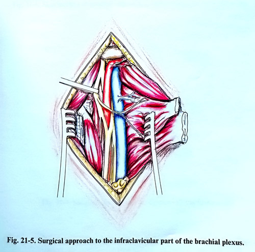
The patient's position on the operating table is the same as in the approach to the plexus in the supraclavicular region, however, the incision of the skin, the subcutaneous tissue, and the fascia is made in a different way. Most practical is the incision that begins from the middle of the clavicle and goes downward along the sulcus between the deltoid and major pectoral muscles (Fig. 21-4). An almost horizontal incision can be made, which begins 5 cm below the middle of the clavicle and proceeding downward at a slight arc and reaches the insertion of the major pectoral muscle. Despite the difference in the skin incisions, after opening the skin and the subcutaneous tissue, the dissection goes into the sulcus between the deltoid and the major pectoral muscle. The cephalic vein is in this sulcus and it can be retracted laterally. The insertion of the major pectoral muscle is elevated under its insertion, and the muscle is divided across near the humerus and lifted. After that it is retracted medially, taking care not to injure the branches of the brachial plexus and vessels, which supply it.
After this surgical step, the deep pectoral fascia and underlaying vessels and nerves are revealed.
If the brachial plexus has to be explored just below the clavicle the minor pectoral muscle is also cut near the coracoid process, leaving only a small part of its insertion, which will serve later for suturing it back. Most medial and most superficially is located the axillary vein which is of large calibre and many tributaries flow into it. An injury of the vein is dangerous due to the possibility of developing air embolism. On this account great attention is necessary in working around it and not using sharp instruments. Laterally and below are exposed the vein and the axillary artery, enveloped by the brachial plexus cords and their branches, (Fig. 21-5).
In traumatic injuries, the nerves appearence is significantly changed that sometimes it is difficult to identify them, and the dissection should start always from anatomically preserved structures and tissues area. After performing everything necessary around the nerve trunks - neurolysis, suture(s) and other techniques, the minor and major pectoral muscles are sutured to their places of attachment. The stitches are placed laterally from the brachial plexus to avoid the development of adjacent scar tissue from these sutures over the nerves.
SURGICAL APPROACH TO THE BRACHIAL PLEXUS IN THE AXILLARY FOSSA
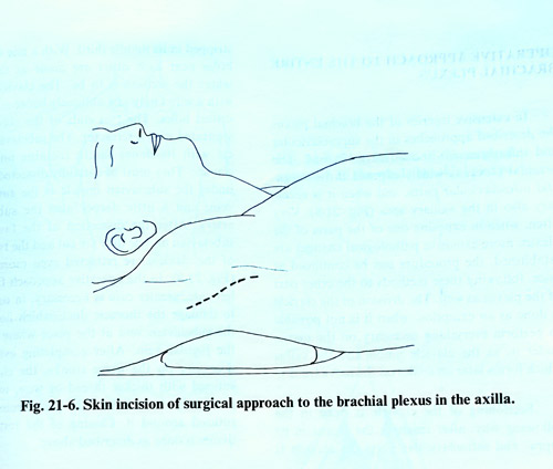
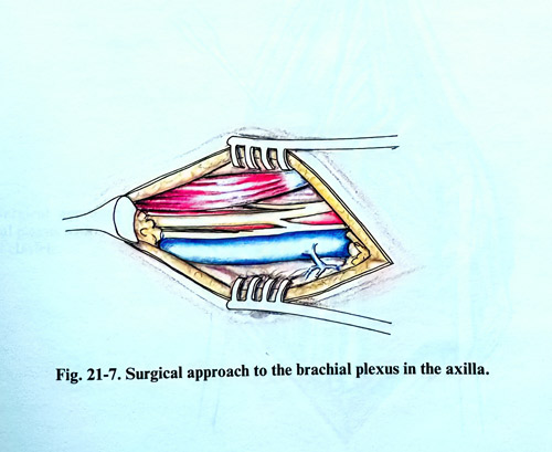
The patient is in a prone position on the operating table with a small cushion being placed under the shoulders. The arm is put on a separate support, in abduction position almost at a right angle. The incision of the skin, the subcutaneous tissue, and the fascia begins from the depth of the axillary fossa and goes distally along the medial sulcus of the biceps muscle (Fig. 21-6). The vessels and nerves are reached after the incision of the fascia. The identification of the individual nerves can be done easily, if their relation to the brachial artery are used as reference. Both initial branches of the median nerve enclose the brachial artery, while the nerve itself is situated along the antero-lateral artery surface. The musculocutaneous nerve is situated laterally, passing through the coracobrachial muscle and innervating it. Medially from the artery are found the ulnar nerve, the medial cutaneous nerves of the arm and forearm (Fig. 21-7). The two latter nerves are considerably thinner than the ulnar nerve. To expose the axillary and radial nerve better, the artery must be slightly retracted, as the nerves are located behind it. Tracing these nerves upward permits exploration of the brachial plexus cords, and if they have to be entirely exposed, it is necessary to cut the major pectoral muscle in proximity with its insertion to the humerus.
OPERATIVE APPROACH TO THE ENTIRE BRACHIAL PLEXUS
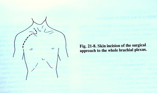
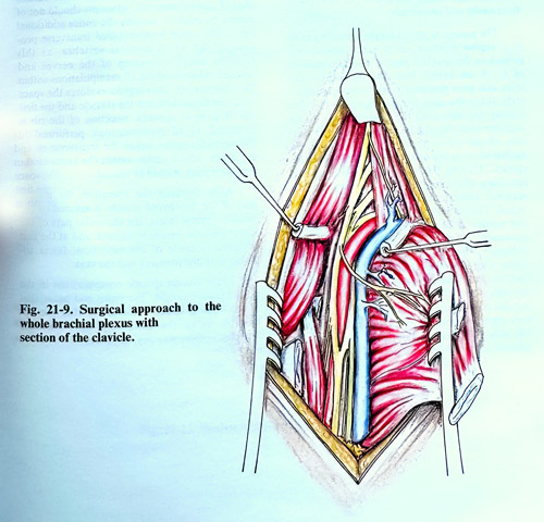
In extensive injuries of the brachial plexus the described approaches in the supraclavicular and infraclavicular fossae can be merged into one approach. The brachial plexus is widely exposed in its supra-and infraclavicular parts, and when it is necessary also in the axillary area (Fig. 21-8). Very often, when exposing one of the parts of the plexus and more extensive pathological changes are established, the procedure can be expanded immediately, following the already described methods to the other parts of the plexus as well. The division of the clavicle is done as an exception, when it is not possible to perform everything necessary on the nerves under it, as the clavicle union and the callus which forms later on can interfere with the nerve trunks.
Sectioning of the clavicle is done in the following way: after reaching the plexus in its supra- and infraclavicular parts, the clavicle is stripped of periosteum in its middle third. With a fine drill two holes near each other are made at the place where the incision is to be placed. The clavicle is cut with a Gigli saw obliquely between the two drilled holes. The two ends of the clavicle are separated with a retractor. The subclavian muscle with its dense fascia remains under the clavicle. They must be carefully dissected, as just under the subclavian muscle is the subclavian vein, and a little deeper also the subclavian artery. After the dissection of the fascia and the subclavian muscle, they are divided and the two parts of the clavicle are retracted even more widely (Fig. 21-9). In the operative approach from the left side, care should be taken in order not to damage the thoracic duct, which flows into the subclavian vein at the place where it joins the jugular vein. After completing everything necessary on the nerve trunks, the clavicle is fixed with a thicker thread or wire, using the previously made holes. The periosteum is also sutured around it. Closing of the rest of the tissues is done in the standard way as described above.
THORACIC OUTLET SYNDROME
Surgical treatment is indicated in symptoms and signs provoked by compression of the vessels and nerves i the thoracic outlet when conservative treatment has proved unsuccessful. Two basic surgical approaches are in use in a case of scalenus anterior syndrome: 1. Transaxillar resection of the first rib accompanied by scalenotomy, and 2. The supraclavicular approach.
The transaxillar approach is performed through a small axillary incision and involves resection of the first rib to decompress the floor of the thoracic outlet. We prefer the supraclavicular approach because the compromised nerve structures and subclavian artery can be exposed more easily and completely.
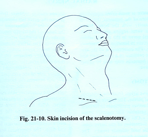
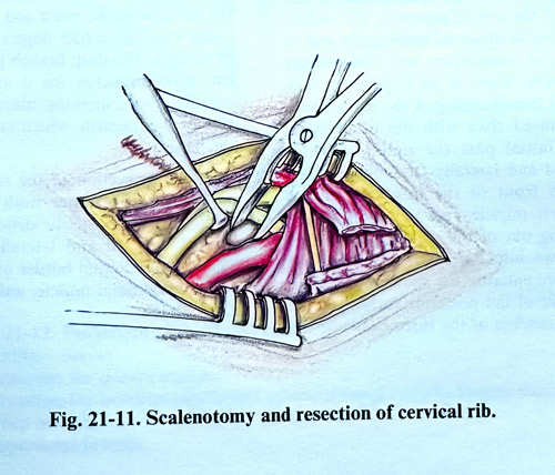
The patient in the supraclavicular approach is placed on the operating table in the supine position, as in the operative approach to the brachial plexus. The skin incision of 6 - 8 cm begins from the sternoclavicular joint and goes posteriorly and obliquely super-iorly along the skin creases (Fig. 21-10). After opening the skin and the subcutaneous tissue together with the platysma, the clavicular portion of the sternomastoid muscle is retracted medial or is transected a 1 cm distance from the clavicle. Cutting the middle cervical fascia and retracting the omohyoid muscle superiorly, the scalenus anterior muscle is reached and dissected. The phrenic nerve goes along the anterior surface of the scalenus anterior muscle in proximity to its medial border and a little obliquely, then inferiorly enters the thoracic cavity. The interscalenic space is penetrated and inspected the subclavian artery passing through it together with the nerves. During this penetration the phrenic nerve and accessory phrenic nerve should be preserved. On the left side the thoracic lymphatic duct and its affluent channels can be seen. They have very thin walls and need protection despite the fact that their damage does not provoke serious complications. In case of obvious stenosis, the anterior scalenus muscle is divided near to its insertion to the first rib. To avoid injury of the anatomical structures, this is done slowly and carefully through the interscalenic space preserving the cupola of the pleura. If a cervical rib or a protruding transverse process of the VIIth cervical vertebra provokes compression on the neural structures and/or subclavian artery, the most prominent bone part is exposed under the periosteum, and nibbled in small pieces (Fig. 21-11). All fibrous bands in the area contributing to the stenotic condition should also be removed. It is not necessary to remove the entire additional rib or the whole hypertrophied transverse process of the VIIth cervical vertebra, as this involves too strong retraction of the nerves and arteries. After completing the work inside the interscalenic space, the space between the clavicle and the first rib is explored. If there is residual stenosis, resection of the first rib is also indicated. Meanwhile, the transverse cervical artery, which passes within the limits of the operative field, should be spared.
After completing the decompression, the middle part of the omohyoid muscle is sutured to the middle cervical fascia, the clavicular part of the sternomastoid muscle is sutured, and at the end a suture is made of the superficial fascia together with the platysma and the skin.
There are no specific complications in the postoperative period. The pain and paraesthesias disappear either immediately after the operation or in the following days. Muscle atrophy, if there was any, improves later on.
RADIAL NERVE
The radial nerve is most often injured on the arm in a fracture of the humerus, due to its location in proximity with the bone. Nerve contusions in the same region are not rare. Injury of the radial nerve in the elbow area most frequently occurs in luxation and fractures of the radial bone. The compression of the arm during sleep or the pressure of a tourniquet and operations on the humerus may also be causes of injury to this nerve.
SURGICAL ANATOMY
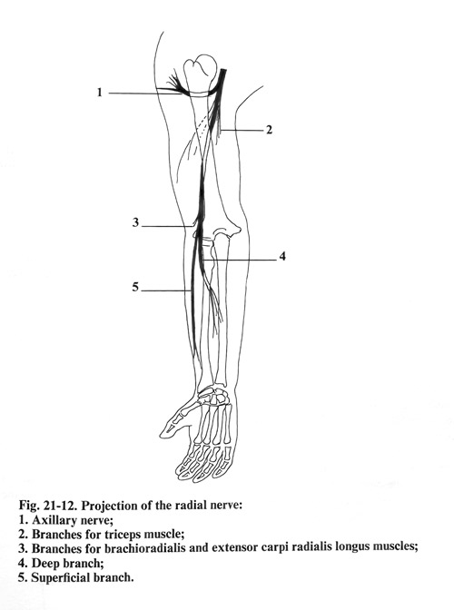
The radial nerve arises from the posterior cord of the brachial plexus. The degree to which the different spinal nerves participate in forming the radial nerve is variable. The superior trunk of the plexus (C5 - C6) plays a considerably greater role in the formation of the nerve compared to the middle and inferior trunks. The level at which the nerve forms varies from the supra-medial to infra-lateral margin of the minor pectoral muscle. From this level to the inferior end of the tendon of the latissimus dorsi muscle, the radial nerve is the thickest branch of the brachial plexus. If the epineurium of the radial nerve of the arm is removed, the individual bundles can be dissected more easily and at a great length in comparison with the median and ulnar nerves. There is an impression that the intraneural interchanging of the nerve fibres is less pronounced than with the other large nerves. This initial part the radial nerve is situated behind and laterally from the axillary artery, just in front of the insertion of the latissimus dorsi and the teres major muscles. Coming out of the axillary cavity, the nerve goes down into the spiral groove of the humerus. Before entering the spiral groove separate the cutaneous nerve of the posterior surface of the arm and the branches of the three heads of the triceps muscle. In the spiral groove the nerve lies immediately on the periosteum of the humerus and is enclosed between the heads of the triceps muscle. Together with it go the deep brachial artery and the accompanying veins. The artery is initially situated lateral to the nerve; lower down, it passes medially. At the boundary between the inferior and middle third of the arm, the nerve, perforates the lateral intermuscular septum, and goes down on the anterior side of the arm and the elbow, where it is found between the brachial and the brachioradial muscles. In the spiral groove of the humerus the cutaneous skin nerve for the forearm posterior surface separates from the radial nerve. After its exit from the canal the nerve gives off branches to the brachioradialis and extensor carpi radialis longus. Anterior to the elbow it is divided into two branches: superficial and deep. The superficial branch continues in a distal direction below the brachioradialis and, lateral to the radial artery, passes below the tendon of this muscle and comes out above the fascia on the boundary between the middle and inferior third of the forearm. Its terminal branches innervate the skin of the radial half of the dorsum of the palm and the dorsum of the radial two and a half fingers without the distal phalanges. The deep branch perforating supinator, gives branches for it and after that also supplies the remaining muscles on the dorsal side of the forearm, which extend the wrist and the fingers.
The projection of the radial nerve on the surface of the upper limb begins from the middle of the posterior deltoid muscle border, goes downward and laterally as a spiral and reaches the internal border of the initial part of the brachioradial muscle, anterior to the elbow (Fig. 21-12).
SURGICAL APPROACHES
SURGICAL APPROACH TO THE RADIAL NERVE IN THE SUPERIOR THIRD OF THE ARM
The surgical approach to the radial nerve in the superior third of the arm as it emerges from the posterior cord of the brachial plexus, has been described in the exploration of the brachial plexus in the axillary cavity. In reaching the vessels and the nerves it should be comsidered that the radial nerve is located behind the brachial artery. It can be dissected from its origin from the posterior cord of the brachial plexus until it enters the spiral groove and even lower by penetrating between the heads of the triceps. At this place are separated from the nerve a skin branch - posterior cutaneous nerve of the arm, and small branches for the heads of the triceps and care should be taken not to injure these branches.
Near the nerve is the origin of the deep brachial artery.
SURGICAL APPROACH TO THE RADIAL NERVE IN THE MIDDLE THIRD OF THE ARM
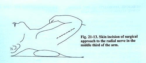
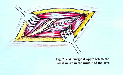
The patient is placed supine on the operating table with the arm adducted at the shoulder, flexed at the elbow and over the thorax. An incision of the skin, the subcutaneous tissue, and the fascia is made about 20 cm long, vertically along the dorsal durface of the arm (Fig. 21-13). After that at the superior end of the incision is penetrated between the long and lateral heads of the triceps and the nerve is exposed over the surface of the humerus. Then the dissected parts of the muscle are split from one another along the muscle fibres, and the nerve is followed distally. In this approach, to reveal the nerve at a greater distance downward is created a tunnel under the lateral head of the triceps. Through its course together with the nerve also run the branches for the heads of the triceps and the deep brachial artery and its branches, whose dissection must be very carefully performed (Fig. 21-14). In the spiral groove also separates the skin nerve (posterior cutaneous nerve of the forearm) and its preservation also requires attention.
SURGICAL APPROACH TO THE RADIAL NERVE IN THE INFERIOR THIRD OF THE ARM AND ELBOW
For the exposure of the radial nerve in the inferior third of the arm the patient is placed in a supine position, with the arm being in adduction.
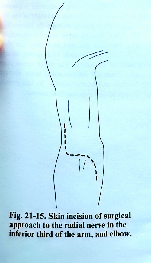
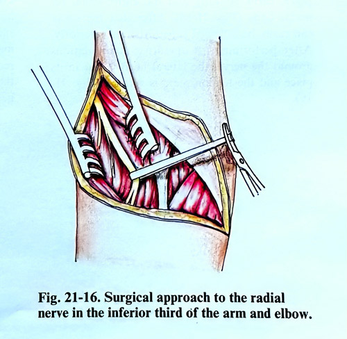
The incision begins 5 - 6 cm above the lateral epicondyle of the humerus and goes downward to the skin creases of the elbow. It follows and, when needed, continues several more centimetres along the lateral side of the forearm (Fig. 21-15). After cutting the skin, the subcutaneous tissue, and the fascia is penetrated between the brachial and brachioradial muscles in depth and in proximity to the bone to the place where the nerve is located (Fig. 21-16). From there downward the nerve is easily followed till the place of its division into superficial and deep branches. At the superior end of this incision do appear usually the small branches to the brachioradial and extensor carpi radialis longus muscles and they deserve attention not to be injured.
SURGICAL APPROACH TO THE RADIAL NERVE ALONG THE ENTIRE ARM
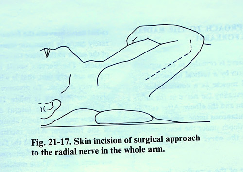
In limited injuries of the radial nerve the described operative approaches to the different levels of the arm are appropriate. If the injury is larger and there is a defect of the nerve, then these incisions are not sufficient to expose the peripheral and the central segment of the nerve, and should be resorted to the merging of the incisions between adjacent local approaches. In these cases we apply a longer skin incision that begins from the middle of the deltoid muscle's posterior border, goes vertically toward the olecranon and 3 cm from it turns laterally, reaching the lateral humerus epicondyle (Fig. 22-17). Along the same incision is also divided the brachial fascia. The surgeon penetrates into the superior part of the incision between the long and the lateral head of the triceps, where the radial nerve is exposed.
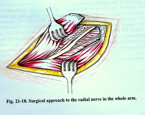
The two heads are separated from one another and reaching downward to where they are attached, they are separated up to the tendon parts attached to the olecranon. After that the lateral head of the triceps is dissected and the tendon parts are cross-sectioned. In this way the lateral triceps head, which encloses the humerus spiral groove from the dorsal side can be turned laterally and the entire canal remains open (Fig. 21-18). After that the nerve can easily be followed superiorly and inferiorly.
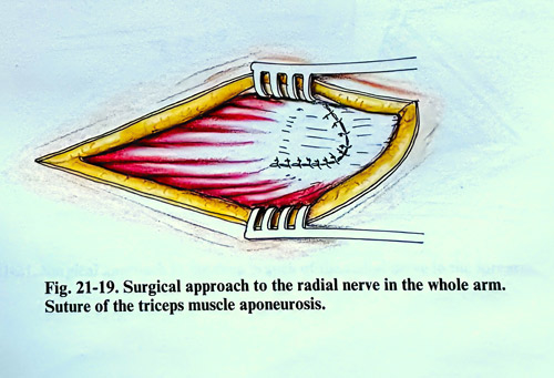
After completeing all operative work related to the nerve, the lateral head is repositioned and the tendon plate is sutured (Fig. 21-19).
TRANSPOSITION OF THE RADIAL NERVE
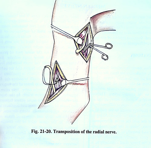
In case of injury of the radial nerve at the arm with large defects, a transpositioning of the nerve is necessary from the posterior to the anterior humerus surface. The decision on transposition of the radial nerve is made during the exploration of the nerve in the entire spiral groove if it is obvious that the nerve defect is large and it is impossible to do an end-to-end suture. For this transposition the nerve is exposed in the superior third of the arm with an incision along the internal sulcus of the biceps muscle distally from the axillary fossa. At this place the central nerve segment is dissected together with neuroma till its emergence from the posterior cord of the brachial plexus, and it is pulled upward. Its small branches going to the triceps are preserved. After that, the distal end of the nerve is revealed in the anterior third of the arm. On both ends of the nerve thicker threads are sutured before being resected, i.e. threads are sutured to the neuroma of the central segment and to the cicatrix of the peripheral end.
After that a tunnel is made with a sufficiently long instrument between the biceps and the brachial muscle. Through this tunnel the long instrument is inserted and the nerve end suture threads on the longer of the two nerve segments is passed through the tunnel (Fig. 21-20). After that the nerve ends are excised till reaching normal nerve structure and sutured. This suture is made in flexion of the arm maximally at the elbow. The nerve suture place remains in a normal intermuscular space between the brachial and biceps muscles. This method can overcome nerve gaps up to 11 cm.
SURGICAL APPROACH TO THE RADIAL NERVE IN THE FOREARM
The radial nerve is exposed in the superior third of the forearm by a vertical incision along the brachioradial muscle as a continuation of the incision for exposure of the nerve in the inferior third of the arm and the elbow. After cutting the skin, the subcutaneous tissue, and the fascia are penetrated under the brachioradial muscle to reveal the nerve on the supinator muscle.
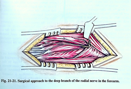
The exposure of the superficial branch of the nerve is achieved by extending the same incision inferiorly along the brachioradial muscle. By the separation of the muscle and its displacement slightly laterally, the nerve is reached. Distally the superficial branch of the radial nerve perforates the fascia laterally to the tendon of brachioradialis muscle and branches in the subcutaneous tissue. The exposure of the deep branch of the radial nerve between the forearm muscles is very rarely made in traumatic lesions or entrapment of the nerve while traversing the supinator muscle. The nerve's small branches in this region are very thin and finding them at times is very difficult. To to expose proper the nerve at this place is made an incision of 10-12 cm long beginning from the lateral epicodyle of the humerus and going down along the dorsal surface of the forearm. After the skin incision, the subcutaneous tissue, and the fascia is penetrated between the extensor digitorum and the extensor carpi ulnaris muscles. These muscles are separated as far as the lateral epicondyle and are retracted (Fig. 21-21). The inferior part of the supinator is revealed an there the deep branch of the radial nerve comes out. In case of entrapment of the deep branch of radial nerve in the canal of the supinator muscle, the approach can be limited. The skin incision is only along the brachioradial muscle, following the radial nerve in the supinator muscle. If there is stenosis in the tendinous entrance into the canal, it is opened along its length.
MEDIAN NERVE
The median nerve is most often damaged in open injuries of the upper limb, especially in injuries of the inferior third of the forearm and wrist. It can be involved also in fractures of the arm and forearm. Gunshot injuries of the median nerve are most frequent at the elbow.
SURGICAL ANATOMY
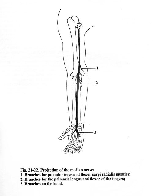
The median nerve is formed from two initial branches, which separate from the medial and lateral cords of the brachial plexus. These two branches join at the level of the inferior border of the minor pectoral muscle immediately anterior to the axillary artery. The two initial branches of the median nerve or the so-called 'fork', can be discovered at different levels from the minor pectoral muscle to the middle of the arm. Sometimes it may be double. Along the whole length of the arm the nerve accompanies the brachial artery, being in the beginning situated anteriorly and laterally to it. In the middle of the arm it goes exactly anteriorly to the artery, while in the lower third - medially to it. In about 10 - 13% of cases, the artery can be discovered over the nerve (superficial brachial artery), or may be double, so that one branch is over the nerve, the other under it. Along the entire arm the nerve has the same thickness, excluding about 40% of cases, in which it is connected with the musculocutaneous nerve. In 5% of cases the musculocutaneous nerve is part of the median nerve for a certain distance or along the entire length for the musculoputaneus nerve. In such cases the branches for the biceps and the brachial muscle come out of the median nerve at the level of the middle third of the arm or at the level of the border between the middle and superior third. Coming down into the elbow pit (cubital fossa), the nerve is situated medially from the artery and the biceps tendon. At this place it lies approximately in the middle between the biceps tendon and the medial humerus epicondyle. In the elbow pit separate the small branches for the fingers and the wrist flexors. Most superiorly separate the small branches for the brachial head of the pronator teres. If one branch comes out of the nerve, it is secondarily divided along its path and not less than two branches enter into the pronator teres muscle. The level of separation of these branches varies within the limits from the superior limit of the inferior third of the arm to the elbow. Somewhat more distally separate the small branches for the ulnar part of pronator teres and for the flexor carpi radialis muscles. At the same place separate the small branches for the superficial flexor of the fingers and the palmaris longus muscles. A little more distally to the elbow separate the small branches for flexor pollicis longus, the deep flexor of the fingers and the anterior interosseous nerve. Very often they separate as a common trunk. In the depth of the elbow pit, the nerve passes between the two heads of pronator teres and continues in the forearm, approximately along its axis, lying between the deep and superficial flexors of the fingers. In the distal third of the forearm the median nerve goes more superficially, being situated between the tendons of the flexor carpi radialis and the palmaris longus muscles. Less frequently, it is between the tendons of the palmaris longus and the flexor digitorum superficialis muscles. Distally the nerve passes together with the tendons of the flexors of the fingers through the carpal tunnel and reaches the palm, where it divides into its terminal branches. This division most often takes place in immediate proximity to the distal end of the transverse carpal ligament. It is divided in two branches, which, on their part, branch out into the thenar and the common nerves for the fingers. The small branches for the thenar muscles are always situated more superficially to the common trunk of the nerve.
The median nerve is projected on the arm along the line, which goes downward along the internal sulcus of the biceps to the middle between the tendon of the same muscle and the medial epicondyle of the humerus. In the forearm the nerve projection is situated exactly along its anterior surface midline (Fig. 21-22).
SURGICAL APPROACHES
When exposing the median nerve in all parts of the upper limb, the patient is placed in a supine position on the operation table, with the arm slightly abducted and placed on a separate small table.
SURGICAL APPROACH TO THE MEDIAN NERVE IN THE ARM
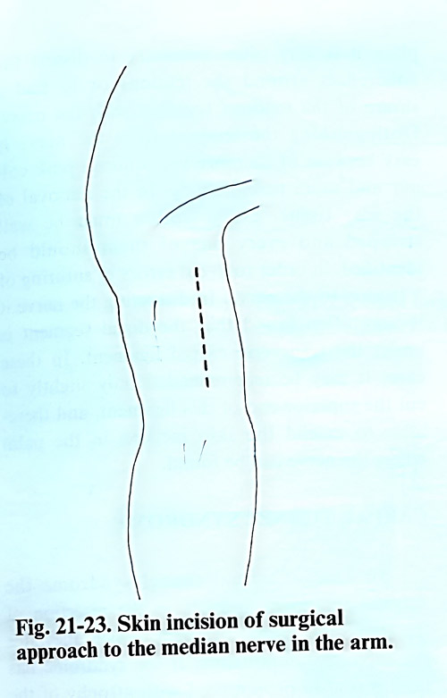
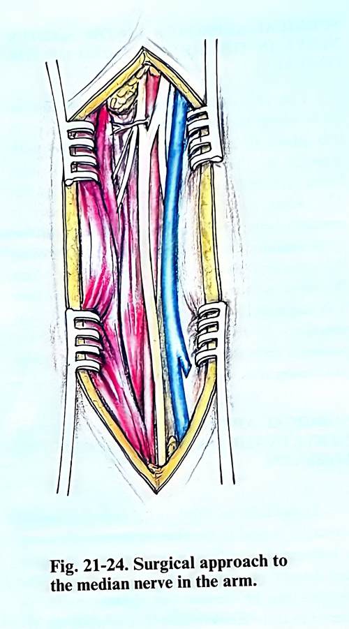
The skin incision is made in a vertical direction 2 - 3 cm anterior or posterior to the internal sulcus of the biceps (Fig. 21-23). Along the same incisioned are divided the subcutaneous tissue and the fascia. Next is penetrated under the fascia a slightly medially to reach the brachial artery and the median nerve. Due to the close proximity of the median nerve to the brachial artery there is very often a combined injury of both of them, and in the separation of the nerve requires a meticulous work (Fig. 21-24). The pulsations of the artery serve as an orientation in exposing the nerve. When closing the surgical approach , it is important that nerve repair and fascia closure are not one over the other to avoid additional scars over the nerve suture.
SURGICAL APPROACH TO THE MEDIAN NERVE IN THE ELBOW
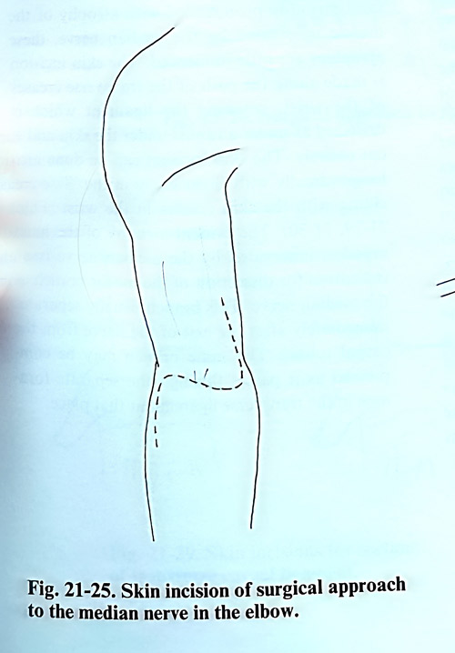
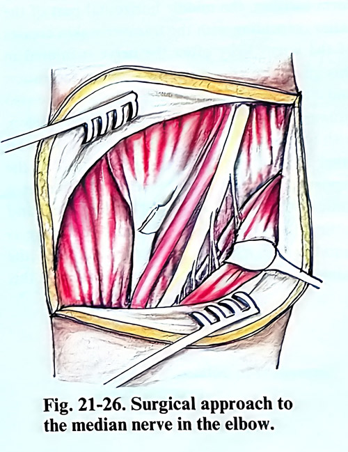
In exposing the median nerve in the elbow it is best to have the skin incision made in the form of an 'S', so that the horizontal part of the letter coincides with the elbow pit crease (Fig. 21-25). In the subcutaneous layer, major veins are usually found, and if an obstacle for the approach, can be coagulated and cut. The fascia is separated and divided with an incision medial to the nerve, so that a flap is made to cover the nerve without I thets sutures be in contact with nerve. The location of the nerve is usually in the middle between the biceps tendon and the medial humerus epicondyle. The nerve can also be located at 1 - 2 cm medial to the artery whose pulsations can easily be felt by palpation. If it is necessary to expose the nerve distally, the pronator teres is retracted medially and distally. At this place, the small branches of the nerve separate for the wrist and the finger flexor muscles and their identification in the scar tissue, and preservation respectively, may be difficult (Fig. 21-26). In case of median nerve entrapment by the lacertus fibrosus, hypertrophic musculus teres major, or the fibrous arch of the superficial flexor of fingers, they may be undercut.
SURGICAL APPROACH TO THE MEDIAN NERVE IN THE MIDDLE THIRD OF THE FOREARM
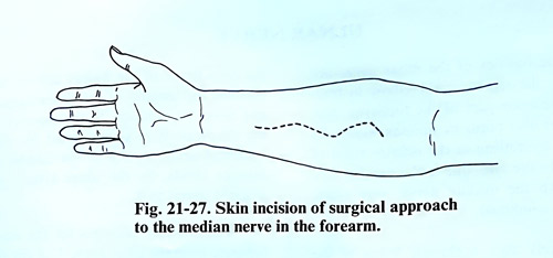
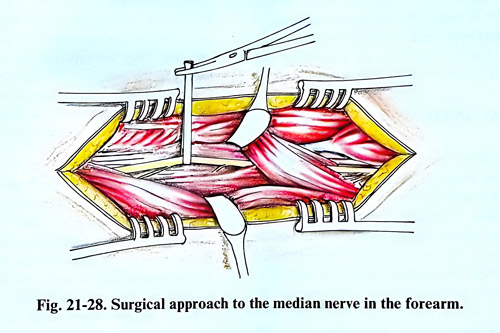
The skin incision in this region can be made along the projection of the nerve, as in this place it is located deep to the surface and is covered by the ventral muscle groups, so that there is no danger of skin scars interfereing with the nerve suture area (Fig. 21- 27). After the fascia is opened, the space between the pronator teres and flexor carpi radialis muscles is dissected. It is penetrated in depth through the superficial flexor of the fingers, under which the nerve is located (Fig. 21-28). From this location it is easily followed in a distal direction. If it is necessary to follow the nerve more proximally, the pronator teres should not be incised, but a tunnel should be created under it.
SURGICAL APPROACH TO THE MEDIAN NERVE IN THE INFERIOR THIRD OF THE FOREARM
In the inferior third of the forearm, special attention is paid to the skin incision as the nerve is between the tendons of the flexors of the wrist and fingers immediately below the fascia. The incision can be longitudinal, but slightly medial or lateral to the nerve projection. It cam be also convenient to make an 'S'-shape incision, the middle horizontal part of the latter coinciding with the transverse skin creases of the wrist. Very often the nerve is injured in this place by transversally running laceration and cut injuries. In these cases, the scars are also situated transversally, and after their excision, with two additional incisions one superiorly and other inferiorly in the form of the letter 'S', the exposure of the nerve is made possible in a sufficiently broad area. After exposing and dividing the fascia of the forearm, the nerve is found in the space between the tendon of the palmaris longus and flexor carpi radialis muscles. At this place it is very often required to dissect the adhesions around the tendons or even to repair tendons together with the nerve repair.
Distinguishing the tendons from the nerve is easy because the nerve has more intense pink colour and different vascularity. In the removal of the scar tissue, every tendon must be well stripped and every one of them should be identified, in order to avoid errors of suturing a tendon to the nerve. In dissecting the nerve it is very often found that the distal segment is under the transverse carpal ligament. In these cases it may be recommended only slightly to cut the superior end of this ligament, and thereafter to extend the skin incision in the palm where the nerve can be exposed.
CARPAL TUNNEL SYNDROME
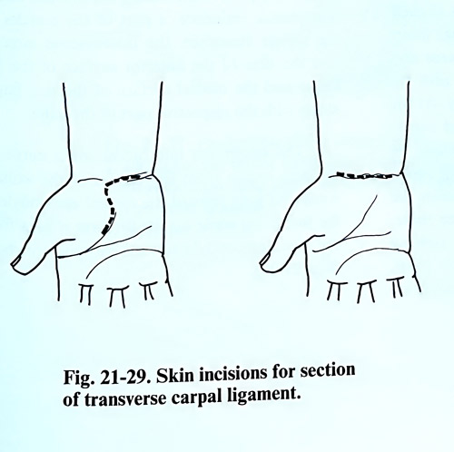
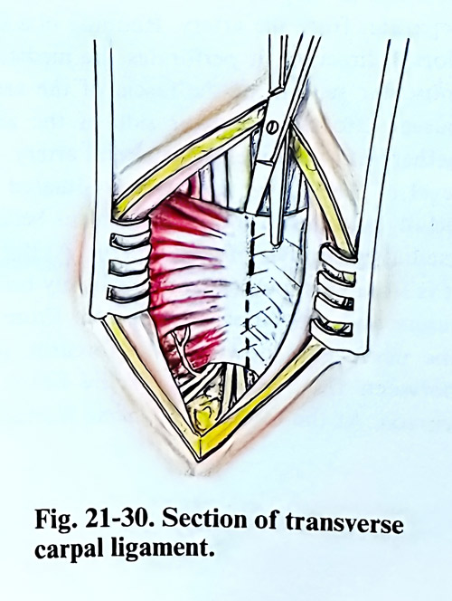
In case of carpal tunnel syndrome the surgical treatment consists in the resection of the transverse carpal ligament, which relieves the subjective complaints. If the syndrome has been in an advance stage with atrophy of the muscles innervated by the median nerve, these atrophies are just a little improved. The skin incision is made along the path of the transverse creases of the wrist, reaching the ligament which is dissected under the skin and is divided completely. The skin incision can be done also longitudinally with slight curve in the 'S', coinciding with the skin creases in the wrist (Figs. 21-29; 21-30). The evident atrophy of the hand muscles, innervated by the median nerve is an indication for dissection of the motor branch of the median nerve. This branch usually separates immediately after the exit of the nerve from the carpal tunnel. The same branch may be compressed if it passes through the separate foramen in the transverse ligament at that place.
ULNAR NERVE
Traumatic injuries of the ulnar nerve are most often in the region of the elbow in fractures of the inferior part of the humerus. Not rarely, injuries also occur in wounds, especially those caused by cutting in the inferior third of the forearm. In the arm, due to the proximity of the ulnar with the median nerve, very often, their injury is combined. Very rarely also, a chronic lesion of the ulnar nerve is obtained, which we designate as ulnar tunnel syndrome' or when it is manifested late after the consequences of a fracture and dislocation of the elbow, as 'ulnar palsy'.
SURGICAL ANATOMY
The ulnar nerve arises from the medial cord of the brachial plexus. It contains fibres from the C8 and Tl spinal nerves. Approximately in half the cases, it obtains a bundle of the lateral cord of the plexus, known as the lateral pedicle of the nerve. At its origin it is situated medially from the axillary artery and continues distally to the middle of the arm together with the brachial artery, preserving the same relation. After that, the nerve separates from the artery. Running in a slightly dorsal direction, it perforates the medial inter-muscular septum of the brachial fascia and passes posteriorly together with the superior collateral artery. At the level of the elbow, the nerve is situated superficially and lies in the vertical sulcus behind the medial epicondyle of the humerus. At this place, it is situated superficially immediately below the fascia and can be easily palpated. From there, the nerve goes down to the forearm, passing between the two heads of the flexor carpi ulnaris. At the forearm, the nerve is situated on the deep flexor of the fingers and in the space between the tendons of the flexor carpi ulnaris and flexor digitorum superficialis muscles. In the proximal third of the forearm, it is accompanied by the ulnar recurrent artery, while in the middle and the distal thirds - by the ulnar artery situated laterally to ithe nerve.
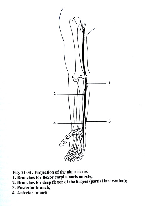
The articular branches for the elbow joint separate from the ulnar nerve 7 - 8 cm above the level of the medial epicondyle and below this level are the branches for the flexor carpi ulnaris muscle. These last muscle branches are superior and inferior, penetrating into the muscle in the superior third of the forearm. The branch innervating the medial part of the deep flexor of the fingers is situated at the level of the medial epicondyle of the humerus.
On the border between the middle and inferior thirds of the forearm, the nerve is divided into two branches. One of the branches - the dorsal branch - innervates the ulnar half of the dorsasl surface of the hand and the dorsal surface of two medial fingers (4th and 5th) and a half, excluding the distal phalanges. The second branch - the palmar branch - passes down along the palmar surface of the hand, innervating the muscles of the hypothenar eminence, part of the muscles of the thenar eminence, the interosseous muscles and the skin of the anterior surface of the 5th finger and the medial surface of the ring finger along with the respective part of the palm.
The projection line of the ulnar nerve on the arm surface begins from the depths of the axillary fossa and goes toward the medial epicondyle of the humerus, while on the forearm it goes from the same epicondyle toward the pisiform bone (Fig. 21-31).
SURGICAL APPROACHES
The patient's position on the operating table is supine with the upper limb abducted on a separate table in supination position.
SURGICAL APPROACH TO THE ULNAR NERVE IN THE ARM
In the upper and middle third of the arm the surgical approach to the ulnar nerve coincides with the approach to the median nerve. The skin incision is made 2 - 3 cm anterior to the internal groove of the arm. The subcutaneous tissue and the fascia are divided along the same incision. The fascia is dissected medially, revealing the biceps muscle and medially from it the vessels and the nerve bundle. The ulnar nerve is situated along the internal side of the brachial artery. When the skin incision is along the projection of the nerve, it is best to divide the fascia 2-3 cm more laterally from the nerve and artery. In this way the nerve suture will be away from the scars of wound healing.
In the exposure of the nerve in the inferior two thirds of the arm, the skin incision passes 2 - 3 cm dorsally to the nerve projection. Through the same incision the subcutaneous layer and the fascia are divided. After that the fascia is dissected anteriorly and the nerve is revealed between the lateral border of the triceps and the internal intermuscular septum.
SURGICAL APPROACH TO THE ULNAR NERVE AT THE ELBOW AND TRANSPOSITION OF THE NERVE
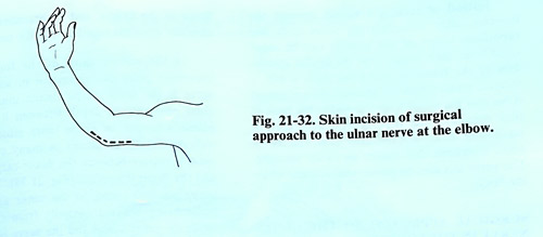
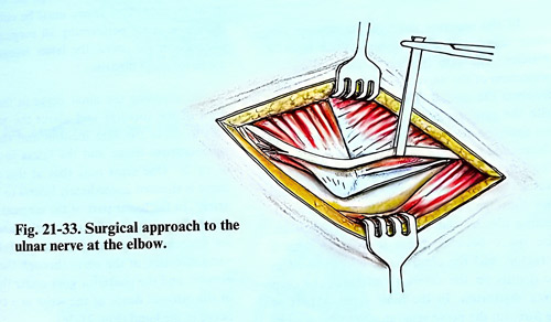
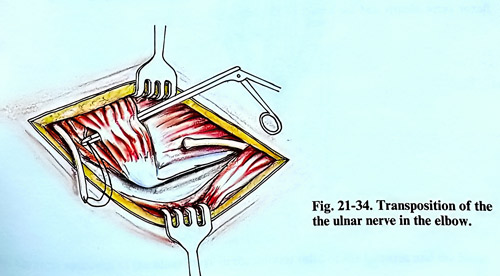
The skin incision is made on the medial surface of the elbow 2 cm anterior to the medial epicondyle of the humerus, beginning 5 - 6 cm above it and reaching 5 - 6 cm under it. Along the same incision are opened the subcutaneous tissue and the fascia. This incision is situated anterior to the projection of the nerve and final stitches are not over the damaged nerve. Separating the fascia slightly posteriorly, the nerve groove situated posterior to the internal epicondyle is reached where the nerve can be dissected very easily (Figs. 21-32; 21-33 ). Small gaps of the nerve can be approximated by fixing the upper limb in extension of the elbow. In traumatic injury of the nerve with large gaps, after exposure of the nerve, it can be transposed and sutured anteriorly to the elbow. The same transposition can also be made in cases of compression of the ulnar nerve in the groove after fracture, luxation or stenosis. In flexing the limb at the elbow, the nerve is fixed to its bone sulcus and is slightly stretched. Transposing the nerve anterior to the elbow joint in flexion position makes it possible to overcome a nerve defect of as much as 11 cm. Transposition of the interrupted ulnar nerve can be done by introducing one of the nerve segments through the muscles just below the medial epicondyle of the humerus, or over the muscle. In the superior part of the incision, the ulnar nerve is dissected between the triceps and the internal medial intermuscular septum and retracted superiorly.
A small part of the internal intermuscular septum should be excised at the place where the proximal segment of the interrupted nerve will be transfered onto the anterior surface of the elbow. The distal segment is revealed on the forearm in the space between the flexor carpi ulnaris muscle and the flexor digitorum superficialis. In dissecting the nerve, the branches for the flexor carpi ulnaris and the flexor digitorum profundus are spared. With a long instrument, a tunnel is made between the superficial and deep muscle groups of the forearm. The instrument is introduced under the pronator teres of the forearm, the flexor digitorum superficialis and the flexor carpi radialis. Depending on which of the two nerve ends - proximal or distal - is longer, it is inserted through the tunnel with the help of a long instrument, after which a suture of the nerve is made. The place of the suture usually remains in the muscle tunnel (Fig. 21-34).
Instead of making a tunnel through the muscles, after dissecting the nerve ends, the superficial layers of the muscles can be separated from their attachment, near the medial epicondyle of the humerus and from them a flap is turned over the sutured nerve. The muscles are sutured to the bone attachment at their original place where was left a small tendinous remnant. The nerve can also be placed just under the fascia.
SURGICAL APPROACH TO THE ULNAR NERVE IN THE FOREARM
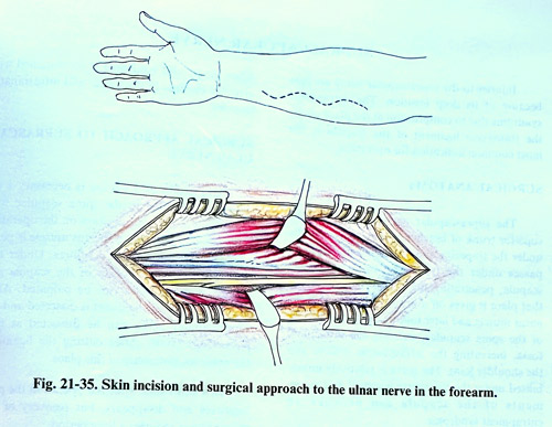
In the superior third of the forearm, the ulnar nerve is situated under a thick layer of muscles. The skin incision is made along the projection of the nerve, i.e. along the line which connects the medial epicondyle of the humerus with the pisiform bone of the wrist. The nerve can easily be discovered between the flexor carpi ulnaris and the flexor digitorum superficialis.
The intermuscular space is not free, as the muscles are adherent among themselves, so that they must be separated exactly along the connecting fibrous plane between each two muscles. The space between them is widened with a retractor, and the ulnar nerve is discovered in the depths on the anterior surface of the deep flexor digitorum. In the most superior part of the forearm the nerve gives branches for the flexor carpi ulnaris and for a part of the deep flexor of the fingers, and its branches must be carefully dissected together with the nerve.
In the middle third of the forearm, an important point of orientation in exposing the nerve is the flexor carpi ulnaris muscle. It should be penetrated between it and the palmaris longus, or if the latter muscle is not well developed, as occurs in many cases, it should be between the flexor carpi ulnaris and the flexor digitorum (Fig. 21-35). The ulnar nerve passes parallel to the ulnar artery, the nerve being situated medially from the artery while the entire vessel and the nerve are under the fascia, which envelops the deep muscles of forearm. This fascia must be divided to reach the nerve. After performing the surgical interventions on the nerve, it will remains under the flexor carpi ulnaris.
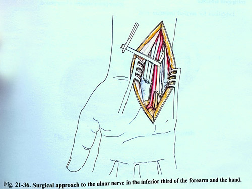
In exposing the ulnar nerve in the inferior third of the forearm and about the wrist, it is best to make the skin, subcutaneous layer and fascia incision in the 'S' shape. At this location the nerve is positioned immediately lateral to the ulnar artery. In the lower part of the surgical field, the ulnar nerve divides into anterior and posterior branches. The anterior branch follows the same direction of the nerve through the canal of Guyon, and the posterior goes under the tendon of the internal flexor of the wrist as a cutaneous nerve to the hand (Fig. 21-36).
SUPRASCAPULAR NERVE
Injuries to the suprascapular nerve are rare because of its deep location. The entrapment syndrome due to compression at the place under the transverse ligament of the scapula is the most common indication for operation.
SURGICAL ANATOMY
The suprascapular nerve arises from the superior trunk of brachial plexus, runs laterally under the trapezius and omohyoid muscles and passes under the transverse ligament of the scapula, penetrating the supraspinate fossa. At that place it gives off a branch for the supraspinatus muscle and later follows the lateral border of the spina scapulae, entering the infraspinal fossa, innervating the infraspinatus muscle and the shoulder joint capsule. The nerve is relatively immobile under the transverse ligament and movements of the scapula can provoke an entrapment syndrome.
Indications for surgical treatment exist when the painful syndrome is accompanied with atrophic changes in the supra- and infraspinatus muscles.
SURGICAL APPROACH TO SUPRASCAPULAR NERVE
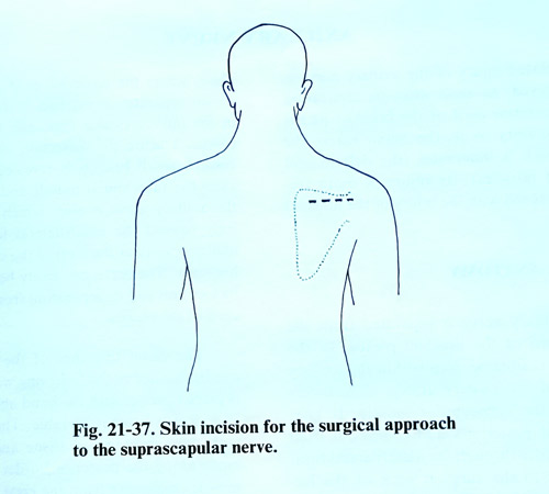
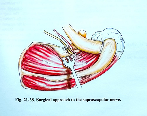
An incision of 5 - 6 cm is necessary 1 cm above and parallel to the spina scapulae with the patient in a lateral position on the operating table (Fig. 21-37). The trapezius muscle is penetrated, splitting the muscle fibres. Under this muscle the superior border of the scapula and supraspinatus muscles can be palpated. After that, the transverse ligament is dissected and the suprascapular nerve can be mobilized at that place (Fig. 21-38). After cutting the ligament, the nerve looks strangulated at this point.
Soon after the operation the pain improves and disappears, but recovery of the motor power requires a longer period.
AXILLARY NERVE
An isolated injury of the axillary nerve is rarely observed, as soon after its separation from the posterior cord of the brachial plexus and in proximity to it, the nerve enters the muscles which it innervates (the deltoid and teres minor muscles). Its injury is more frequently combined with the injury of the brachial plexus.
SURGICAL ANATOMY
The axillary nerve is separated from the posterior cord of the brachial plexus. In the beginning it is located deep inside the axillary fossa, behind the axillary artery, immediately anterior to the subscapular muscle. It goes inferiorly and posteriorly and comes out from the axillary fossa through the quadrilateral foramen, medial to the surgical neck of the humerus. Exiting from the axillary cavity, the nerve gives off a branch for the teres minor, while the greater part of the branches go to the deltoid muscle. One of its branches - the upper lateral cutaneous nerve of the arm - innervates the skin on the external surface of the arm. Together with the nerve are found the posterior circumflex artery and the veins accompanying it.
SURGICAL APPROACH
The initial part of the axillary nerve at the place, where the posterior cord of the brachial plexus separates, is reached in the same way as in the infraclavicular approach to the brachial plexus. During the dissection of the posterior cord, a small branch is exposed that innervates the subscapular muscle and a little distally the axillary nerve emerges. It passes posteriorly toward the quadrilateral foramen of the axillary cavity at the level of the surgical neck of humerus. The nerve can easily be recognised by its location and its separation from the posterior cord of the plexus.
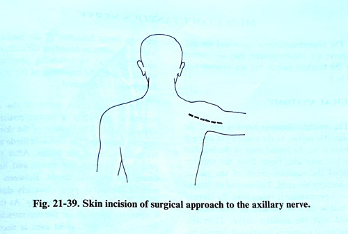
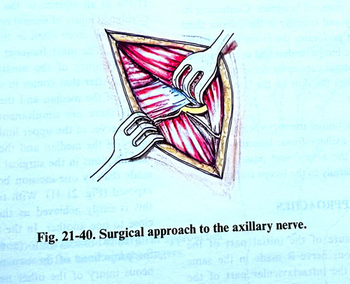
To expose the distal branches of the axillary nerve the patient is placed prone, with the hand abducted slightly laterally on a separate table. The skin incision with the subcutaneous tissue and the fascia is made along the posterior border of the deltoid muscle, beginning from the lateral part of spina scapulae and running to the humerus where the muscle is inserted (Fig. 21-39 ). The posterior border of the deltoid muscle is dissected and retracted cranially and laterally. The long head of the triceps is retracted medially. Penetrating carefully in the depths toward the surgical neck of the humerus is encountered fibrous and fatty tissue, in which the axillary nerve and the posterior circumflex artery are situated together with the accompanying veins (Fig. 21-40).
MUSCULOCUTANEOUS NERVE
The musculocutaneous nerve and the axillary nerve are seldom injured; they are isolated from the brachial plexus, but are situated very close to it.
SURGICAL ANATOMY
The musculocutaneous nerve is one of the most variable nerves. In the majority of cases, it originates from the lateral cord of the brachial plexus but may also begin from the anterior branch of the superior trunk of the plexus before forming the cord. The level of its appearance is not permanent: it varies from the clavicle to the middle third of the arm. In some cases it is joined with the median nerve. Then the coracobrachial muscle is innervated directly by bundles from the lateral cord of the brachial plexus. In the beginning, the nerve goes inferiorly and slightly laterally to the brachial artery, perforating the coracobrachial muscle, and at the same time giving off small branches for its innervation. After that, it passes through the space between the biceps and brachialis, giving off branches for these muscles. Continuing its way inferiorly and laterally in the inferior third of the arm, it perforates the fascia just laterally to the biceps tendon and as a cutaneous nerve - the lateral cutaneous nerve of the forearm, it supplies the skin on the lateral surface of the forearm.
The projection of the nerve begins from the depths of the axillary fossa, laterally from the pulsations of the artery, and passes inferiorly and slightly laterally to the biceps tendon.
SURGICAL APPROACHES
The patient is placed in a supine position, and stretched aside. The exposure of the initial part of the musculocutaneous nerve is made in the same way as to the infraclavicular part of the brachial plexus in the axillary fossa.
The nerve is exposed relatively easily, revealing first the two initial branches of the median nerve that are found on the axillary artery, and after that, in following proximally the lateral branch, the beginning of the musculocutaneous nerve is reached.
For the distal parts approach, the incision of the skin, subcutaneous tissue and the fascia, is made along the internal border of the biceps. After that is penetrated between the biceps and brachialis, where the nerve is located. Between the two muscles the nerve runs obliquely downwards and slightly medially to laterally. At this place, it separates branches for the adjacent two muscles and as they are very thin, great care is needed for their preservation.
SIMULTANEOUS LESIONS OF DIFFERENT NERVES
Injuries of more than one nerve of the upper limb are very frequently encountered. According to our investigations they constitute 11,7% of all injuries to the peripheral nerves.
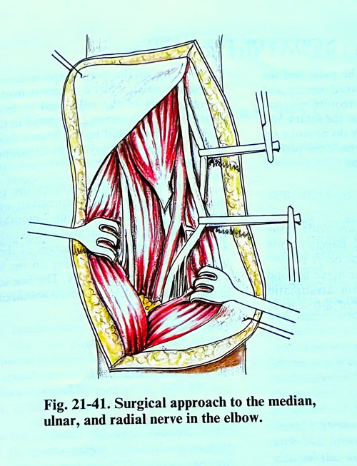
Combined injury of several nerves of the upper limb is observed more often in the arm than in the forearm. The most frequent combination consists of injury of the median and the ulnar nerves. After that comes in order of frequency injury of the median and the radial, and more rarely there are simultaneous injuries of the three nerves of the upper limb. In simultaneous injury of the median and the ulnar nerves, the skin incisions in the surgical approaches are to expose both nerves by one incision. (Fig. 21-41). At the arm, combined approaches are easily achieved as the nerves run very close to one another. In the forearm it is most convenient to make curved incision between the projections of the two nerves. In some simultaneous nerve injuries of the upper limb, the surgical approaches must be planned as described above, independently of one another.
NERVES OF THE HAND
Injuries of the nerves of the palm and the fingers, do not cause wide-spread motor and sensory disturbances. They require surgical treatment in many cases when the specific motor and sensory disturbances on the palm and the fingers can impede the performance of a number of professions.
OPERATIVE TECHNIQUE
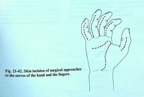
On the palm, the incisions are made along the path of its creases, and on the fingers on the lateral surface in proximity to the nerve projection (Fig. 21-42). Suturing of nerves is done under a microscope with a thin atraumatic needle and sutures of the order of 8-0 to 10-0.
Overcoming of the nerve gaps is achieved by flexing the wrist, when a suture is made in the proximal part of the palm and in flexing the fingers, when the nerves are sutured in the distal part of the palm and in the fingers themselves. In case of bigger gaps can be used grafting from the sural nerve.
After suturing the nerve in the proximal part of the palm, the restoration of rough sensation takes place in about 7 months, and if the suture is in the distal part of the palm in 3 . 4 months. In suturing the nerves of the fingers this term is shorter, depending on the level (on which phalanx) the injury is. The fine sensation in the fingers usually is restored within one year.