BRACHIAL PLEXUS
Injuries of the brachial plexus most often occur after open or closed trauma or gunshot injuries below and above the clavicle, first rib, and scapular fractures, or dislocation of the shoulder joint. A frequent type injury of the brachial plexus is by its traction, which leads to avulsion of its roots from the spinal cord. This injury occurs as a result of strong and sudden traction of the upper limb or impacts on the shoulder region.
Injuries of the brachial plexus occur also during delivery, when the newborn has been pulled out by the upper limb.
Aneurysms of the subclavian artery and tumours in the subclavian region are considered comparatively rare causes of injury to the brachial plexus. Sometimes the brachial plexus is damaged by a cervical rib, by stenosis between the scalene muscles, through which the plexus roots pass or by compression by the subclavian muscle below the clavicle.
SURGICAL ANATOMY
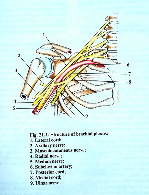
The brachial plexus is formed by the anterior branches of C5 - C8 and Tl spinal nerves which are roots of the brachial plexus. In approximately half of the cases, the C4 spinal nerve is included, while in others - it is the T2 nerve. In this way the brachial plexus is in some cases formed by inclusion of C4 - T1, in others by C5 - T2. The first is designated as of plexus superior type, while the second - as of inferior type. The anterior branch of the C5 spinal nerve joins the C6 at a sharp angle, forming the superior trunk of the plexus. The anterior branch of the C7 spinal nerve remains independent and forms the middle trunk, while the C8, and Tl merge in the inferior plexus trunk. The three trunks come out of interscalenus slit and continue downward anteriorly and laterally, reaching the superior surface of the first rib (Fig. 21-1). Every trunk is divided into an anterior and posterior branch, which pass below the middle part of the clavicle and enter the axillary cavity. The anterior and posterior branches of the three trunks form three cords in the axillary cavity: the lateral, medial and posterior cords, which envelop the axillary artery.
The lateral cord is formed by the union of the anterior branches of the superior and middle trunks and contains fibres from the CS, C6, and C7 spinal nerves. The medial cord is a continuation of the anterior branch of the inferior trunk and therefore contains fibres from the C7 and Tl spinal nerves. The posterior cord is formed from the posterior branches of the three trunks and consequently contains fibres of all nerves, which form the brachial plexus. There are variations in the formation of the plexus. Sometimes the brachial plexus is formed simply - without the formation of trunks. The roots of the plexus are divided laterally from the spinal column into anterior and posterior branches. The posterior branches of all plexus roots are united in the posterior cord. The anterior branches of the CS, Co and C7 roots are merged in the lateral cord, while those of the C8 and Tl merge in the medial cord.
The entire brachial plexus extends from the cervical spine to the distal region of the axillary cavity, i.e. to the level of the “surgical” neck of the humerus. The clavicle divides the plexus into the two parts: supraclavicular and infraclavicular. The first is situated in the supraclavicular fossa and comprises the roots, the trunks of the plexus, and their division into anterior and posterior branches. The infraclavicular part, which consists of the plexus cords, is situated in the axillary cavity.
At their point of exit from the spinal canal the plexus roots are situated in the sulci of the transverse processes of the cervical vertebrae. Anterior to the roots in a vertical direction are situated the vertebral artery and vein. The first thoracic nerve is situated immediately posterior to the stellate ganglion of the sympathetic chain. Just lateral from the spinal column, the brachial plexus is situated in the slit between the anterior and medial scalene muscles. The subclavian artery is situated in the inferior part of this slit anterior to the C8 and Tl nerves and their unification occurs in the inferior plexus trunk. If there is a well-developed scalenus minimus muscle, the artery remains separated from the plexus by it. In the supraclavicular fossa the brachial plexus is situated posterior to the sternomastoid muscle, omohyoid muscle, and the superior and middle fascias of the neck. The suprascapular artery goes anterior to the plexus and immediately above the clavicle, while the cervical transverse artery passes between the middle and superior plexus trunk.
The cords of the infraclavicular part of the brachial plexus are situated around the axillary artery in the axillary fossa. Lateral from the artery is found the lateral cord. The musculocutaneous nerve and the lateral branch of the median nerve separate from it. Medial from the artery is located the medial cord, from which separate the medial cutaneous nerve of the arm, the medial cutaneous nerve of the forearm, the ulnar nerve, and the medial branch of the median nerve. Posterior to the artery is the posterior cord of the brachial plexus, which is the thickest, and which divides at the level of the quadrangular foramen of the axilla into the axillary and radial nerves.
SURGICAL INDICATIONS
Traumatic lesions of brachial plexus do not always need surgical treatment. Open injuries, due to a sharp traumatic agent need urgent exploration and primary suturing. Lacerated wounds and gunshot injuries should be kept under observation. The surgical treatment is indicated after some weeks or 2 - 3 months, when clinically and electrophysiological investigations reveal any serious lesion. The closed injuries of brachial plexus also need surgical exploration after some period of observation. Surgical treatment is useless in a case of avulsion of brachial plexus roots from the spinal cord. When end to end suture is impossible, autotransplantation with sural nerve or neurotisation with intercostal nerves is indicated.
SURGICAL APPROACHES
Surgical approaches to the brachial plexus differ, depending on which part of the plexus must be reached. For limited injuries approaches of the plexus are used exposures to only pre-determined locations. In extensive injuries, the entire plexus must be exposed from its roots, situated lateral from the spine, to the origin of all nerves which separate from the plexus to the upper limb.
SURGICAL APPROACH TO THE BRACHIAL PLEXUS IN THE SUPRACLAVICULAR FOSSA
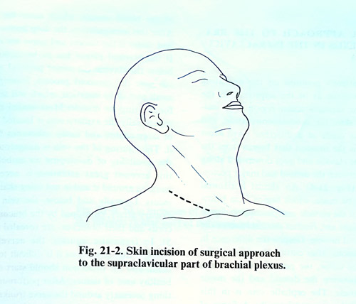
The patient is placed in a supine position on the operating table, a cushion being put under the shoulders, and the head is turned slightly to the opposite side of the operation site. The most suitable incision is the one coinciding with the skin creases in this region, i.e. the incision is oblique regarding the clavicle or almost to the half of the angle formed by the posterior border of the sternomastoid muscle and the clavicle (Fig. 21-2). Together with the skin and the subcutaneous tissue, the platysma, and the superficial cervical fascia are divided. The superficial jugular vein, which goes under the skin is also divided between ligatures or is retracted. After that the middle cervical fascia is exposed where the sleeve of the omohyoid muscle can be seen. This muscle is situated under the clavicle upon the brachial plexus; it should be freely retracted laterally and superiorly; the middle cervical fascia must be incised inferior to the muscle. If this does not ensure a sufficiently wide operative field, the muscle can be divided in the middle, at its tendon between its two bellies. The brachial plexus is revealed in the loose fatty and fibrous tissues under the fascia.
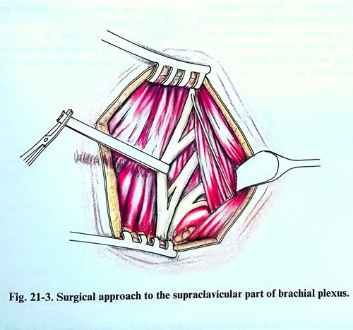
In seeking the nerve trunks it is best to reach the surface of the scalenus anterior muscle. The phrenic nerve is exposed on its anterior surface and should be preserved. Laterally from the scalenus muscle the C5, C6, C7, C8 and the T1 nerves are exposed, which are the roots of the brachial plexus. The anterior branches of the C5 and C6 spinal nerves go downward and forward and join into one common trunk (superior trunk). Immediately below them the anterior branch of C7 nerve is separated and it continues laterally and inferiorly as the middle trunk of the plexus. Deep behind the clavicle, the subclavian artery and vein are revealed, and behind them the anterior branches of C8 and TI nerves, which soon after their exit from the spinal canal join together as the inferior trunk of the plexus. Owing to their deep location their dissection is more difficult and it is necessary that the patient's shoulder be lowered as much as possible (Fig. 21-3).
In dissecting the roots and the trunks of the plexus one must have in mind that, superficially on the nerve trunks and parallel to the clavicle, pass the suprascapular artery and the superficial artery of the neck, while deep between the C6 and C7 nerves may pass the transverse artery of the neck. These arteries must be preserved, especially when the plexus injury is accompanied by injury of the subclavian or axillary arteries, as they serve the collateral blood circulation of the upper limb. Utmost attention is given to the preservation of the pleural cupola, internal jugular vein, and subclavian artery situated within the limits of the operative field, as even small damage leads to haemorrhage, which is difficult to stop due to the depth of the blood vessels. In this region the injury of veins may provoke an air embolism, if the body is too elevated. On this account the surgical manipulations should be performed under visual control and without the use of sharp instruments.
Depending on the pathological changes found, neurolysis or resection of the neuroma is made with subsequent suturing of the nerve trunks. The technique of neurolysis and suturing of the nerves does not differ from those on other peripheral nerves. For overcoming the nerve gaps when suturing, the patients head has to be turned to the injured side, and the shoulder must be elevated as high as possible.
SURGICAL APPROACH TO THE BRACHIAL PLEXUS IN THE INFRACLAVICULAR FOSSA
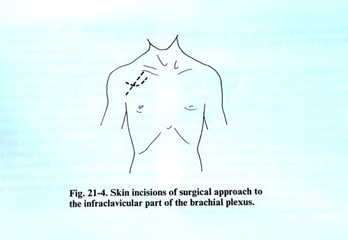
The patient's position on the operating table is the same as in the approach to the plexus in the supraclavicular region, however, the incision of the skin, the subcutaneous tissue, and the fascia is made in a different way. Most practical is the incision that begins from the middle of the clavicle and goes downward along the sulcus between the deltoid and major pectoral muscles (Fig. 21-4). An almost horizontal incision can be made, which begins 5 cm below the middle of the clavicle and proceeding downward at a slight arc and reaches the insertion of the major pectoral muscle. Despite the difference in the skin incisions, after opening the skin and the subcutaneous tissue, the dissection goes into the sulcus between the deltoid and the major pectoral muscle. The cephalic vein is in this sulcus and it can be retracted laterally. The insertion of the major pectoral muscle is elevated under its insertion, and the muscle is divided across near the humerus and lifted. After that it is retracted medially, taking care not to injure the branches of the brachial plexus and vessels, which supply it.
After this surgical step, the deep pectoral fascia and underlaying vessels and nerves are revealed.
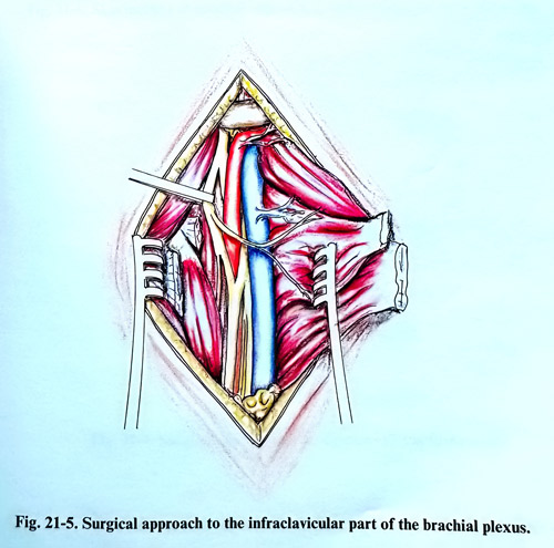
If the brachial plexus has to be explored just below the clavicle the minor pectoral muscle is also cut near the coracoid process, leaving only a small part of its insertion, which will serve later for suturing it back. Most medial and most superficially is located the axillary vein which is of large calibre and many tributaries flow into it. An injury of the vein is dangerous due to the possibility of developing air embolism. On this account great attention is necessary in working around it and not using sharp instruments. Laterally and below are exposed the vein and the axillary artery, enveloped by the brachial plexus cords and their branches, (Fig. 21-5).
In traumatic injuries, the nerves appearence is significantly changed that sometimes it is difficult to identify them, and the dissection should start always from anatomically preserved structures and tissues area. After performing everything necessary around the nerve trunks - neurolysis, suture(s) and other techniques, the minor and major pectoral muscles are sutured to their places of attachment. The stitches are placed laterally from the brachial plexus to avoid the development of adjacent scar tissue from these sutures over the nerves.
SURGICAL APPROACH TO THE BRACHIAL PLEXUS IN THE AXILLARY FOSSA
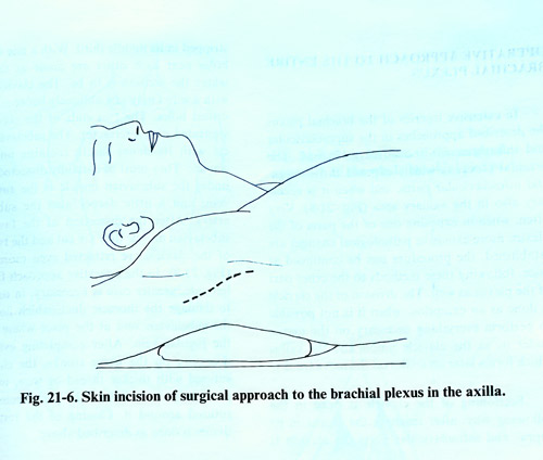
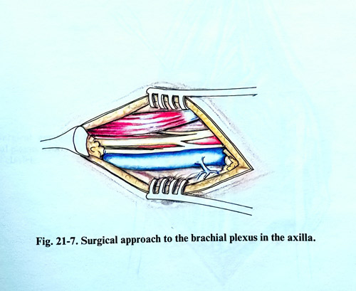
The patient is in a prone position on the operating table with a small cushion being placed under the shoulders. The arm is put on a separate support, in abduction position almost at a right angle. The incision of the skin, the subcutaneous tissue, and the fascia begins from the depth of the axillary fossa and goes distally along the medial sulcus of the biceps muscle (Fig. 21-6). The vessels and nerves are reached after the incision of the fascia. The identification of the individual nerves can be done easily, if their relation to the brachial artery are used as reference. Both initial branches of the median nerve enclose the brachial artery, while the nerve itself is situated along the antero-lateral artery surface. The musculocutaneous nerve is situated laterally, passing through the coracobrachial muscle and innervating it. Medially from the artery are found the ulnar nerve, the medial cutaneous nerves of the arm and forearm (Fig. 21-7). The two latter nerves are considerably thinner than the ulnar nerve. To expose the axillary and radial nerve better, the artery must be slightly retracted, as the nerves are located behind it. Tracing these nerves upward permits exploration of the brachial plexus cords, and if they have to be entirely exposed, it is necessary to cut the major pectoral muscle in proximity with its insertion to the humerus.
OPERATIVE APPROACH TO THE ENTIRE BRACHIAL PLEXUS
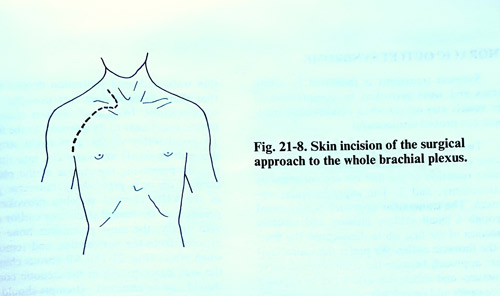
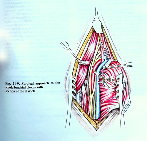
In extensive injuries of the brachial plexus the described approaches in the supraclavicular and infraclavicular fossae can be merged into one approach. The brachial plexus is widely exposed in its supra-and infraclavicular parts, and when it is necessary also in the axillary area (Fig. 21-8). Very often, when exposing one of the parts of the plexus and more extensive pathological changes are established, the procedure can be expanded immediately, following the already described methods to the other parts of the plexus as well. The division of the clavicle is done as an exception, when it is not possible to perform everything necessary on the nerves under it, as the clavicle union and the callus which forms later on can interfere with the nerve trunks.
Sectioning of the clavicle is done in the following way: after reaching the plexus in its supra- and infraclavicular parts, the clavicle is stripped of periosteum in its middle third. With a fine drill two holes near each other are made at the place where the incision is to be placed. The clavicle is cut with a Gigli saw obliquely between the two drilled holes. The two ends of the clavicle are separated with a retractor. The subclavian muscle with its dense fascia remains under the clavicle. They must be carefully dissected, as just under the subclavian muscle is the subclavian vein, and a little deeper also the subclavian artery. After the dissection of the fascia and the subclavian muscle, they are divided and the two parts of the clavicle are retracted even more widely (Fig. 21-9). In the operative approach from the left side, care should be taken in order not to damage the thoracic duct, which flows into the subclavian vein at the place where it joins the jugular vein. After completing everything necessary on the nerve trunks, the clavicle is fixed with a thicker thread or wire, using the previously made holes. The periosteum is also sutured around it. Closing of the rest of the tissues is done in the standard way as described above.