THORACIC OUTLET SYNDROME
Surgical treatment is indicated in symptoms and signs provoked by compression of the vessels and nerves i the thoracic outlet when conservative treatment has proved unsuccessful. Two basic surgical approaches are in use in a case of scalenus anterior syndrome: 1. Transaxillar resection of the first rib accompanied by scalenotomy, and 2. The supraclavicular approach.
The transaxillar approach is performed through a small axillary incision and involves resection of the first rib to decompress the floor of the thoracic outlet. We prefer the supraclavicular approach because the compromised nerve structures and subclavian artery can be exposed more easily and completely.
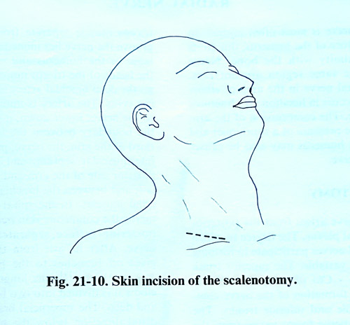
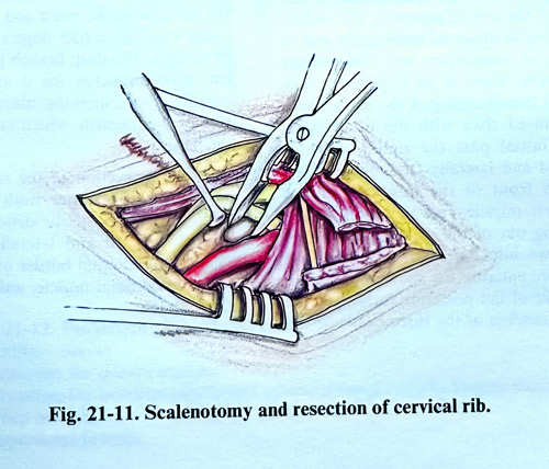
The patient in the supraclavicular approach is placed on the operating table in the supine position, as in the operative approach to the brachial plexus. The skin incision of 6 - 8 cm begins from the sternoclavicular joint and goes posteriorly and obliquely super-iorly along the skin creases (Fig. 21-10). After opening the skin and the subcutaneous tissue together with the platysma, the clavicular portion of the sternomastoid muscle is retracted medial or is transected a 1 cm distance from the clavicle. Cutting the middle cervical fascia and retracting the omohyoid muscle superiorly, the scalenus anterior muscle is reached and dissected. The phrenic nerve goes along the anterior surface of the scalenus anterior muscle in proximity to its medial border and a little obliquely, then inferiorly enters the thoracic cavity. The interscalenic space is penetrated and inspected the subclavian artery passing through it together with the nerves. During this penetration the phrenic nerve and accessory phrenic nerve should be preserved. On the left side the thoracic lymphatic duct and its affluent channels can be seen. They have very thin walls and need protection despite the fact that their damage does not provoke serious complications. In case of obvious stenosis, the anterior scalenus muscle is divided near to its insertion to the first rib. To avoid injury of the anatomical structures, this is done slowly and carefully through the interscalenic space preserving the cupola of the pleura. If a cervical rib or a protruding transverse process of the VIIth cervical vertebra provokes compression on the neural structures and/or subclavian artery, the most prominent bone part is exposed under the periosteum, and nibbled in small pieces (Fig. 21-11). All fibrous bands in the area contributing to the stenotic condition should also be removed. It is not necessary to remove the entire additional rib or the whole hypertrophied transverse process of the VIIth cervical vertebra, as this involves too strong retraction of the nerves and arteries. After completing the work inside the interscalenic space, the space between the clavicle and the first rib is explored. If there is residual stenosis, resection of the first rib is also indicated. Meanwhile, the transverse cervical artery, which passes within the limits of the operative field, should be spared.
After completing the decompression, the middle part of the omohyoid muscle is sutured to the middle cervical fascia, the clavicular part of the sternomastoid muscle is sutured, and at the end a suture is made of the superficial fascia together with the platysma and the skin.
There are no specific complications in the postoperative period. The pain and paraesthesias disappear either immediately after the operation or in the following days. Muscle atrophy, if there was any, improves later on.