
MEDIAN NERVE
The median nerve is most often damaged in open injuries of the upper limb, especially in injuries of the inferior third of the forearm and wrist. It can be involved also in fractures of the arm and forearm. Gunshot injuries of the median nerve are most frequent at the elbow.
SURGICAL ANATOMY
The median nerve is formed from two initial branches, which separate from the medial and lateral cords of the brachial plexus. These two branches join at the level of the inferior border of the minor pectoral muscle immediately anterior to the axillary artery. The two initial branches of the median nerve or the so-called 'fork', can be discovered at different levels from the minor pectoral muscle to the middle of the arm. Sometimes it may be double. Along the whole length of the arm the nerve accompanies the brachial artery, being in the beginning situated anteriorly and laterally to it. In the middle of the arm it goes exactly anteriorly to the artery, while in the lower third - medially to it. In about 10 - 13% of cases, the artery can be discovered over the nerve (superficial brachial artery), or may be double, so that one branch is over the nerve, the other under it. Along the entire arm the nerve has the same thickness, excluding about 40% of cases, in which it is connected with the musculocutaneous nerve. In 5% of cases the musculocutaneous nerve is part of the median nerve for a certain distance or along the entire length for the musculoputaneus nerve. In such cases the branches for the biceps and the brachial muscle come out of the median nerve at the level of the middle third of the arm or at the level of the border between the middle and superior third. Coming down into the elbow pit (cubital fossa), the nerve is situated medially from the artery and the biceps tendon. At this place it lies approximately in the middle between the biceps tendon and the medial humerus epicondyle. In the elbow pit separate the small branches for the fingers and the wrist flexors. Most superiorly separate the small branches for the brachial head of the pronator teres. If one branch comes out of the nerve, it is secondarily divided along its path and not less than two branches enter into the pronator teres muscle. The level of separation of these branches varies within the limits from the superior limit of the inferior third of the arm to the elbow. Somewhat more distally separate the small branches for the ulnar part of pronator teres and for the flexor carpi radialis muscles. At the same place separate the small branches for the superficial flexor of the fingers and the palmaris longus muscles. A little more distally to the elbow separate the small branches for flexor pollicis longus, the deep flexor of the fingers and the anterior interosseous nerve. Very often they separate as a common trunk. In the depth of the elbow pit, the nerve passes between the two heads of pronator teres and continues in the forearm, approximately along its axis, lying between the deep and superficial flexors of the fingers. In the distal third of the forearm the median nerve goes more superficially, being situated between the tendons of the flexor carpi radialis and the palmaris longus muscles. Less frequently, it is between the tendons of the palmaris longus and the flexor digitorum superficialis muscles. Distally the nerve passes together with the tendons of the flexors of the fingers through the carpal tunnel and reaches the palm, where it divides into its terminal branches. This division most often takes place in immediate proximity to the distal end of the transverse carpal ligament. It is divided in two branches, which, on their part, branch out into the thenar and the common nerves for the fingers. The small branches for the thenar muscles are always situated more superficially to the common trunk of the nerve.
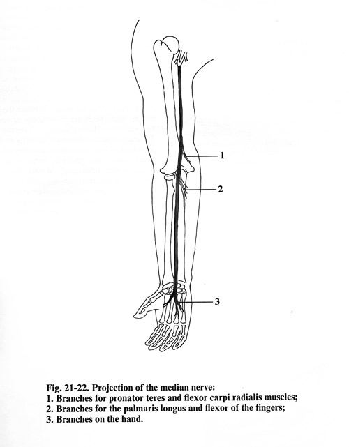
The median nerve is projected on the arm along the line, which goes downward along the internal sulcus of the biceps to the middle between the tendon of the same muscle and the medial epicondyle of the humerus. In the forearm the nerve projection is situated exactly along its anterior surface midline (Fig. 21-22).
SURGICAL APPROACHES
When exposing the median nerve in all parts of the upper limb, the patient is placed in a supine position on the operation table, with the arm slightly abducted and placed on a separate small table.
SURGICAL APPROACH TO THE MEDIAN NERVE IN THE ARM
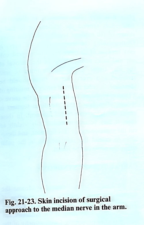
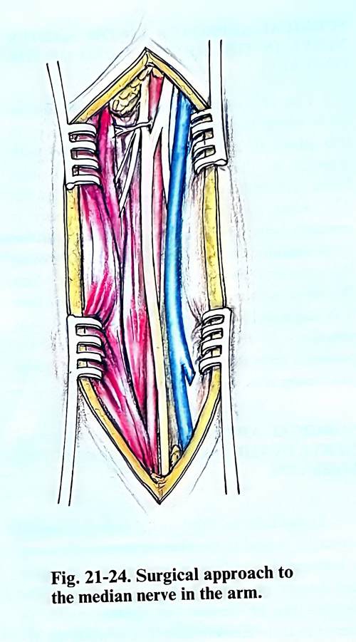
The skin incision is made in a vertical direction 2 - 3 cm anterior or posterior to the internal sulcus of the biceps (Fig. 21-23). Along the same incisioned are divided the subcutaneous tissue and the fascia. Next is penetrated under the fascia a slightly medially to reach the brachial artery and the median nerve. Due to the close proximity of the median nerve to the brachial artery there is very often a combined injury of both of them, and in the separation of the nerve requires a meticulous work (Fig. 21-24). The pulsations of the artery serve as an orientation in exposing the nerve. When closing the surgical approach , it is important that nerve repair and fascia closure are not one over the other to avoid additional scars over the nerve suture.
SURGICAL APPROACH TO THE MEDIAN NERVE IN THE ELBOW
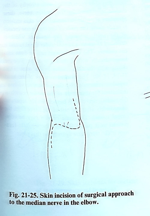
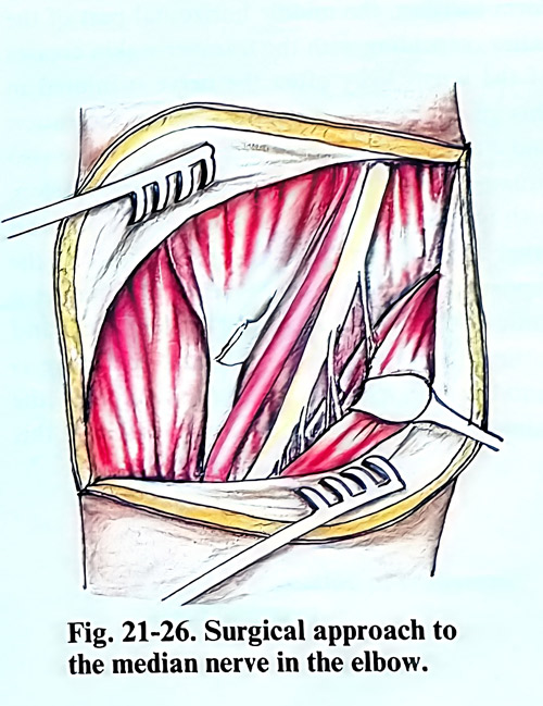
In exposing the median nerve in the elbow it is best to have the skin incision made in the form of an 'S', so that the horizontal part of the letter coincides with the elbow pit crease (Fig. 21-25). In the subcutaneous layer, major veins are usually found, and if an obstacle for the approach, can be coagulated and cut. The fascia is separated and divided with an incision medial to the nerve, so that a flap is made to cover the nerve without I thets sutures be in contact with nerve. The location of the nerve is usually in the middle between the biceps tendon and the medial humerus epicondyle. The nerve can also be located at 1 - 2 cm medial to the artery whose pulsations can easily be felt by palpation. If it is necessary to expose the nerve distally, the pronator teres is retracted medially and distally. At this place, the small branches of the nerve separate for the wrist and the finger flexor muscles and their identification in the scar tissue, and preservation respectively, may be difficult (Fig. 21-26). In case of median nerve entrapment by the lacertus fibrosus, hypertrophic musculus teres major, or the fibrous arch of the superficial flexor of fingers, they may be undercut.
SURGICAL APPROACH TO THE MEDIAN NERVE IN THE MIDDLE THIRD OF THE FOREARM
The skin incision in this region can be made along the projection of the nerve, as in this place it is located deep to the surface and is covered by the ventral muscle groups, so that there is no danger of skin scars interfereing with the nerve suture area (Fig. 21- 27). After the fascia is opened, the space between the pronator teres and flexor carpi radialis muscles is dissected. It is penetrated in depth through the superficial flexor of the fingers, under which the nerve is located (Fig. 21-28). From this location it is easily followed in a distal direction. If it is necessary to follow the nerve more proximally, the pronator teres should not be incised, but a tunnel should be created under it.
SURGICAL APPROACH TO THE MEDIAN NERVE IN THE INFERIOR THIRD OF THE FOREARM
In the inferior third of the forearm, special attention is paid to the skin incision as the nerve is between the tendons of the flexors of the wrist and fingers immediately below the fascia. The incision can be longitudinal, but slightly medial or lateral to the nerve projection. It cam be also convenient to make an 'S'-shape incision, the middle horizontal part of the latter coinciding with the transverse skin creases of the wrist. Very often the nerve is injured in this place by transversally running laceration and cut injuries. In these cases, the scars are also situated transversally, and after their excision, with two additional incisions one superiorly and other inferiorly in the form of the letter 'S', the exposure of the nerve is made possible in a sufficiently broad area. After exposing and dividing the fascia of the forearm, the nerve is found in the space between the tendon of the palmaris longus and flexor carpi radialis muscles. At this place it is very often required to dissect the adhesions around the tendons or even to repair tendons together with the nerve repair.
Distinguishing the tendons from the nerve is easy because the nerve has more intense pink colour and different vascularity. In the removal of the scar tissue, every tendon must be well stripped and every one of them should be identified, in order to avoid errors of suturing a tendon to the nerve. In dissecting the nerve it is very often found that the distal segment is under the transverse carpal ligament. In these cases it may be recommended only slightly to cut the superior end of this ligament, and thereafter to extend the skin incision in the palm where the nerve can be exposed.