ULNAR NERVE
Traumatic injuries of the ulnar nerve are most often in the region of the elbow in fractures of the inferior part of the humerus. Not rarely, injuries also occur in wounds, especially those caused by cutting in the inferior third of the forearm. In the arm, due to the proximity of the ulnar with the median nerve, very often, their injury is combined. Very rarely also, a chronic lesion of the ulnar nerve is obtained, which we designate as ulnar tunnel syndrome' or when it is manifested late after the consequences of a fracture and dislocation of the elbow, as 'ulnar palsy'.
SURGICAL ANATOMY
The ulnar nerve arises from the medial cord of the brachial plexus. It contains fibres from the C8 and Tl spinal nerves. Approximately in half the cases, it obtains a bundle of the lateral cord of the plexus, known as the lateral pedicle of the nerve. At its origin it is situated medially from the axillary artery and continues distally to the middle of the arm together with the brachial artery, preserving the same relation. After that, the nerve separates from the artery. Running in a slightly dorsal direction, it perforates the medial inter-muscular septum of the brachial fascia and passes posteriorly together with the superior collateral artery. At the level of the elbow, the nerve is situated superficially and lies in the vertical sulcus behind the medial epicondyle of the humerus. At this place, it is situated superficially immediately below the fascia and can be easily palpated. From there, the nerve goes down to the forearm, passing between the two heads of the flexor carpi ulnaris. At the forearm, the nerve is situated on the deep flexor of the fingers and in the space between the tendons of the flexor carpi ulnaris and flexor digitorum superficialis muscles. In the proximal third of the forearm, it is accompanied by the ulnar recurrent artery, while in the middle and the distal thirds - by the ulnar artery situated laterally to ithe nerve.
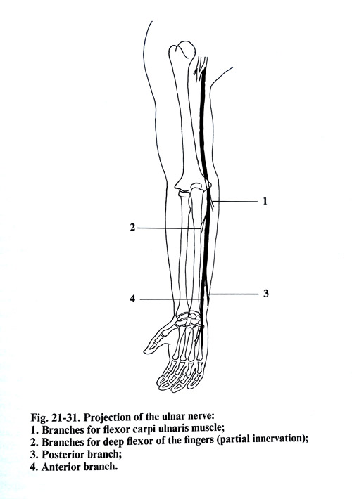
The articular branches for the elbow joint separate from the ulnar nerve 7 - 8 cm above the level of the medial epicondyle and below this level are the branches for the flexor carpi ulnaris muscle. These last muscle branches are superior and inferior, penetrating into the muscle in the superior third of the forearm. The branch innervating the medial part of the deep flexor of the fingers is situated at the level of the medial epicondyle of the humerus.
On the border between the middle and inferior thirds of the forearm, the nerve is divided into two branches. One of the branches - the dorsal branch - innervates the ulnar half of the dorsasl surface of the hand and the dorsal surface of two medial fingers (4th and 5th) and a half, excluding the distal phalanges. The second branch - the palmar branch - passes down along the palmar surface of the hand, innervating the muscles of the hypothenar eminence, part of the muscles of the thenar eminence, the interosseous muscles and the skin of the anterior surface of the 5th finger and the medial surface of the ring finger along with the respective part of the palm.
The projection line of the ulnar nerve on the arm surface begins from the depths of the axillary fossa and goes toward the medial epicondyle of the humerus, while on the forearm it goes from the same epicondyle toward the pisiform bone (Fig. 21-31).
SURGICAL APPROACHES
The patient's position on the operating table is supine with the upper limb abducted on a separate table in supination position.
SURGICAL APPROACH TO THE ULNAR NERVE IN THE ARM
In the upper and middle third of the arm the surgical approach to the ulnar nerve coincides with the approach to the median nerve. The skin incision is made 2 - 3 cm anterior to the internal groove of the arm. The subcutaneous tissue and the fascia are divided along the same incision. The fascia is dissected medially, revealing the biceps muscle and medially from it the vessels and the nerve bundle. The ulnar nerve is situated along the internal side of the brachial artery. When the skin incision is along the projection of the nerve, it is best to divide the fascia 2-3 cm more laterally from the nerve and artery. In this way the nerve suture will be away from the scars of wound healing.
In the exposure of the nerve in the inferior two thirds of the arm, the skin incision passes 2 - 3 cm dorsally to the nerve projection. Through the same incision the subcutaneous layer and the fascia are divided. After that the fascia is dissected anteriorly and the nerve is revealed between the lateral border of the triceps and the internal intermuscular septum.
SURGICAL APPROACH TO THE ULNAR NERVE AT THE ELBOW AND TRANSPOSITION OF THE NERVE
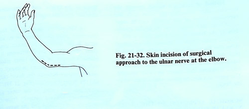
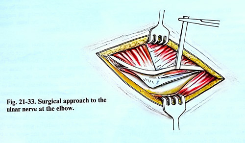
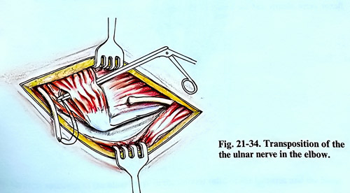
The skin incision is made on the medial surface of the elbow 2 cm anterior to the medial epicondyle of the humerus, beginning 5 - 6 cm above it and reaching 5 - 6 cm under it. Along the same incision are opened the subcutaneous tissue and the fascia. This incision is situated anterior to the projection of the nerve and final stitches are not over the damaged nerve. Separating the fascia slightly posteriorly, the nerve groove situated posterior to the internal epicondyle is reached where the nerve can be dissected very easily (Figs. 21-32; 21-33 ). Small gaps of the nerve can be approximated by fixing the upper limb in extension of the elbow. In traumatic injury of the nerve with large gaps, after exposure of the nerve, it can be transposed and sutured anteriorly to the elbow. The same transposition can also be made in cases of compression of the ulnar nerve in the groove after fracture, luxation or stenosis. In flexing the limb at the elbow, the nerve is fixed to its bone sulcus and is slightly stretched. Transposing the nerve anterior to the elbow joint in flexion position makes it possible to overcome a nerve defect of as much as 11 cm. Transposition of the interrupted ulnar nerve can be done by introducing one of the nerve segments through the muscles just below the medial epicondyle of the humerus, or over the muscle. In the superior part of the incision, the ulnar nerve is dissected between the triceps and the internal medial intermuscular septum and retracted superiorly.
A small part of the internal intermuscular septum should be excised at the place where the proximal segment of the interrupted nerve will be transfered onto the anterior surface of the elbow. The distal segment is revealed on the forearm in the space between the flexor carpi ulnaris muscle and the flexor digitorum superficialis. In dissecting the nerve, the branches for the flexor carpi ulnaris and the flexor digitorum profundus are spared. With a long instrument, a tunnel is made between the superficial and deep muscle groups of the forearm. The instrument is introduced under the pronator teres of the forearm, the flexor digitorum superficialis and the flexor carpi radialis. Depending on which of the two nerve ends - proximal or distal - is longer, it is inserted through the tunnel with the help of a long instrument, after which a suture of the nerve is made. The place of the suture usually remains in the muscle tunnel (Fig. 21-34).
Instead of making a tunnel through the muscles, after dissecting the nerve ends, the superficial layers of the muscles can be separated from their attachment, near the medial epicondyle of the humerus and from them a flap is turned over the sutured nerve. The muscles are sutured to the bone attachment at their original place where was left a small tendinous remnant. The nerve can also be placed just under the fascia.
SURGICAL APPROACH TO THE ULNAR NERVE IN THE FOREARM
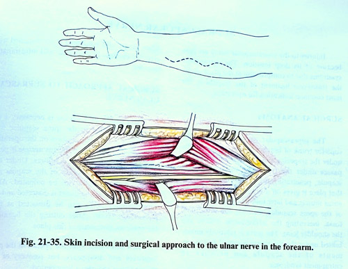
In the superior third of the forearm, the ulnar nerve is situated under a thick layer of muscles. The skin incision is made along the projection of the nerve, i.e. along the line which connects the medial epicondyle of the humerus with the pisiform bone of the wrist. The nerve can easily be discovered between the flexor carpi ulnaris and the flexor digitorum superficialis.
The intermuscular space is not free, as the muscles are adherent among themselves, so that they must be separated exactly along the connecting fibrous plane between each two muscles. The space between them is widened with a retractor, and the ulnar nerve is discovered in the depths on the anterior surface of the deep flexor digitorum. In the most superior part of the forearm the nerve gives branches for the flexor carpi ulnaris and for a part of the deep flexor of the fingers, and its branches must be carefully dissected together with the nerve.
In the middle third of the forearm, an important point of orientation in exposing the nerve is the flexor carpi ulnaris muscle. It should be penetrated between it and the palmaris longus, or if the latter muscle is not well developed, as occurs in many cases, it should be between the flexor carpi ulnaris and the flexor digitorum (Fig. 21-35). The ulnar nerve passes parallel to the ulnar artery, the nerve being situated medially from the artery while the entire vessel and the nerve are under the fascia, which envelops the deep muscles of forearm. This fascia must be divided to reach the nerve. After performing the surgical interventions on the nerve, it will remains under the flexor carpi ulnaris.
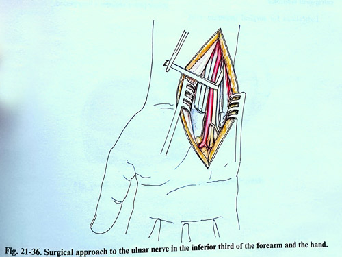
In exposing the ulnar nerve in the inferior third of the forearm and about the wrist, it is best to make the skin, subcutaneous layer and fascia incision in the 'S' shape. At this location the nerve is positioned immediately lateral to the ulnar artery. In the lower part of the surgical field, the ulnar nerve divides into anterior and posterior branches. The anterior branch follows the same direction of the nerve through the canal of Guyon, and the posterior goes under the tendon of the internal flexor of the wrist as a cutaneous nerve to the hand (Fig. 21-36).