RADIAL NERVE
The radial nerve is most often injured on the arm in a fracture of the humerus, due to its location in proximity with the bone. Nerve contusions in the same region are not rare. Injury of the radial nerve in the elbow area most frequently occurs in luxation and fractures of the radial bone. The compression of the arm during sleep or the pressure of a tourniquet and operations on the humerus may also be causes of injury to this nerve.
SURGICAL ANATOMY
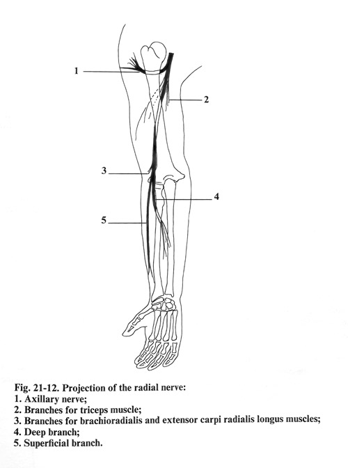
The radial nerve arises from the posterior cord of the brachial plexus. The degree to which the different spinal nerves participate in forming the radial nerve is variable. The superior trunk of the plexus (C5 - C6) plays a considerably greater role in the formation of the nerve compared to the middle and inferior trunks. The level at which the nerve forms varies from the supra-medial to infra-lateral margin of the minor pectoral muscle. From this level to the inferior end of the tendon of the latissimus dorsi muscle, the radial nerve is the thickest branch of the brachial plexus. If the epineurium of the radial nerve of the arm is removed, the individual bundles can be dissected more easily and at a great length in comparison with the median and ulnar nerves. There is an impression that the intraneural interchanging of the nerve fibres is less pronounced than with the other large nerves. This initial part the radial nerve is situated behind and laterally from the axillary artery, just in front of the insertion of the latissimus dorsi and the teres major muscles. Coming out of the axillary cavity, the nerve goes down into the spiral groove of the humerus. Before entering the spiral groove separate the cutaneous nerve of the posterior surface of the arm and the branches of the three heads of the triceps muscle. In the spiral groove the nerve lies immediately on the periosteum of the humerus and is enclosed between the heads of the triceps muscle. Together with it go the deep brachial artery and the accompanying veins. The artery is initially situated lateral to the nerve; lower down, it passes medially. At the boundary between the inferior and middle third of the arm, the nerve, perforates the lateral intermuscular septum, and goes down on the anterior side of the arm and the elbow, where it is found between the brachial and the brachioradial muscles. In the spiral groove of the humerus the cutaneous skin nerve for the forearm posterior surface separates from the radial nerve. After its exit from the canal the nerve gives off branches to the brachioradialis and extensor carpi radialis longus. Anterior to the elbow it is divided into two branches: superficial and deep. The superficial branch continues in a distal direction below the brachioradialis and, lateral to the radial artery, passes below the tendon of this muscle and comes out above the fascia on the boundary between the middle and inferior third of the forearm. Its terminal branches innervate the skin of the radial half of the dorsum of the palm and the dorsum of the radial two and a half fingers without the distal phalanges. The deep branch perforating supinator, gives branches for it and after that also supplies the remaining muscles on the dorsal side of the forearm, which extend the wrist and the fingers.
The projection of the radial nerve on the surface of the upper limb begins from the middle of the posterior deltoid muscle border, goes downward and laterally as a spiral and reaches the internal border of the initial part of the brachioradial muscle, anterior to the elbow (Fig. 21-12).
SURGICAL APPROACHES
SURGICAL APPROACH TO THE RADIAL NERVE IN THE SUPERIOR THIRD OF THE ARM
The surgical approach to the radial nerve in the superior third of the arm as it emerges from the posterior cord of the brachial plexus, has been described in the exploration of the brachial plexus in the axillary cavity. In reaching the vessels and the nerves it should be comsidered that the radial nerve is located behind the brachial artery. It can be dissected from its origin from the posterior cord of the brachial plexus until it enters the spiral groove and even lower by penetrating between the heads of the triceps. At this place are separated from the nerve a skin branch - posterior cutaneous nerve of the arm, and small branches for the heads of the triceps and care should be taken not to injure these branches.
Near the nerve is the origin of the deep brachial artery.
SURGICAL APPROACH TO THE RADIAL NERVE IN THE MIDDLE THIRD OF THE ARM
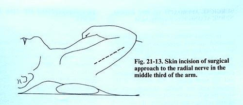
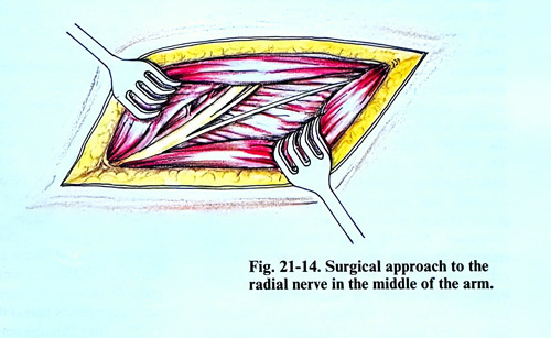
The patient is placed supine on the operating table with the arm adducted at the shoulder, flexed at the elbow and over the thorax. An incision of the skin, the subcutaneous tissue, and the fascia is made about 20 cm long, vertically along the dorsal durface of the arm (Fig. 21-13). After that at the superior end of the incision is penetrated between the long and lateral heads of the triceps and the nerve is exposed over the surface of the humerus. Then the dissected parts of the muscle are split from one another along the muscle fibres, and the nerve is followed distally. In this approach, to reveal the nerve at a greater distance downward is created a tunnel under the lateral head of the triceps. Through its course together with the nerve also run the branches for the heads of the triceps and the deep brachial artery and its branches, whose dissection must be very carefully performed (Fig. 21-14). In the spiral groove also separates the skin nerve (posterior cutaneous nerve of the forearm) and its preservation also requires attention.
SURGICAL APPROACH TO THE RADIAL NERVE IN THE INFERIOR THIRD OF THE ARM AND ELBOW
For the exposure of the radial nerve in the inferior third of the arm the patient is placed in a supine position, with the arm being in adduction.
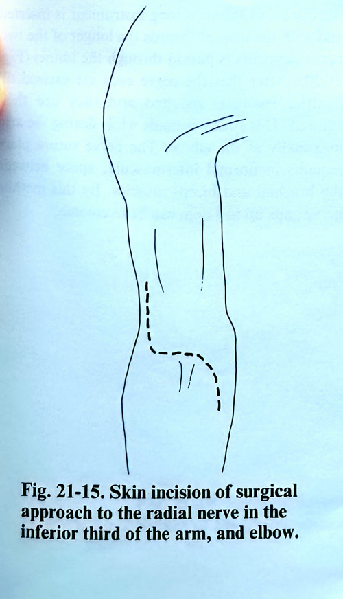
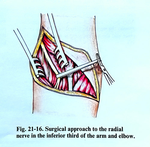
The incision begins 5 - 6 cm above the lateral epicondyle of the humerus and goes downward to the skin creases of the elbow. It follows and, when needed, continues several more centimetres along the lateral side of the forearm (Fig. 21-15). After cutting the skin, the subcutaneous tissue, and the fascia is penetrated between the brachial and brachioradial muscles in depth and in proximity to the bone to the place where the nerve is located (Fig. 21-16). From there downward the nerve is easily followed till the place of its division into superficial and deep branches. At the superior end of this incision do appear usually the small branches to the brachioradial and extensor carpi radialis longus muscles and they deserve attention not to be injured.
SURGICAL APPROACH TO THE RADIAL NERVE ALONG THE ENTIRE ARM
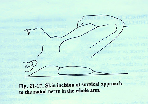
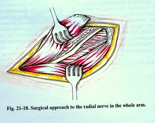
In limited injuries of the radial nerve the described operative approaches to the different levels of the arm are appropriate. If the injury is larger and there is a defect of the nerve, then these incisions are not sufficient to expose the peripheral and the central segment of the nerve, and should be resorted to the merging of the incisions between adjacent local approaches. In these cases we apply a longer skin incision that begins from the middle of the deltoid muscle's posterior border, goes vertically toward the olecranon and 3 cm from it turns laterally, reaching the lateral humerus epicondyle (Fig. 22-17). Along the same incision is also divided the brachial fascia. The surgeon penetrates into the superior part of the incision between the long and the lateral head of the triceps, where the radial nerve is exposed.
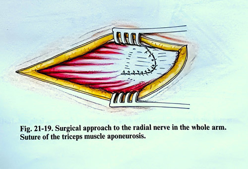
The two heads are separated from one another and reaching downward to where they are attached, they are separated up to the tendon parts attached to the olecranon. After that the lateral head of the triceps is dissected and the tendon parts are cross-sectioned. In this way the lateral triceps head, which encloses the humerus spiral groove from the dorsal side can be turned laterally and the entire canal remains open (Fig. 21-18). After that the nerve can easily be followed superiorly and inferiorly.
After completeing all operative work related to the nerve, the lateral head is repositioned and the tendon plate is sutured (Fig. 21-19).
TRANSPOSITION OF THE RADIAL NERVE
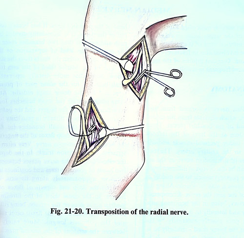
In case of injury of the radial nerve at the arm with large defects, a transpositioning of the nerve is necessary from the posterior to the anterior humerus surface. The decision on transposition of the radial nerve is made during the exploration of the nerve in the entire spiral groove if it is obvious that the nerve defect is large and it is impossible to do an end-to-end suture. For this transposition the nerve is exposed in the superior third of the arm with an incision along the internal sulcus of the biceps muscle distally from the axillary fossa. At this place the central nerve segment is dissected together with neuroma till its emergence from the posterior cord of the brachial plexus, and it is pulled upward. Its small branches going to the triceps are preserved. After that, the distal end of the nerve is revealed in the anterior third of the arm. On both ends of the nerve thicker threads are sutured before being resected, i.e. threads are sutured to the neuroma of the central segment and to the cicatrix of the peripheral end.
After that a tunnel is made with a sufficiently long instrument between the biceps and the brachial muscle. Through this tunnel the long instrument is inserted and the nerve end suture threads on the longer of the two nerve segments is passed through the tunnel (Fig. 21-20). After that the nerve ends are excised till reaching normal nerve structure and sutured. This suture is made in flexion of the arm maximally at the elbow. The nerve suture place remains in a normal intermuscular space between the brachial and biceps muscles. This method can overcome nerve gaps up to 11 cm.
SURGICAL APPROACH TO THE RADIAL NERVE IN THE FOREARM
The radial nerve is exposed in the superior third of the forearm by a vertical incision along the brachioradial muscle as a continuation of the incision for exposure of the nerve in the inferior third of the arm and the elbow. After cutting the skin, the subcutaneous tissue, and the fascia are penetrated under the brachioradial muscle to reveal the nerve on the supinator muscle.
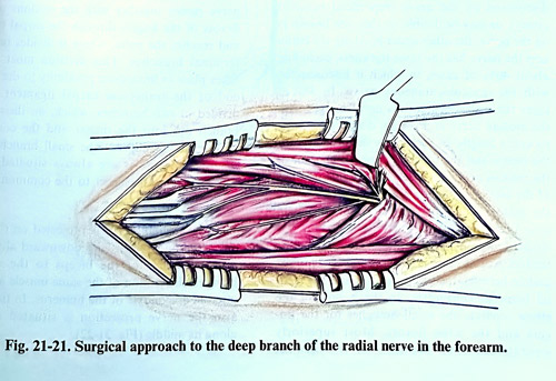
The exposure of the superficial branch of the nerve is achieved by extending the same incision inferiorly along the brachioradial muscle. By the separation of the muscle and its displacement slightly laterally, the nerve is reached. Distally the superficial branch of the radial nerve perforates the fascia laterally to the tendon of brachioradialis muscle and branches in the subcutaneous tissue. The exposure of the deep branch of the radial nerve between the forearm muscles is very rarely made in traumatic lesions or entrapment of the nerve while traversing the supinator muscle. The nerve's small branches in this region are very thin and finding them at times is very difficult. To to expose proper the nerve at this place is made an incision of 10-12 cm long beginning from the lateral epicodyle of the humerus and going down along the dorsal surface of the forearm. After the skin incision, the subcutaneous tissue, and the fascia is penetrated between the extensor digitorum and the extensor carpi ulnaris muscles. These muscles are separated as far as the lateral epicondyle and are retracted (Fig. 21-21). The inferior part of the supinator is revealed an there the deep branch of the radial nerve comes out. In case of entrapment of the deep branch of radial nerve in the canal of the supinator muscle, the approach can be limited. The skin incision is only along the brachioradial muscle, following the radial nerve in the supinator muscle. If there is stenosis in the tendinous entrance into the canal, it is opened along its length.