AXILLARY NERVE
An isolated injury of the axillary nerve is rarely observed, as soon after its separation from the posterior cord of the brachial plexus and in proximity to it, the nerve enters the muscles which it innervates (the deltoid and teres minor muscles). Its injury is more frequently combined with the injury of the brachial plexus.
SURGICAL ANATOMY
The axillary nerve is separated from the posterior cord of the brachial plexus. In the beginning it is located deep inside the axillary fossa, behind the axillary artery, immediately anterior to the subscapular muscle. It goes inferiorly and posteriorly and comes out from the axillary fossa through the quadrilateral foramen, medial to the surgical neck of the humerus. Exiting from the axillary cavity, the nerve gives off a branch for the teres minor, while the greater part of the branches go to the deltoid muscle. One of its branches - the upper lateral cutaneous nerve of the arm - innervates the skin on the external surface of the arm. Together with the nerve are found the posterior circumflex artery and the veins accompanying it.
SURGICAL APPROACH
The initial part of the axillary nerve at the place, where the posterior cord of the brachial plexus separates, is reached in the same way as in the infraclavicular approach to the brachial plexus. During the dissection of the posterior cord, a small branch is exposed that innervates the subscapular muscle and a little distally the axillary nerve emerges. It passes posteriorly toward the quadrilateral foramen of the axillary cavity at the level of the surgical neck of humerus. The nerve can easily be recognised by its location and its separation from the posterior cord of the plexus.
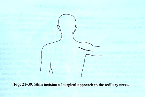
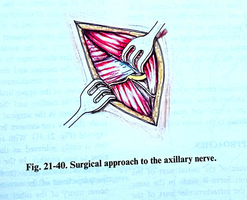
To expose the distal branches of the axillary nerve the patient is placed prone, with the hand abducted slightly laterally on a separate table. The skin incision with the subcutaneous tissue and the fascia is made along the posterior border of the deltoid muscle, beginning from the lateral part of spina scapulae and running to the humerus where the muscle is inserted (Fig. 21-39 ). The posterior border of the deltoid muscle is dissected and retracted cranially and laterally. The long head of the triceps is retracted medially. Penetrating carefully in the depths toward the surgical neck of the humerus is encountered fibrous and fatty tissue, in which the axillary nerve and the posterior circumflex artery are situated together with the accompanying veins (Fig. 21-40).