Legacy: "Atlast of Neurosurgery" / L.Karaguiosov, A. Ramadan, K.Karaguiosov / Kiwait/ 1998
PREV>13. Tumors of The Spine and Spinal Cord
NEXT<15. Malformations of The Spine and Spinal Cord
14. OPERATIONS ON INTERVERTEBRAL DISCS
CERVICAL DISC OPERATIONS
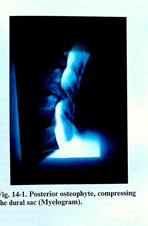
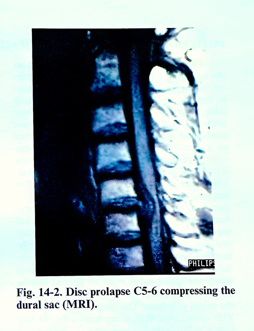
Disc prolapses or osteophytes developing in the cervical spine and compressing the content of the spinal canal have three typical localizations. The median prolapses cause a compression of the spinal cord, the anterolaterally located exert a compression on one side of the spinal cord, and the lateral prolapses or osteophytes provoke only root compressions.
The disc prolapses and osteophytes are located at different levels, most frequently at C 4-5, C 5-6, and C 6-7. Laterally developed osteophytes are able to compromise the vertebral arteries and in some rare cases large anterior osteophytes induce dysphagia (Fig. 14-1; 14-2).In cases of lateral compression causing painful syndromes or radiculopathy, surgery is indicated after failure of conservative treatment.
It is difficult to determine the duration of the conservative treatment, but 3 - 4 weeks are usually sufficient for improvement to be demonstrated. Surgical treatment may be indicated earlier when the pain is intolerable and/or neurological deficit deteriorates quickly. The profession of the patient is the most important consideration, as even mild weakness may affect professional activities.
Spondylotic myelopathy requires surgical decompression when it is progressive and does not respond to conservative treatment. Advanced age is not a contraindication for surgical treatment if the patient's condition permits general anaesthesia for 2 - 3 hours.
The surgical approaches used for the different types of degenerative cervical disc lesions can be posterior and anterior. There are no absolutely strict rules on the indications for these approaches. Large series operated by one approach, either anterior or posterior, when compared, show very little difference in the postoperative success. In many occasions the application of one or other approach depends upon the preference and the experience of the surgeon. Irrespective of this, there are many factors which influence the choice of approach.
Laminectomy remains the preferred option for cases of canal stenosis. It is also appropriate where the myelopathy is mainly due to posterior compression caused by bulging and thickness of the flavum ligaments.
Hemilaminectomy and posterior foraminotomy are particularly appropriate in lateral soft disc prolapses, when there is radiculopathy. They are also indicated when the intervertebral foramen is narrowed by facetal osteophytes or intraforaminal disc prolapses.
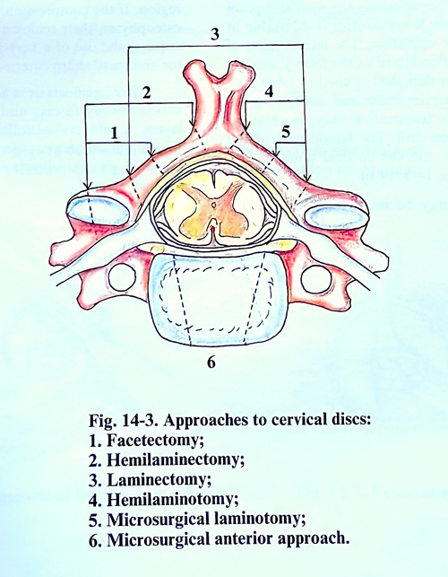
The anterior transdiscal or transcorporal approach, combined with fusion or without it, is indicated where central or anterolateral compression of nerve roots or the spinal cord exists due to disc prolapse or osteophytes (Fig. 14-3).
Multiple level disease is very common in cervical spondylosis, causing radiculopathy or myelopathy. In some cases a decision has to be made about the number of levels which require surgical intervention. In most cases it is possible to decide radiologically which single level is most responsible for the disease by comparing the clinical manifestations with the most severely affected levels. In cases with myelopathy and, less frequently in radiculopathy, the radiologically involved level is often between one and three segments cranial to that which is estrablished from the clinical presentation.
In some cases the extent of the affection may be so widespread that a single responsible level cannot be identified with any degree of certainty. This is particularly valid in cases with canal stenosis. In such a situation all abnormal levels require decompression and therefore demand laminectomy.
POSTERIOR APPROACH IN RADICULOPATHY
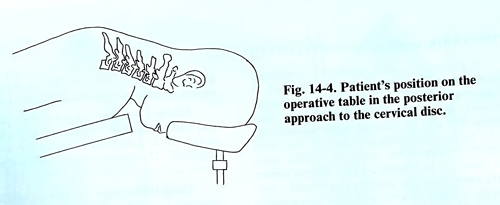
The patient is placed in a prone position on the operating table as in craniectomy of posterior fossa (Fig. 14-4). The sitting position can also be used. To avoid air embolism, the operating table is angled in such a way that the lower limbs are in flexion at both the hip and knee joints, and the head is slightly flexed and fixed on a special frame. Before the operation, X-ray control of the level is advisable using a needle introduced at the supposed level as a marker. This X-ray control may also be done later after reaching the vertebral laminae.
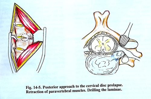
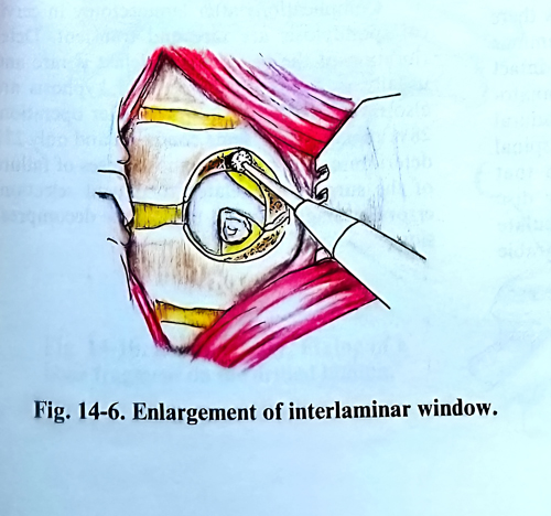
The skin incision is in the midline, 5 cm in length. Penetration follows along the midline and the nuchal septum and the spinous processes. The laminae and the articular processes of the two neighbouring vertebrae at the level of the disc prolapse are stripped. Confirmation of the level where the spinal canal must be opened can be done at this stage of the operation by palpating the spinal processes. The spinous process of the VIth cervical vertebra is recognised by the fact that it is bifid, whereas on the underlying VIIth is not. At the same time it is the last vertebra to which the nuchal septum is attached. New X-ray control is advisable at this stage of the operation. The lateral parts of the laminae and one third of the facet joints are drilled, leaving a thin shell of cortical bone over the intervertebral foramen and the underlying the nerve root. This fenestration may be enlarged with a high-speed drill. The lateral part of the flavum ligament and the cortical bone is then easily lifted (Figs. 14-5; 14-6).
The root may be distended if an underlying anterior disc fragment or osteophyte is present, and care must be taken not to injure the stretched root. According to the size and exact location of the compression, this initial foraminotomy may be enlarged to a facetectomy, hemilaminotomy or hemilaminectomy. If venous bleeding occurs from the epidural space, it is easily controlled with bipolar coagulation.
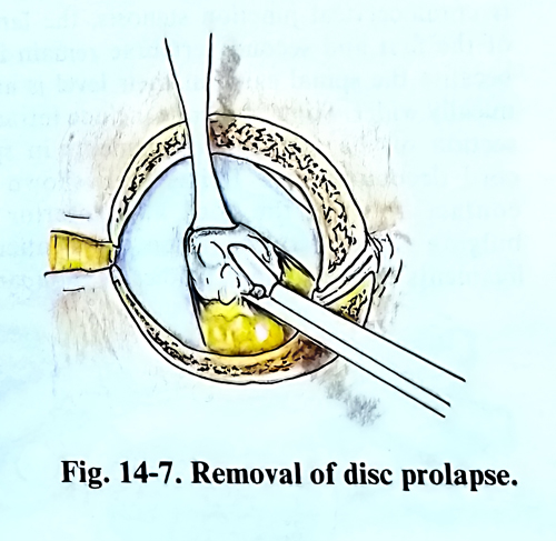
Exploration is initially carried out in the axilla of the root. A blunt hook is an excellent instrument for this purpose. The disc fragments are usually extruded and are easily removed (Fig 14-7). Not infrequently, the disc bulging is within the posterior ligament or annulus and an incision must be made to remove the fragment(s). It is unnecessary to remove large amounts of disc material or to curette the disc space as would be done with a lumbar discectomy, and the surgeon must be satisfied that no compressive fragments are left behind. Exploration with a blunt hook should be made also above, below, and medial to the root to be certain that all fragments are removed. They generally are small in comparison with those in the lumbar region. If the compression of the roots is due to osteophytes, their removal is more difficult and requires the use of a very fine periosteal elevator and small sharp curettes.
After haemostasis is achieved, the muscles, subcutaneous fascia, and skin are closed in layers. A soft cervical collar is generally applied for the immediate postoperative period, but it is removed on the following days, and rehabilitation is started.
POSTERIOR APPROACH IN MYELOPATHY
A posterior surgical approach is commonly employed to decompress the spinal cord; it consists of laminectomy with or without foraminotomy and section of the denticulate ligaments. The aim of decompressive laminectomy is to enlarge the spinal canal by removing posterior compressing structures, which allow the dural sac and spinal cord to migrate posteriorly away from the compressing ventral osteophytes. It also relieves posterior compression caused by enfolding and hypertrophy of the flavum ligaments.
The spinal cord in myelopathic patients is highly vulnerable and requires protection. Extreme flexion or extension during intubation for general anaesthesia can cause at times irreparable ischaemic cord damage. Determining the limit of neck movements before induction of anaesthesia and not exceeding them is most important.
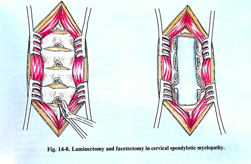
The surgical technique has no differences from the general rules of laminectomy. The patient is placed in a prone position on the operating table with the head fixed in a special headrest with a pin fixator as in operation of posterior fossa. The sitting position is convenient for the surgeon, but there is a risk in elderly patients, and in patients with unstable circulation. The laminae are resected laterally up to the lateral dimension of the dural sac. If it is necessary, the medial parts of joint facets can be also removed (Fig. 14-8). Complete facetectomy can be done in case of stenosis of the intervertebral foramen and there is evidence of radiculopathy corresponding to that level. Unless there is craniocervical junction stenosis, the laminae of the first and second vertebrae remain intact because the spinal canal at their level is anatomically wider. Some surgeons include intradural section of the denticulate ligaments in spinal cord decompression. It has been shown that contact between the cord and anterior disc bulging is not reduced when the denticulate ligaments are sectioned. Studies of comparable groups of patients with and without denticulate section revealed no difference in surgical results.
Epidural bleeding is not a serious problem and haemostasis can be achieved using bipolar coagulation, pieces of Gelfoam or oxidised cellulose. The muscles, fascia, subcutaneous tissue, and skin are closed in layers.
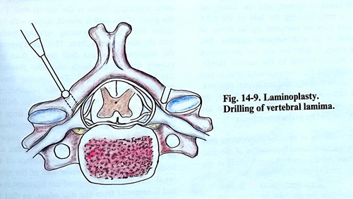
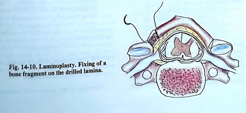
By using laminoplasty, the spinal canal can be enlarged by opening the laminae on one side to create an 'open door'. After dissecting the posterior apophyses, the laminae and the facet joints, the drill is used to open through at the lamina-facet junction in the same fashion as for 'en bloc' laminectomy. The inner cortex and the flavum ligaments are completely removed only on one side (usually the side with the most significant pathology). On the other side, the drilling is completed through the outer cortex only (Fig. 14-9). The bloc of the laminae is carefully elevated 10 - 15 mm from the dura. The tips of the spinous processes that were removed previously are placed in the trough at each level.The sutures are passed through the border of cutted surface of the laminae, the bone pieces and capsules of the facet joint at each level, ensuring the new position of the laminae (Fig.14-10).
The patient can get out of bed the next day after the operation. A soft collar is applied for one or two weeks, and after that exercises start.n Later, X-films are obtained in flexion and extension to check for postoperative instability.
Complications after laminectomy in cervical spondylosis are rare and transient. Deterioration of the neurological deficit is rare and usually transient. Instability and kyphosis are also rare. About 70% improve after operation, 28% remain in the same condition and only 2% deteriorate. The most common causes of failure of the surgery are related to patient selection, errors in diagnosis, and inadequate decompression.
ANTERIOR APPROACH TO THE CERVICAL DISCS
The patient is placed on the operating table in a supine position, the nuchal region being slightly elevated with an appropriate cushion. Many authors apply traction on the skull with a weight about 3 kg. The shoulders are lowered maximally to allow X-ray control during the operation.
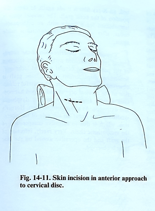
When one level should be approached, the skin incision goes transversally or slightly obliquely along the course of the skin creases with preference for the left side to preserve the recurrent branch of the vagus nerve (Fig. 14-11). In case of an approach to more than one level, the skin incision is located along the anterior border of the sternocleidomastoid muscle. The platysma is divided parallel to the course of its fibres with the superficial cervical fascia. The underlying sternocleidomastoid muscle is then visible. The anterior border of the sternocleidomastoid muscle is dissected and is retracted laterally to expose the middle cervical fascia. The middle cervical fascia is opened, and the omohyoid muscle is seen, crossing the field at the C5-6 level, and may be retracted up or down or transected at its midtendinous segment. The carotid artery is identified and the dissection continues medial to the carotid sheath. After retraction the sternomastoid muscle and carotid artery laterally, and the larynx, pharynx, trachea and oesophagus from the medial side, the anterior surface of the cervical vertebrae is exposed, covered by prevertebral fascia.
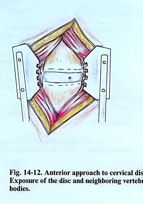
The existence of anterior osteophytes may facilitate the anatomical localisation of the disc level of the intervention. X-ray control to confirm the affected level is usually needed, and is facilitated by inserting a lumbar needle into the supposed disc. After the needle is removed, the opening remains as a referent point or, alternatively, this may be marked with electrocoagulation. Applying electrocoagulation, the anterior longitudinal ligament is cut along the midline, after which the disc, that is to be intervened on, and the neighbouring vertebral bodies are uncovered from the ligament and the deep prevertebral muscles, using a periosteal elevator. If bleeding occurs, it is easily controlled by coagulation or bone wax. The separated anterior longitudinal ligament and the deep prevertebral muscles are retracted with a special retractor (Fig. 14-12). The protruded anterior osteophytes at the disc level are removed with a bone nibbler.
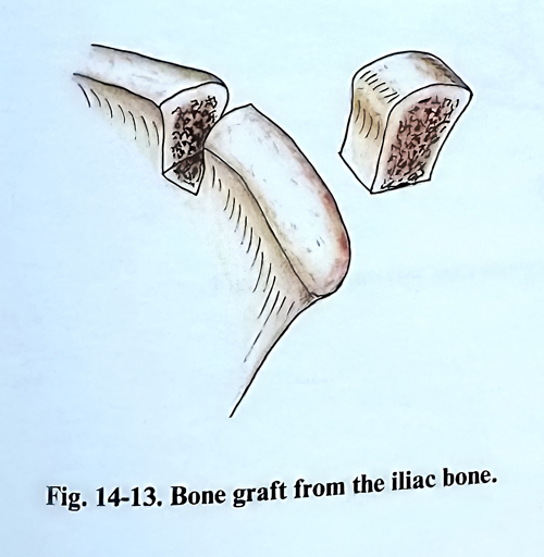
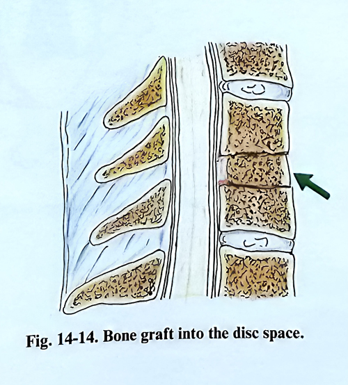
The surgeon’s maniplations that follow depend on the operative technique to be applied. According to the technique of Smith-Robinson, the disc must be removed with a curette together with the cartilage plates on the vertebral bodies, reaching their cancellous bone tissue. The removal continues until the posterior longitudinal ligament is revealed and all parts of the disc are removed from behind the vertebral bodies together with all osteophytes. After that the respective bone graft, taken by another team from the iliac bone crest is placed. (Figs. 14-13, 14-14)
In Cloward’s technique, the most important tool is the special trephine drill. First is placed the guide of the drill, fixed with 4 spikes to the adjacent vertebral bodies, leaving the disc for intervention between them. This guide prevents the drilling to penetrate deeper than the prescribed for the level, avoiding also lateral displacement. The removed by the drilling includes the disc and the adjacent parts of the vertebral bodies. Drilling stops at the cortical plate of the vertebrae and disc/posterior ligament layer. With curettes is removed the remaing layer exposing the anterior wall of the spinal canal. After decompression, the produced defect is measured and a bone graft from the iliac crest is adjusted, both anteriorly and posteriorly.
According to the microsurgical technique of Hankinson and Wilson, the posterior parts of the disc with osteophytes and disc prolapses are removed through the small window at the anterior part of the disc, which is 1 cm square. The lateral parts of the disc remain intact as two columns, sufficiently strong to support the cervical spine. Changing the position of the microscope to the right and to the left allows removal through this small window of all posterior borders of the two vertebrae and disc prolapses.
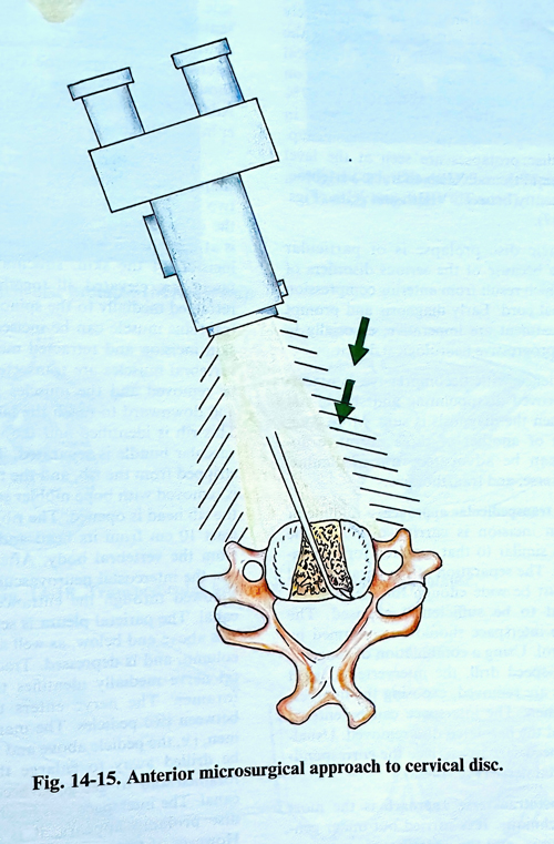
In such a discectomy it is not necessary to insert a bone graft, and there is also no need for postoperative immobilisation. This partial discectomy can be done up to 2 - 3 levels. After operation collapse of the disc space is rare and the mobility is not affected. We prefer this technique (Fig. 14-15).
No special care is taken during the postoperative period. When the patient is put to bed, an X-ray film is made again, to check the position of the bone graft, if such was placed. The cervical spine is immobilised for 6 weeks with an orthopaedic collar, after which an X-ray control is made again.
Complications. Deterioration of the neurological deficit is avoided with a proper selection of indications for the different methods and careful surgical technique. A displacement of the bone graft is very rare. The careful retraction of the cervical structures and a good haemostasis protect against postoperative dysphagia and dyspnoea. As a rare complication, Horner's syndrome may occur on the side of the operation, which is due to excessive compression of the sympathetic chain. Very rarely, infection of the graft or the operative wound on the iliac bone can be observed.
THORACIC DISC OPERATIONS
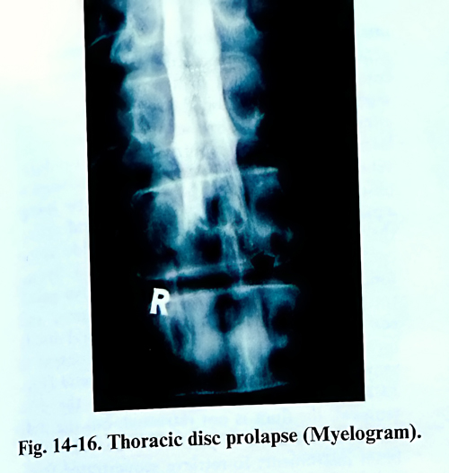
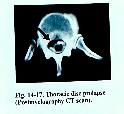
Herniations of thoracic discs, compressing the content of the spinal canal are relatively uncommon, compared to those involving the cervical and lumbar segments. In many statistical studies, it has been shown that operations on thoracic disc prolapses comprise only 0,2 - 0,7% of all disc operations. They are usually in patients from the 35 to 55 year age group. Thoracic disc prolapses are seen at the level between the IVth and XIIth thoracic vertebrae, most frequently between VIIIth and XIth (Figs.14-16; 14-17).
Thoracic disc prolapse is of particular importance because of the serious disorders of function which result from anterior compression of the spinal cord. Early diagnosis and prompt surgical treatment are imperative, especially in cases with progressive neurological deficit.
Experience with decompressive laminectomy was proved disappointing and it should not be used when the diagnosis is comfirmed. More commonly one of the three following below operative approaches, can be recommended: transpedicular, costotransversectomy, and transthoracic.
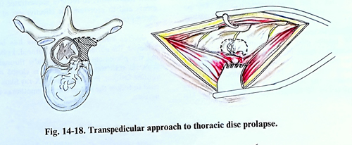
In the transpedicular approach, a midline or paramedian incision is carried out, and the exposure is similar to that required for hemilaminectomy. The separation of the paravertebral muscles must be wide enough for the intervertebral joint to be sufficiently exposed. The appropriate interspace should be confirmed by X-ray control. Using a combination of rongeurs and a high-speed drill, the intervertebral joint and pedicle are removed, exposing the intervertebral foramen. The interspace can be entered laterally and the herniated disc removed. Usually it is not necessary to sacrifice the corresponding intercostal nerve (Fig. 14-18).
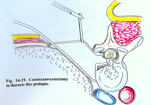
The costotransverse approach is the most common technique. It is carried out under general anaesthesia, and the anaesthesiologist must be able to control lung ventillation if the pleural cavity is entered inadvertently. The patient lies in a semiprone lateral position. A pad is placed in the axilla and below the shoulders. To avoid increased venous pressure the abdomen should be kept free, with support carefully placed under the chest and iliac crests. A pillow is placed between the legs and uppermost knee is flexed. The approach can be made from either side. The choice depends from the clinical presentation and radiological data. In a case of central disc prolapse and with an absence of lateralizing findings, the right-side approach should be used, because the important artery of Adamkiewich usually originates from a left lower intercostal artery (Fig. 14-19).
The skin incision follows a long crescent-shape course, extending along the length of at least two or three vertebral bodies above and below the affected disc space. The apex of the curve is at least 10 cm lateral to the midline. After the incision of the skin, the subcutaneous tissue and fascia are elevated all together as a flap and retracted medially to the spinous processes. The trapezius muscle can be incised in line with the skin incision and retracted medially. The para-vertebral muscles are transected over the rib to be removed and the muscles retracted upward and downward to reach the lamina and the rib.
This rib is identified and the intercostal neuro-vascular bundle is separated. The periosteum is stripped from the rib, and the transverse process is removed with bone nibbler so that the joint of the rib head is opened. The rib is cut laterally at least 10 cm from its head and is disarticulated from the vertebral body. After removal of the rib, the intercostal neurovascular bundle can be followed through the entrance into the spinal canal. The parietal pleura is separated from the ribs above and below, as well as from the spinal column, and is retracted. Following the intercostal nerve medially identifies the intervertebral foramen, where the nerve enters the spinal canal between two pedicles. The margins of the foramen, i.e. the pedicle above and below, must then be drilled away to enlarge the intervertebral foramen and to get good access to the spinal canal. The interspace can be entered and once the disc prolapse appears, it is easily removed. However, if there are bony ridges, osteophytes or free fragments, more bone must be removed until the spinal canal can be entered. The high-speed drill or a sharp curette is used to remove the bony ridges after the disc space has been emptied of disc material. After complete haemostasis, the pleura is checked for inadvertent damage through positive ventilation pressure. If it has been violated, an extrapleural chest tube may be put in place before completing the closure. A chest X-ray film taken in the recovery room will help if there are concerns about a pneumothorax.
The transthoracic approach provides direct access to the anterior and lateral portion of the disc. A thoracic surgeon is required at least during early experiences with this approach.
The operation is carried out with the patient under general anaesthesia and intubated with a Carlens tube so that each lung may be ventilated separately for better control of complications.
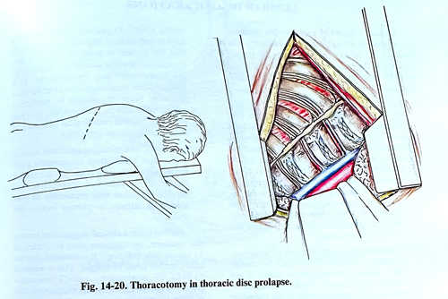
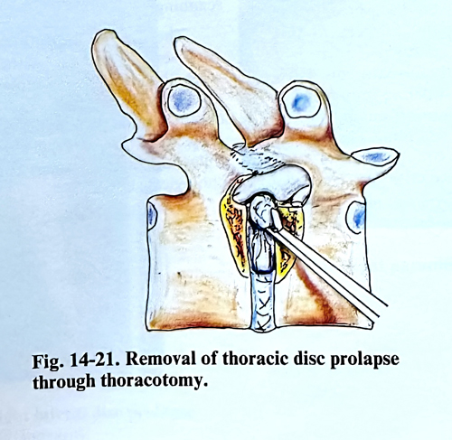
The chest is entered through a standard right posterior thoracotomy incision. The right side is chosen because the artery of Adamkiewich usually enters on the left side and because the heart and great vessels pose a problem through a left transthoracic incision. The patient is positioned on the left lateral side. At least 15 cm of rib, including the rib head, should be removed to gain complete exposure to the lateral part of the spinal canal. A linear incision is made in the parietal pleura to expose the intervertebral vessels, and next the sympathetic chain (Fig. 14-20). Usually, ligating or dividing the segmental artery and vein above and below the affected disc is not necessary. The intercostal nerve is followed to its foramen and gently retracted to expose the pedicle above and below. A high-speed drill is used to resect a segment of pedicle and the vertebral bodies on either side the disk as to gain access to the lateral and ventral epidural space. Epidural venous bleeding can be troublesome because it obscures the anatomic structures of the spinal canal. By exposing the lateral epidural space, the surgeon can coagulate and divide some epidural veins to expose the ventral dura. Symptomatic thoracic disc herniation always penetrates the posterior longitudinal ligament and can be easily removed. The intervertebral disc is incised over the protrusion and the content is removed by pituitary forceps and curette (Fig 14-21). During all manipulations in the disc removal, the dura is not retracted, but the wall of the spinal canal is palpated repeatedly with blunt instruments to retrieve sequestrated fragments and to ascertain the effectiveness of the decompression.
After complete haemostasis has been achieved, the parietal pleura is sutured. Following the placement of a chest tube away of the main incision, the chest is closed in a routine fashion. Postoperatively, X-ray films are used to follow the size of the pneumothorax if it persists.
LUMBAR DISC OPERATIONS
Surgical removal of a lumbar disc prolapse is indicated in several different conditions. The result of the surgery depends upon the correct indications for surgery. Patients who are candidates for surgical treatment can be divided into four groups:
1.Immediate surgical intervention is mandatory in acute massive disc herniation presenting with cauda equina syndrome (drop foot and sphincter troubles). Except in situations where marked spinal canal stenosis exists, a huge sequestrated fragment will be found severely compressing the roots of the cauda equina. An immediate open operation with bony decompression to allow the removal of the sequestrated fragments is required but, even when performed without delay, the results of sphincter recovery can be very disappointing.
2. In patients with lesser degrees of neurological deficit, surgery should also be considered. There is usually motor deficit of the calf muscles, weak dorsiflexion of the foot and sensory deficit in the respective dermatomes.
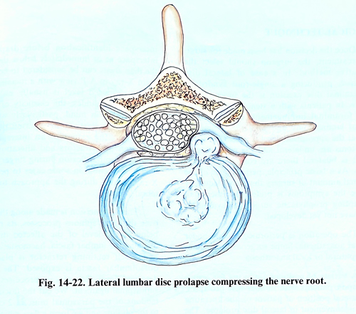
3. The most frequent indication for surgical treatment is the pain (Fig. 14-22). In some of these cases there is a mild neurological deficit, which cannot be discovered in the presence of severe pain. Surgery in this group of patients is indicated if there is a failure of conservative treatment. How much time should waited for improvement from conservative treatment is uncertain. The key to measuring the success of conservative treatment is not only the patient's relief from the leg pain but also improvement in straight leg raising ability. In patients with three to five days in bed without improvement, it is expected that the patient is going to have a protracted recovery course, for which a surgical intervention may be indicated. It is proposed that surgical treatment in the acute radicular syndrome should be performed within three months of symptoms, to try to avoid the chronic pathological changes that can occur within the nerve root.
4. In patients with recurring attacks of sciatica, that benefited from conservative treatment, discussion with them on the further course of management is appropriate, and surgical treatment may reasonably be considered to attempt to prevent further episodes.
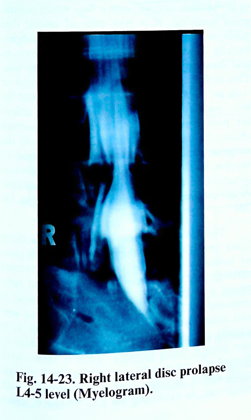
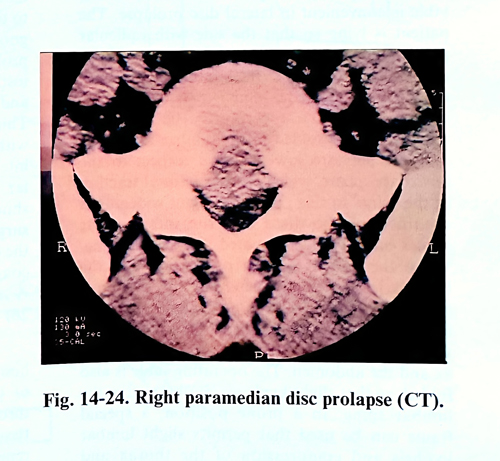
A clinical diagnosis of lumbar disc prolapse must be confirmed by appropriate radiology. It is essential that the entire lumbar canal be imaged. This is achieved by MRI, CT, myelography, and CT postmyelography (Figs. 14-23; 14-24). In many cases, two of these investigations are enough for a correct diagnosis. These investigations should be preceded by plain radiography, as abnormalities in lumbosacral segmentation may not be discerned on the MRI and CT scanning.
The surgical indications should be decided very cautiously with neurotic patients, when the subjective complaints dominate over objective information. The presence of a non-objective deficit findings reduces the success of surgery, and the surgery should proceed only following careful patient counselling with psychological support.
SURGICAL TECHNIQUE
Once the decision has been made for surgical treatment, the surgeon should select the optimal operative method. In a case of lateral disc prolapse, producing a single-root syndrome, interlaminal disc excision is the procedure of choice. In patients with a stenotic spinal canal with foraminal root compression, decompressive partial laminectomy and bilateral foraminotomy at the appropriate level is necessary. If the symptoms are unilateral and the myelographic defect is bilateral there are controversial opinions. As a basic principle, performance of the minimum surgical intervention that can adequately relieve the symptoms is preferred rather than attempting prophylactic surgery for symptoms that have not yet developed.
The operation is performed usually under general anaesthesia, although it can be perfomed under lumbar, epidural or local anaesthesia and a variety of surgical patient positions are used.
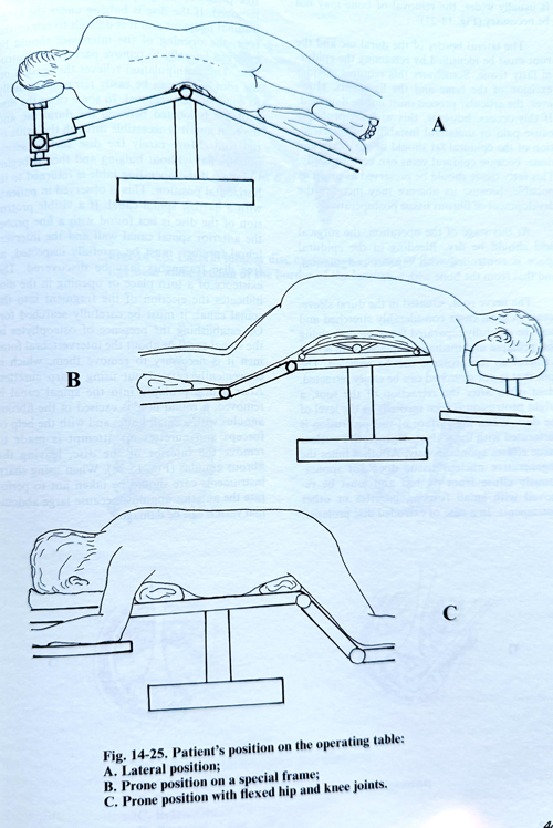
The lateral position on the operating table is convenient in lateral disc prolapse. The side with radicular pain remains on top (Fig. 14-25). A hard cushion is placed under the lumbar part to form a slight lateral bending of the lumbar part of the spine with its convexity upwards. The operating table is slightly bended in the lumbar region, so that the legs are lowered with respect to the horizontal plane, which ensures mild natural traction of the lumbar spine. In this position provides some widening of the discs spaces and the interlaminar spaces on the upper side of the spine.
When the patient is in a prone position, the head is turned slightly laterally and lies on a separate support, while an elongated hard cushion is put to each side supporting the shoulder and in the pelvis region, and avoid compression of the thorax and the abdomen. The operating table is also folded, so that slight flexion be obtained in the lumbar spine. Occasionally there is a special frame to obtain this position properly. A modified prone position can be obtained by flexing the hips and knees 90 degrees, and the lumbar spine is better flexed in comparison with the other positions.
It is necessary to perform surface indication markers for interspace identification before draping. The interspace at or immediately below the level of the iliac crests can be considered from pre-existing lumbar X-rays. A plain X-ray in A-P view with a metallic marker at the supposed level is usually taken. When using surface markers, the elasticity of the skin must be kept in mind, particularly with obese patients. This elasticity may permit the surface to shift to the extent of one interspace from distortion by retractors and alterations in the degree of lumbar flexion during surgery. That is why it is essential in some cases to perform an X-ray control with a needle at the beginning of the approach to the vertebral laminae.
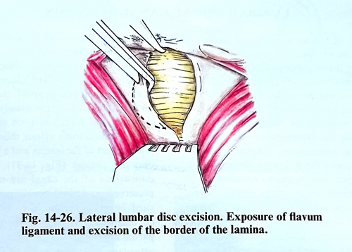
The skin incision is made along the midline over about three spinous processes, its mid-point being at the level of the affected disc. After reaching the lumbar fascia, haemostasis is made and a self retaining retractor is placed. The dorsal lumbar fascia is exposed. The fascia is detached from the spinous processes and the supraspinal ligament. The fascia and the tendons of the paraspinal muscles 2 cm lateral to the midline are divided, producing a flap, which facilitates later good closure. If it is a case of lateral disc prolapse, the paravertebal muscles are subperiostally dissected from the two spinal processes and the corresponding laminae only on the side of the affection. This is done with a sharp periosteal elevator or with an electric knife. The two laminae and the interspace are exposed together with the articular capsule. In this dissection the sacrum should be carefully identified, as it can confirm to the surgeon the correct level localisation. The bleeding is stopped by electro-coagulation and packing for a few minutes, and a self-retaining retractor is placed (Fig. 14-26).
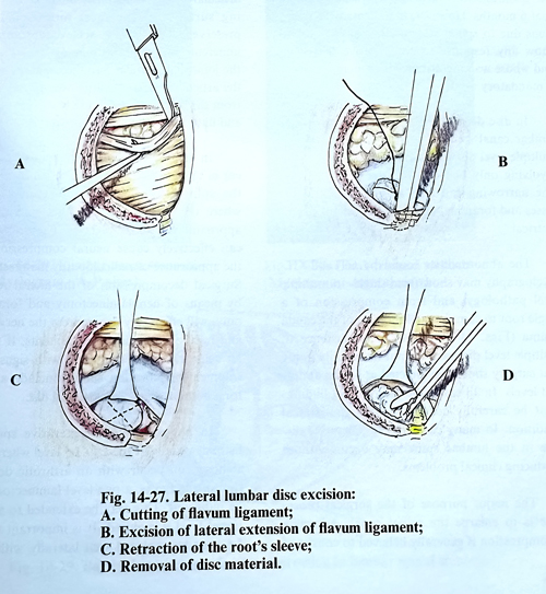
The flavum ligament is lifted with a sharp hook and is divided near the midline. A small cottonoid, tied to the thread, is inserted through the hole to separate the epidural fatty tissue and the dura from the spinal canal wall. The flavum ligament is removed by punches, paying special attention to its lateral extensions. At L5 is usually necessary to remove the inferior edge of the higher lying lamina. This is done with Kerrison pumches or with a small burr electric drill, and if bleeding occurs, it is stopped with bone wax. In exploring the L5-S1 space, which is usually wider, the removal of bone may even not be necessary (Fig. 14-27).
The lateral border of the dural sac and the root must be identified by retracting the epidural fatty tissue. Sometimes this requires a wider excision of the bone and the ligaments. How-ever, the articular process must not be damaged. If this occurs, however, this will not probably cause pain or unilateral instability. The retraction of the epidural fat should be very carefully done, because epidural veins can be torn easily. This fatty tissue should be preserved as much as possible, because its absence may increase the development of fibrous tissue postoperatively. At this stage of the operation, the surgical field should be dry. Bleeding in the epidural space is controlled with bipolar coagulation, and that from the bone with wax.
The nerve root, situated in the dural sleeve, seems in most cases considerably stretched and must be carefully separated from the underlying disc prolapse and medially displaced with a root retractor so the prolapse can be revealed. The root that is not stretched can be easily retracted. Most often after the retraction of the root, a bright protrusion is seen medially at the level of the disc. When the surface of this protrusion is perforated with the scalpel, degenerative nuclear tissue extrudes spontaneously. At other times the degenerative nucleus tissue does not spontaneously extrude from its bed and must be removed with small forceps, curettes or other instruments. In a case of extruded disc prolapse, free pieces of the pulpous tissue can be easily removed. If the disc is bulging under the longitudinal ligament and it is difficult to retract the root, the opening of the interspace should be enlarged laterally to remove part of the protrusion. This manipulation relieves the tension on the root and it can be easily retracted medially to complete the removal. In some cases the disc prolapse is located between the dural sac and the root and it is accessible through the axilla of the root. More rarely the disc surface seems initially flat without bulging and then it begins to bulge, if the operating table is returned to its horizontal position. This is observed in patients with a narrow spinal canal. If a visible protrusion of the disc is not found with a fine probe, the anterior spinal canal wall and the intervertebral foramen must be carefully inspected, as free disc fragments may be discovered. The existence of a rupture or any opening in the disc indicates the ejection of a fragment into the spinal canal and it must be carefully searched for it.
On establishing the presence of osteophytes in the spinal canal or within the intervertebral foramen it is necessary to remove them, which is conveniently carried out using sharp curettes.
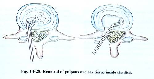
After a disc prolapse into the spinal canal is removed, a round hole is excised in the fibrous annulus with a small knife, and with the help of forceps and curettes an attempt is made to remove the remaining nucleus parts of the disc, leaving the fibrous annulus (Fig. 15-28). When using sharp instruments care should be taken not to perforate the anterior annulus, because large abdominal vessels can be damaged.
LUMBAR SPINAL CANAL STENOSIS
Surgery of lumbar spinal canal stenosis is one of the most common procedures in the repertoire of the neurosurgeon. Indications for surgical decompression are based on the clinical manifestations and the results of diagnostic tests demonstrating compression of the conus medullaris and/or roots of the cauda equina, which correlate with the symptomatic level and the side of neural compression. Surgical treatment is indicated under the following circumstances: sphincteric disturbances due to compression of the conus medullaris or cauda equina; severe radicular symptoms with a monoradicular or polyradicular motor deficit; and increasing neurogenic intermittent claudication, especially if the manifestations have been present for more that 6 months. However, in patients with symptoms due to spinal stenosis that have failed to show any response to conservative treatment and whose walking distance is reducing, surgery is mandatory.
In disc disease, the most common form of lumbar canal stenosis occurs as a generalised, multiple level disorder, or as a segmental lesion involving only one or two adjacent vertebrae. The narrowing can also affect the lateral recesses and foraminal areas, and is usually asymmetrical.
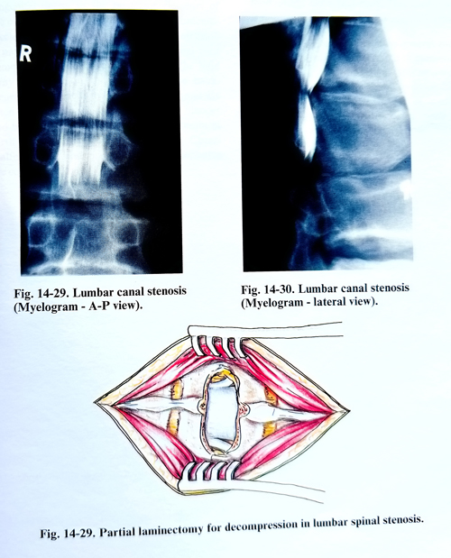
The abnormalities found by CT and CT-myelography may show from single- to multiple level pathology, and from compression of a single root to complete obliteration of the cauda equina (Figs. 14-29; 14-30). The existence of multiple level disease does not necessarily imply that surgery should be directed at all the abnormal levels. In all cases, the radiological findings must be carefully correlated with the clinical condition. In many cases the degenerative disease in the lumbar spine may occur without producing clinical problems.
The major purpose of the surgical procedure is to enlarge the spinal canal. Adequate decompression is generally believed to consist of total laminectomy with removal of the medial one half of the facet joints on both sides at each level of decompression. Laminectomy with partial facetectomy provides adequate decompression while maintaining spinal stability. Because lumbar spinal stenosis is usually at the level of the discs and facet joints, on many occasions, partial laminectomy of one third or half of the inferior part of the lamina and a partial facetectomy are enough (Fig. 14-31). If the midline dimensions of the canal are reasonably well preserved, and the stenotic process is mainly sited laterally, the nerve roots may often be decompressed through fenestrations. Where partial laminectomy is performed the pars interarticularis and the majority of the weight-bearing surfaces of the facet joints should be preserved. This is best achieved by using an undercutting technique to remove the portion of the joint that intrudes into the canal, preserving the articular surfaces. Large osteophytes arising from the vertebral bodies are best tamped down and flattened with a small-bore punch.
In degenerative scoliosis, stenosis may occur in the lateral recesses on the concave side of the spine beneath enlarged, arthritic facets where the foramina are further narrowed by approximation of the pedicles. These alterations can effectively cause neural compression with the appearance of radiculopathy manifestations.
Surgical decompression of the lateral recesses, by means of hemilaminectomy and foraminotomy, will effectively decompress the nerve roots and relieve the radicular complaints. If there is an acute radicular syndrome with signs of entrapment, exploration should include a search for a herniated and sequestrated disc, if it has not been visualized on the preoperative images
In patients with degenerative spondylolisthesis usually at the L4 - L5 level where severe stenosis can occur with an arthrotic degeneration of the facets, a two level laminectomy may be indicated. This may be extended to an additional level if required. It is important that the LS root be explored well laterally with adequate foraminal decompression using an undercutting technique. The nerve root dural sleeve is followed from its axilla as it winds laterally over the ridge formed at the level of subluxation/vertebral body translation toward the foramen. Sometimes this ridge may need to be tamped down if it distorts the nerve root. Part of the pedicle may also be removed if kinking is present. If the findings are more evident on one side, only a limited decompression needs to be done on the opposite side, not to affect stability.
If preoperative X-ray studies indicate abnormal mobility, the operation should end with stabilisation. It is not uncommon to find that the vertebrae involved have been fused by the spondylotic process. The nerve roots at the level above may also require decompression if adjacent levels are involved by the stenotic process.