LUMBAR SPINAL CANAL STENOSIS
Surgery of lumbar spinal canal stenosis is one of the most common procedures in the repertoire of the neurosurgeon. Indications for surgical decompression are based on the clinical manifestations and the results of diagnostic tests demonstrating compression of the conus medullaris and/or roots of the cauda equina, which correlate with the symptomatic level and the side of neural compression. Surgical treatment is indicated under the following circumstances: sphincteric disturbances due to compression of the conus medullaris or cauda equina; severe radicular symptoms with a monoradicular or polyradicular motor deficit; and increasing neurogenic intermittent claudication, especially if the manifestations have been present for more that 6 months. However, in patients with symptoms due to spinal stenosis that have failed to show any response to conservative treatment and whose walking distance is reducing, surgery is mandatory.
In disc disease, the most common form of lumbar canal stenosis occurs as a generalised, multiple level disorder, or as a segmental lesion involving only one or two adjacent vertebrae. The narrowing can also affect the lateral recesses and foraminal areas, and is usually asymmetrical.
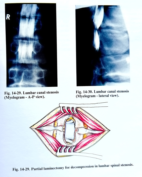
The abnormalities found by CT and CT-myelography may show from single- to multiple level pathology, and from compression of a single root to complete obliteration of the cauda equina (Figs. 14-29; 14-30). The existence of multiple level disease does not necessarily imply that surgery should be directed at all the abnormal levels. In all cases, the radiological findings must be carefully correlated with the clinical condition. In many cases the degenerative disease in the lumbar spine may occur without producing clinical problems.
The major purpose of the surgical procedure is to enlarge the spinal canal. Adequate decompression is generally believed to consist of total laminectomy with removal of the medial one half of the facet joints on both sides at each level of decompression. Laminectomy with partial facetectomy provides adequate decompression while maintaining spinal stability. Because lumbar spinal stenosis is usually at the level of the discs and facet joints, on many occasions, partial laminectomy of one third or half of the inferior part of the lamina and a partial facetectomy are enough (Fig. 14-31). If the midline dimensions of the canal are reasonably well preserved, and the stenotic process is mainly sited laterally, the nerve roots may often be decompressed through fenestrations. Where partial laminectomy is performed the pars interarticularis and the majority of the weight-bearing surfaces of the facet joints should be preserved. This is best achieved by using an undercutting technique to remove the portion of the joint that intrudes into the canal, preserving the articular surfaces. Large osteophytes arising from the vertebral bodies are best tamped down and flattened with a small-bore punch.
In degenerative scoliosis, stenosis may occur in the lateral recesses on the concave side of the spine beneath enlarged, arthritic facets where the foramina are further narrowed by approximation of the pedicles. These alterations can effectively cause neural compression with the appearance of radiculopathy manifestations.
Surgical decompression of the lateral recesses, by means of hemilaminectomy and foraminotomy, will effectively decompress the nerve roots and relieve the radicular complaints. If there is an acute radicular syndrome with signs of entrapment, exploration should include a search for a herniated and sequestrated disc, if it has not been visualized on the preoperative images
In patients with degenerative spondylolisthesis usually at the L4 - L5 level where severe stenosis can occur with an arthrotic degeneration of the facets, a two level laminectomy may be indicated. This may be extended to an additional level if required. It is important that the LS root be explored well laterally with adequate foraminal decompression using an undercutting technique. The nerve root dural sleeve is followed from its axilla as it winds laterally over the ridge formed at the level of subluxation/vertebral body translation toward the foramen. Sometimes this ridge may need to be tamped down if it distorts the nerve root. Part of the pedicle may also be removed if kinking is present. If the findings are more evident on one side, only a limited decompression needs to be done on the opposite side, not to affect stability.
If preoperative X-ray studies indicate abnormal mobility, the operation should end with stabilisation. It is not uncommon to find that the vertebrae involved have been fused by the spondylotic process. The nerve roots at the level above may also require decompression if adjacent levels are involved by the stenotic process.