POSTERIOR APPROACH IN RADICULOPATHY
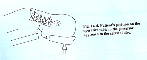
The patient is placed in a prone position on the operating table as in craniectomy of posterior fossa (Fig. 14-4). The sitting position can also be used. To avoid air embolism, the operating table is angled in such a way that the lower limbs are in flexion at both the hip and knee joints, and the head is slightly flexed and fixed on a special frame. Before the operation, X-ray control of the level is advisable using a needle introduced at the supposed level as a marker. This X-ray control may also be done later after reaching the vertebral laminae.
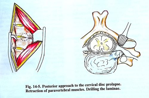
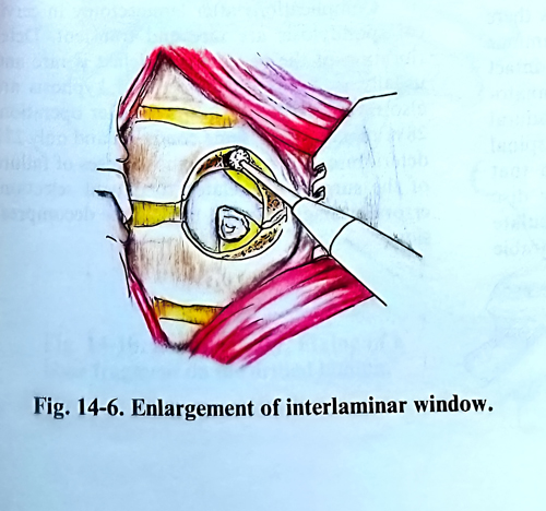
The skin incision is in the midline, 5 cm in length. Penetration follows along the midline and the nuchal septum and the spinous processes. The laminae and the articular processes of the two neighbouring vertebrae at the level of the disc prolapse are stripped. Confirmation of the level where the spinal canal must be opened can be done at this stage of the operation by palpating the spinal processes. The spinous process of the VIth cervical vertebra is recognised by the fact that it is bifid, whereas on the underlying VIIth is not. At the same time it is the last vertebra to which the nuchal septum is attached. New X-ray control is advisable at this stage of the operation. The lateral parts of the laminae and one third of the facet joints are drilled, leaving a thin shell of cortical bone over the intervertebral foramen and the underlying the nerve root. This fenestration may be enlarged with a high-speed drill. The lateral part of the flavum ligament and the cortical bone is then easily lifted (Figs. 14-5; 14-6).
The root may be distended if an underlying anterior disc fragment or osteophyte is present, and care must be taken not to injure the stretched root. According to the size and exact location of the compression, this initial foraminotomy may be enlarged to a facetectomy, hemilaminotomy or hemilaminectomy. If venous bleeding occurs from the epidural space, it is easily controlled with bipolar coagulation.
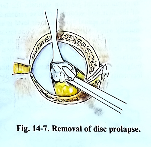
Exploration is initially carried out in the axilla of the root. A blunt hook is an excellent instrument for this purpose. The disc fragments are usually extruded and are easily removed (Fig 14-7). Not infrequently, the disc bulging is within the posterior ligament or annulus and an incision must be made to remove the fragment(s). It is unnecessary to remove large amounts of disc material or to curette the disc space as would be done with a lumbar discectomy, and the surgeon must be satisfied that no compressive fragments are left behind. Exploration with a blunt hook should be made also above, below, and medial to the root to be certain that all fragments are removed. They generally are small in comparison with those in the lumbar region. If the compression of the roots is due to osteophytes, their removal is more difficult and requires the use of a very fine periosteal elevator and small sharp curettes.
After haemostasis is achieved, the muscles, subcutaneous fascia, and skin are closed in layers. A soft cervical collar is generally applied for the immediate postoperative period, but it is removed on the following days, and rehabilitation is started.