POSTERIOR APPROACH IN MYELOPATHY
A posterior surgical approach is commonly employed to decompress the spinal cord; it consists of laminectomy with or without foraminotomy and section of the denticulate ligaments. The aim of decompressive laminectomy is to enlarge the spinal canal by removing posterior compressing structures, which allow the dural sac and spinal cord to migrate posteriorly away from the compressing ventral osteophytes. It also relieves posterior compression caused by enfolding and hypertrophy of the flavum ligaments.
The spinal cord in myelopathic patients is highly vulnerable and requires protection. Extreme flexion or extension during intubation for general anaesthesia can cause at times irreparable ischaemic cord damage. Determining the limit of neck movements before induction of anaesthesia and not exceeding them is most important.
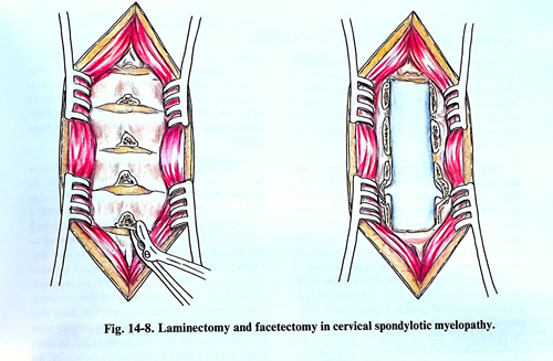
The surgical technique has no differences from the general rules of laminectomy. The patient is placed in a prone position on the operating table with the head fixed in a special headrest with a pin fixator as in operation of posterior fossa. The sitting position is convenient for the surgeon, but there is a risk in elderly patients, and in patients with unstable circulation. The laminae are resected laterally up to the lateral dimension of the dural sac. If it is necessary, the medial parts of joint facets can be also removed (Fig. 14-8). Complete facetectomy can be done in case of stenosis of the intervertebral foramen and there is evidence of radiculopathy corresponding to that level. Unless there is craniocervical junction stenosis, the laminae of the first and second vertebrae remain intact because the spinal canal at their level is anatomically wider. Some surgeons include intradural section of the denticulate ligaments in spinal cord decompression. It has been shown that contact between the cord and anterior disc bulging is not reduced when the denticulate ligaments are sectioned. Studies of comparable groups of patients with and without denticulate section revealed no difference in surgical results.
Epidural bleeding is not a serious problem and haemostasis can be achieved using bipolar coagulation, pieces of Gelfoam or oxidised cellulose. The muscles, fascia, subcutaneous tissue, and skin are closed in layers.
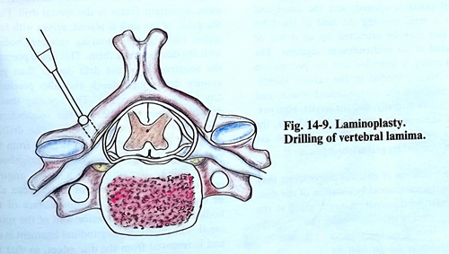
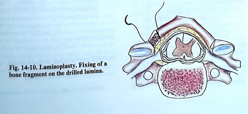
By using laminoplasty, the spinal canal can be enlarged by opening the laminae on one side to create an 'open door'. After dissecting the posterior apophyses, the laminae and the facet joints, the drill is used to open through at the lamina-facet junction in the same fashion as for 'en bloc' laminectomy. The inner cortex and the flavum ligaments are completely removed only on one side (usually the side with the most significant pathology). On the other side, the drilling is completed through the outer cortex only (Fig. 14-9). The bloc of the laminae is carefully elevated 10 - 15 mm from the dura. The tips of the spinous processes that were removed previously are placed in the trough at each level.The sutures are passed through the border of cutted surface of the laminae, the bone pieces and capsules of the facet joint at each level, ensuring the new position of the laminae (Fig.14-10).
The patient can get out of bed the next day after the operation. A soft collar is applied for one or two weeks, and after that exercises start.n Later, X-films are obtained in flexion and extension to check for postoperative instability.
Complications after laminectomy in cervical spondylosis are rare and transient. Deterioration of the neurological deficit is rare and usually transient. Instability and kyphosis are also rare. About 70% improve after operation, 28% remain in the same condition and only 2% deteriorate. The most common causes of failure of the surgery are related to patient selection, errors in diagnosis, and inadequate decompression.