THORACIC DISC OPERATIONS
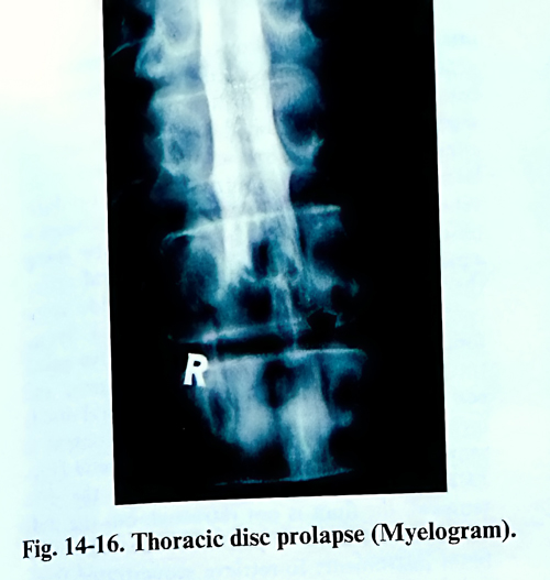
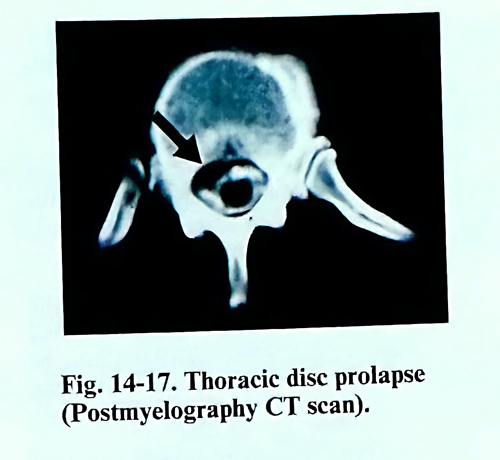
Herniations of thoracic discs, compressing the content of the spinal canal are relatively uncommon, compared to those involving the cervical and lumbar segments. In many statistical studies, it has been shown that operations on thoracic disc prolapses comprise only 0,2 - 0,7% of all disc operations. They are usually in patients from the 35 to 55 year age group. Thoracic disc prolapses are seen at the level between the IVth and XIIth thoracic vertebrae, most frequently between VIIIth and XIth (Figs.14-16; 14-17).
Thoracic disc prolapse is of particular importance because of the serious disorders of function which result from anterior compression of the spinal cord. Early diagnosis and prompt surgical treatment are imperative, especially in cases with progressive neurological deficit.
Experience with decompressive laminectomy was proved disappointing and it should not be used when the diagnosis is comfirmed. More commonly one of the three following below operative approaches, can be recommended: transpedicular, costotransversectomy, and transthoracic.
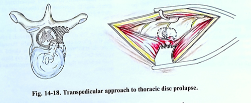
In the transpedicular approach, a midline or paramedian incision is carried out, and the exposure is similar to that required for hemilaminectomy. The separation of the paravertebral muscles must be wide enough for the intervertebral joint to be sufficiently exposed. The appropriate interspace should be confirmed by X-ray control. Using a combination of rongeurs and a high-speed drill, the intervertebral joint and pedicle are removed, exposing the intervertebral foramen. The interspace can be entered laterally and the herniated disc removed. Usually it is not necessary to sacrifice the corresponding intercostal nerve (Fig. 14-18).
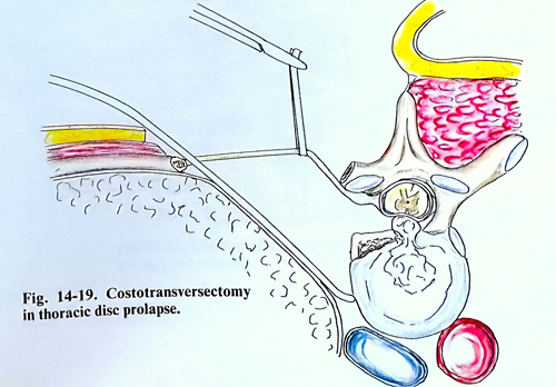
The costotransverse approach is the most common technique. It is carried out under general anaesthesia, and the anaesthesiologist must be able to control lung ventillation if the pleural cavity is entered inadvertently. The patient lies in a semiprone lateral position. A pad is placed in the axilla and below the shoulders. To avoid increased venous pressure the abdomen should be kept free, with support carefully placed under the chest and iliac crests. A pillow is placed between the legs and uppermost knee is flexed. The approach can be made from either side. The choice depends from the clinical presentation and radiological data. In a case of central disc prolapse and with an absence of lateralizing findings, the right-side approach should be used, because the important artery of Adamkiewich usually originates from a left lower intercostal artery (Fig. 14-19).
The skin incision follows a long crescent-shape course, extending along the length of at least two or three vertebral bodies above and below the affected disc space. The apex of the curve is at least 10 cm lateral to the midline. After the incision of the skin, the subcutaneous tissue and fascia are elevated all together as a flap and retracted medially to the spinous processes. The trapezius muscle can be incised in line with the skin incision and retracted medially. The para-vertebral muscles are transected over the rib to be removed and the muscles retracted upward and downward to reach the lamina and the rib.
This rib is identified and the intercostal neuro-vascular bundle is separated. The periosteum is stripped from the rib, and the transverse process is removed with bone nibbler so that the joint of the rib head is opened. The rib is cut laterally at least 10 cm from its head and is disarticulated from the vertebral body. After removal of the rib, the intercostal neurovascular bundle can be followed through the entrance into the spinal canal. The parietal pleura is separated from the ribs above and below, as well as from the spinal column, and is retracted. Following the intercostal nerve medially identifies the intervertebral foramen, where the nerve enters the spinal canal between two pedicles. The margins of the foramen, i.e. the pedicle above and below, must then be drilled away to enlarge the intervertebral foramen and to get good access to the spinal canal. The interspace can be entered and once the disc prolapse appears, it is easily removed. However, if there are bony ridges, osteophytes or free fragments, more bone must be removed until the spinal canal can be entered. The high-speed drill or a sharp curette is used to remove the bony ridges after the disc space has been emptied of disc material. After complete haemostasis, the pleura is checked for inadvertent damage through positive ventilation pressure. If it has been violated, an extrapleural chest tube may be put in place before completing the closure. A chest X-ray film taken in the recovery room will help if there are concerns about a pneumothorax.
The transthoracic approach provides direct access to the anterior and lateral portion of the disc. A thoracic surgeon is required at least during early experiences with this approach.
The operation is carried out with the patient under general anaesthesia and intubated with a Carlens tube so that each lung may be ventilated separately for better control of complications.
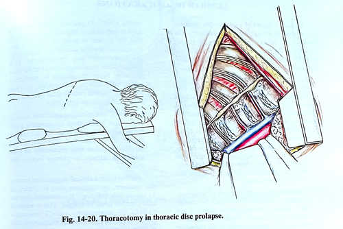
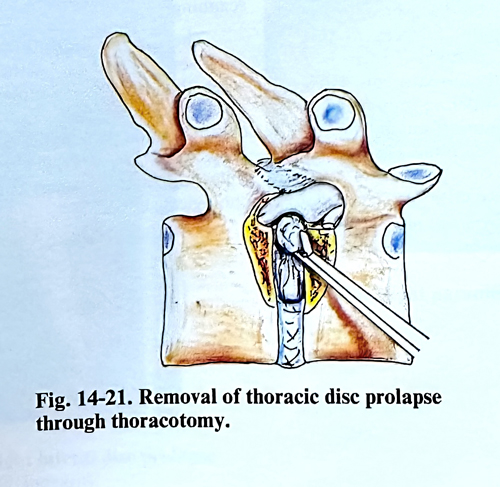
The chest is entered through a standard right posterior thoracotomy incision. The right side is chosen because the artery of Adamkiewich usually enters on the left side and because the heart and great vessels pose a problem through a left transthoracic incision. The patient is positioned on the left lateral side. At least 15 cm of rib, including the rib head, should be removed to gain complete exposure to the lateral part of the spinal canal. A linear incision is made in the parietal pleura to expose the intervertebral vessels, and next the sympathetic chain (Fig. 14-20). Usually, ligating or dividing the segmental artery and vein above and below the affected disc is not necessary. The intercostal nerve is followed to its foramen and gently retracted to expose the pedicle above and below. A high-speed drill is used to resect a segment of pedicle and the vertebral bodies on either side the disk as to gain access to the lateral and ventral epidural space. Epidural venous bleeding can be troublesome because it obscures the anatomic structures of the spinal canal. By exposing the lateral epidural space, the surgeon can coagulate and divide some epidural veins to expose the ventral dura. Symptomatic thoracic disc herniation always penetrates the posterior longitudinal ligament and can be easily removed. The intervertebral disc is incised over the protrusion and the content is removed by pituitary forceps and curette (Fig 14-21). During all manipulations in the disc removal, the dura is not retracted, but the wall of the spinal canal is palpated repeatedly with blunt instruments to retrieve sequestrated fragments and to ascertain the effectiveness of the decompression.
After complete haemostasis has been achieved, the parietal pleura is sutured. Following the placement of a chest tube away of the main incision, the chest is closed in a routine fashion. Postoperatively, X-ray films are used to follow the size of the pneumothorax if it persists.