ANTERIOR APPROACH TO THE CERVICAL DISCS
The patient is placed on the operating table in a supine position, the nuchal region being slightly elevated with an appropriate cushion. Many authors apply traction on the skull with a weight about 3 kg. The shoulders are lowered maximally to allow X-ray control during the operation.
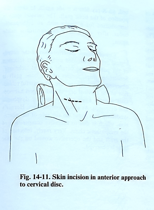
When one level should be approached, the skin incision goes transversally or slightly obliquely along the course of the skin creases with preference for the left side to preserve the recurrent branch of the vagus nerve (Fig. 14-11). In case of an approach to more than one level, the skin incision is located along the anterior border of the sternocleidomastoid muscle. The platysma is divided parallel to the course of its fibres with the superficial cervical fascia. The underlying sternocleidomastoid muscle is then visible. The anterior border of the sternocleidomastoid muscle is dissected and is retracted laterally to expose the middle cervical fascia. The middle cervical fascia is opened, and the omohyoid muscle is seen, crossing the field at the C5-6 level, and may be retracted up or down or transected at its midtendinous segment. The carotid artery is identified and the dissection continues medial to the carotid sheath. After retraction the sternomastoid muscle and carotid artery laterally, and the larynx, pharynx, trachea and oesophagus from the medial side, the anterior surface of the cervical vertebrae is exposed, covered by prevertebral fascia.
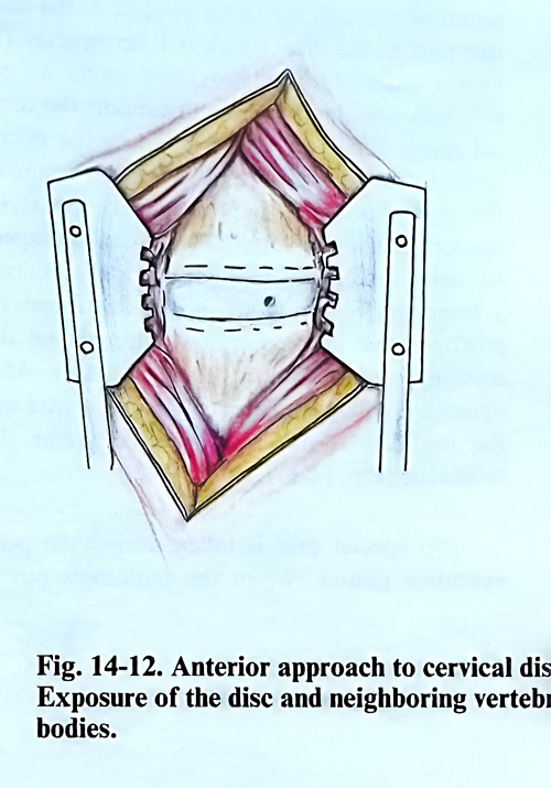
The existence of anterior osteophytes may facilitate the anatomical localisation of the disc level of the intervention. X-ray control to confirm the affected level is usually needed, and is facilitated by inserting a lumbar needle into the supposed disc. After the needle is removed, the opening remains as a referent point or, alternatively, this may be marked with electrocoagulation. Applying electrocoagulation, the anterior longitudinal ligament is cut along the midline, after which the disc, that is to be intervened on, and the neighbouring vertebral bodies are uncovered from the ligament and the deep prevertebral muscles, using a periosteal elevator. If bleeding occurs, it is easily controlled by coagulation or bone wax. The separated anterior longitudinal ligament and the deep prevertebral muscles are retracted with a special retractor (Fig. 14-12). The protruded anterior osteophytes at the disc level are removed with a bone nibbler.
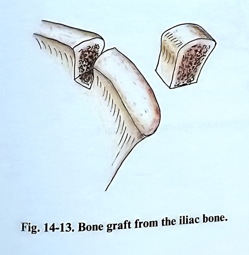
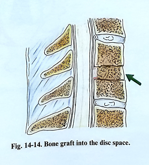
The surgeon’s maniplations that follow depend on the operative technique to be applied. According to the technique of Smith-Robinson, the disc must be removed with a curette together with the cartilage plates on the vertebral bodies, reaching their cancellous bone tissue. The removal continues until the posterior longitudinal ligament is revealed and all parts of the disc are removed from behind the vertebral bodies together with all osteophytes. After that the respective bone graft, taken by another team from the iliac bone crest is placed. (Figs. 14-13, 14-14)
In Cloward’s technique, the most important tool is the special trephine drill. First is placed the guide of the drill, fixed with 4 spikes to the adjacent vertebral bodies, leaving the disc for intervention between them. This guide prevents the drilling to penetrate deeper than the prescribed for the level, avoiding also lateral displacement. The removed by the drilling includes the disc and the adjacent parts of the vertebral bodies. Drilling stops at the cortical plate of the vertebrae and disc/posterior ligament layer. With curettes is removed the remaing layer exposing the anterior wall of the spinal canal. After decompression, the produced defect is measured and a bone graft from the iliac crest is adjusted, both anteriorly and posteriorly.
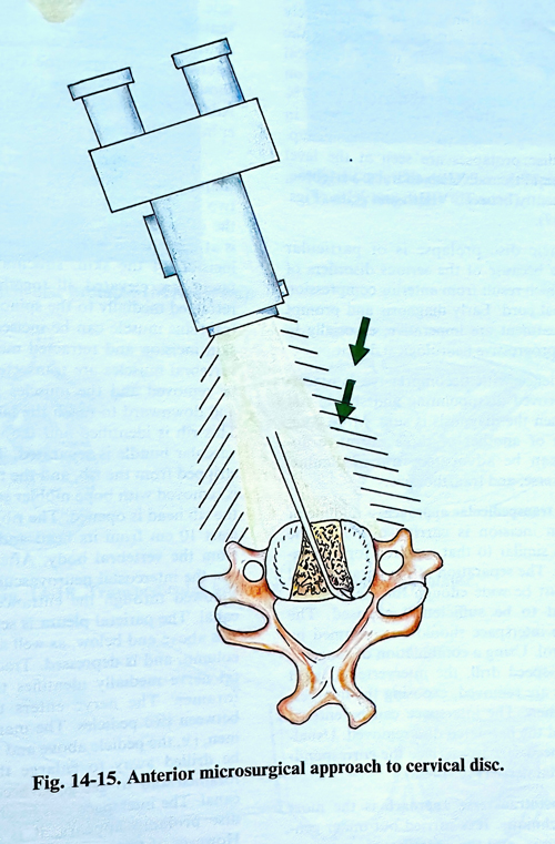
According to the microsurgical technique of Hankinson and Wilson, the posterior parts of the disc with osteophytes and disc prolapses are removed through the small window at the anterior part of the disc, which is 1 cm square. The lateral parts of the disc remain intact as two columns, sufficiently strong to support the cervical spine. Changing the position of the microscope to the right and to the left allows removal through this small window of all posterior borders of the two vertebrae and disc prolapses.
In such a discectomy it is not necessary to insert a bone graft, and there is also no need for postoperative immobilisation. This partial discectomy can be done up to 2 - 3 levels. After operation collapse of the disc space is rare and the mobility is not affected. We prefer this technique (Fig. 14-15).
No special care is taken during the postoperative period. When the patient is put to bed, an X-ray film is made again, to check the position of the bone graft, if such was placed. The cervical spine is immobilised for 6 weeks with an orthopaedic collar, after which an X-ray control is made again.
Complications. Deterioration of the neurological deficit is avoided with a proper selection of indications for the different methods and careful surgical technique. A displacement of the bone graft is very rare. The careful retraction of the cervical structures and a good haemostasis protect against postoperative dysphagia and dyspnoea. As a rare complication, Horner's syndrome may occur on the side of the operation, which is due to excessive compression of the sympathetic chain. Very rarely, infection of the graft or the operative wound on the iliac bone can be observed.