SURGICAL TECHNIQUE
Once the decision has been made for surgical treatment, the surgeon should select the optimal operative method. In a case of lateral disc prolapse, producing a single-root syndrome, interlaminal disc excision is the procedure of choice. In patients with a stenotic spinal canal with foraminal root compression, decompressive partial laminectomy and bilateral foraminotomy at the appropriate level is necessary. If the symptoms are unilateral and the myelographic defect is bilateral there are controversial opinions. As a basic principle, performance of the minimum surgical intervention that can adequately relieve the symptoms is preferred rather than attempting prophylactic surgery for symptoms that have not yet developed.
The operation is performed usually under general anaesthesia, although it can be perfomed under lumbar, epidural or local anaesthesia and a variety of surgical patient positions are used.
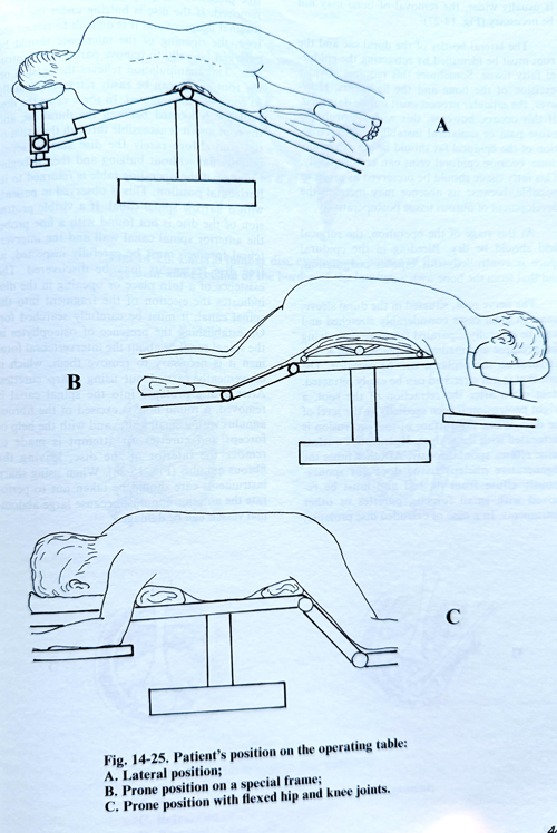
The lateral position on the operating table is convenient in lateral disc prolapse. The side with radicular pain remains on top (Fig. 14-25). A hard cushion is placed under the lumbar part to form a slight lateral bending of the lumbar part of the spine with its convexity upwards. The operating table is slightly bended in the lumbar region, so that the legs are lowered with respect to the horizontal plane, which ensures mild natural traction of the lumbar spine. In this position provides some widening of the discs spaces and the interlaminar spaces on the upper side of the spine.
When the patient is in a prone position, the head is turned slightly laterally and lies on a separate support, while an elongated hard cushion is put to each side supporting the shoulder and in the pelvis region, and avoid compression of the thorax and the abdomen. The operating table is also folded, so that slight flexion be obtained in the lumbar spine. Occasionally there is a special frame to obtain this position properly. A modified prone position can be obtained by flexing the hips and knees 90 degrees, and the lumbar spine is better flexed in comparison with the other positions.
It is necessary to perform surface indication markers for interspace identification before draping. The interspace at or immediately below the level of the iliac crests can be considered from pre-existing lumbar X-rays. A plain X-ray in A-P view with a metallic marker at the supposed level is usually taken. When using surface markers, the elasticity of the skin must be kept in mind, particularly with obese patients. This elasticity may permit the surface to shift to the extent of one interspace from distortion by retractors and alterations in the degree of lumbar flexion during surgery. That is why it is essential in some cases to perform an X-ray control with a needle at the beginning of the approach to the vertebral laminae.
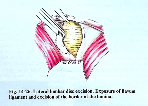
The skin incision is made along the midline over about three spinous processes, its mid-point being at the level of the affected disc. After reaching the lumbar fascia, haemostasis is made and a self retaining retractor is placed. The dorsal lumbar fascia is exposed. The fascia is detached from the spinous processes and the supraspinal ligament. The fascia and the tendons of the paraspinal muscles 2 cm lateral to the midline are divided, producing a flap, which facilitates later good closure. If it is a case of lateral disc prolapse, the paravertebal muscles are subperiostally dissected from the two spinal processes and the corresponding laminae only on the side of the affection. This is done with a sharp periosteal elevator or with an electric knife. The two laminae and the interspace are exposed together with the articular capsule. In this dissection the sacrum should be carefully identified, as it can confirm to the surgeon the correct level localisation. The bleeding is stopped by electro-coagulation and packing for a few minutes, and a self-retaining retractor is placed (Fig. 14-26).
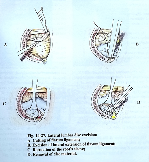
The flavum ligament is lifted with a sharp hook and is divided near the midline. A small cottonoid, tied to the thread, is inserted through the hole to separate the epidural fatty tissue and the dura from the spinal canal wall. The flavum ligament is removed by punches, paying special attention to its lateral extensions. At L5 is usually necessary to remove the inferior edge of the higher lying lamina. This is done with Kerrison pumches or with a small burr electric drill, and if bleeding occurs, it is stopped with bone wax. In exploring the L5-S1 space, which is usually wider, the removal of bone may even not be necessary (Fig. 14-27).
The lateral border of the dural sac and the root must be identified by retracting the epidural fatty tissue. Sometimes this requires a wider excision of the bone and the ligaments. How-ever, the articular process must not be damaged. If this occurs, however, this will not probably cause pain or unilateral instability. The retraction of the epidural fat should be very carefully done, because epidural veins can be torn easily. This fatty tissue should be preserved as much as possible, because its absence may increase the development of fibrous tissue postoperatively. At this stage of the operation, the surgical field should be dry. Bleeding in the epidural space is controlled with bipolar coagulation, and that from the bone with wax.
The nerve root, situated in the dural sleeve, seems in most cases considerably stretched and must be carefully separated from the underlying disc prolapse and medially displaced with a root retractor so the prolapse can be revealed. The root that is not stretched can be easily retracted. Most often after the retraction of the root, a bright protrusion is seen medially at the level of the disc. When the surface of this protrusion is perforated with the scalpel, degenerative nuclear tissue extrudes spontaneously. At other times the degenerative nucleus tissue does not spontaneously extrude from its bed and must be removed with small forceps, curettes or other instruments. In a case of extruded disc prolapse, free pieces of the pulpous tissue can be easily removed. If the disc is bulging under the longitudinal ligament and it is difficult to retract the root, the opening of the interspace should be enlarged laterally to remove part of the protrusion. This manipulation relieves the tension on the root and it can be easily retracted medially to complete the removal. In some cases the disc prolapse is located between the dural sac and the root and it is accessible through the axilla of the root. More rarely the disc surface seems initially flat without bulging and then it begins to bulge, if the operating table is returned to its horizontal position. This is observed in patients with a narrow spinal canal. If a visible protrusion of the disc is not found with a fine probe, the anterior spinal canal wall and the intervertebral foramen must be carefully inspected, as free disc fragments may be discovered. The existence of a rupture or any opening in the disc indicates the ejection of a fragment into the spinal canal and it must be carefully searched for it.
On establishing the presence of osteophytes in the spinal canal or within the intervertebral foramen it is necessary to remove them, which is conveniently carried out using sharp curettes.
After a disc prolapse into the spinal canal is removed, a round hole is excised in the fibrous annulus with a small knife, and with the help of forceps and curettes an attempt is made to remove the remaining nucleus parts of the disc, leaving the fibrous annulus (Fig. 15-28). When using sharp instruments care should be taken not to perforate the anterior annulus, because large abdominal vessels can be damaged.