Legacy: "Atlast of Neurosurgery" / L.Karaguiosov, A. Ramadan, K.Karaguiosov / Kiwait/ 1998
Read PREVIOUS: 3. Tumors - Chapter 1
Read NEXT: 4. Vascular Diseases of the Brain
TUMOURS OF LATERAL VENTRICLES
Surgery of the lateral ventricular tumours is not very common in current practice, as they account for less than 1% of all intracranial tumours. Very often they are benign or of a low degree of malignancy. Their growth inside the lateral ventricle is usually silent due to the abundance of space and they present themselves at a stage characterised by ventriculomegaly and increased intracranial pressure. The intraventricular mass lesion's growth causes additional displacement and isolation of part or the whole of the lateral ventricle. These features determine the specific surgical management of these lesions. Manipulation of these tumours requires incision of apparently intact brain, as access to them is at a very significant depth. Furthermore, visibility of the tumour border is poor and very often the arterial feeders are not accessible at the beginning of the surgery. As their volume is relatively large, space for manipulation must be gained by tumour removal. Tumours inside the ventricle can have different locations, which therefore require a different approach technique. The most common place for a meningioma is the trigonum. Meningiomas and papillomas can be found in parts of the ventricle containing choroid plexus. Other tumours such as ependymomas, subependymomas, astrocytomas, dermoids and epidermoids are also found elsewhere inside the ventricle, but most often they are near the midline at the foramen of Monro and the septum pellucidum.
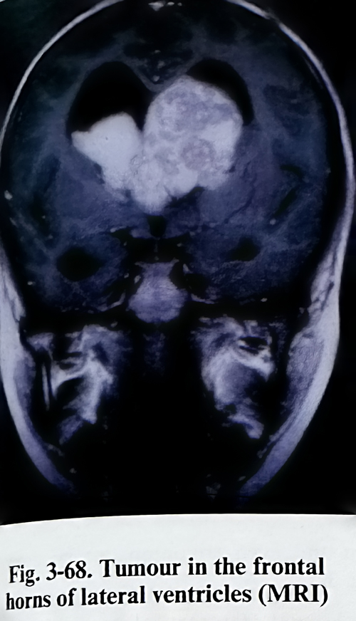
Investigations are based on tridimensional imaging to establish the precise location of the lesion. CT scans - enhanced and non-enhanced in axial, direct coronal and reconstructed images are needed for detailed delineation of the lesion's borders (Fig.3-68). MRI, also Gd enhanced, in at least Tl and T2 weighted images will give additional information on the tumour's texture and location, and on perifocal oedema and the tumour's relationship with important neural and vascular structures. Angiography, studying both the carotid and vertebrobasilar systems, must be used to demonstrate the vascular supply, relationship with choroidal arteries, the major deep venous collectors and their displacement. For the needs of a transcallosal approach, of utmost importance is the knowledge of the position of the big venous cortical parasagittal collectors. Another important area of the investigations is aimed at determining cerebral dominance and predicting the possibility of deficit after cortical incisions in preselected areas. As for the extra requirements of epilepsy surgery, the Wada test must on occasion be considered.
Four main locations inside the lateral ventricle have been specified for clinical surgical needs: frontal horn-Monro area, trigonum, body of the ventricle, and temporal horn. All approaches have been designed to reach the lesion by the shortest path with the largest possible incision of the cerebral mantle, not affecting functionally important (eloquent) cortex and pathways. Cortical incisions are used to reach the lesion, with some locations requiring callosotomies and rarely polar resections.
Every incision or resection gives a specific view and access for manipulation of a limited part of the lateral ventricle. Therefore the approach must be decided after the location of the lesion has been defined in detail. The resections are done following the same rules as in glioma surgery, but corpus callosum divisions require some particular elements in the technique. Isolated parts of the ventricles are dilated and this gives sufficient space for manipulation during the approach to the lesion. Craniotomies are performed over the cortical incision site and are relatively small.
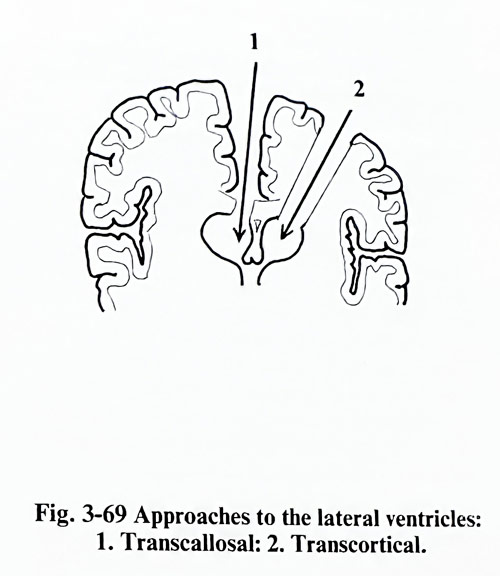
The frontal horn and the ventricular body are approached either by a frontal transcortical approach or by a transcallosal route (Fig. 3-69).
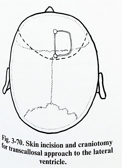
Transcallosal route. The patient is placed in the pin headholder with the head straight and elevated about 20 degrees. A curvilinear incision is made parallel to and about 2 cm behind the coronal suture across the midline (Fig. 3-70).
The incision should be long enough to allow reflection of the skin flap at least 6 cm anterior to the coronal suture. The scalp flap is reflected anteriorly and the sagittal and coronal sutures should be identified on the skull surface.
The bone flap should be made with its medial margin at the midline, using at least two burr holes. One of them is usually made with its medial margin at the midline just behind the junction of the coronal suture with the sagittal suture. The second burr hole should be made with its medial margin at the midline approximately 7 cm anterior to the first burr hole. If the tumour is inside the ventricular body it is helpful to place the bone flap slightly anteriorly. The craniotomy can be preplanned on the midsagittal MR image, properly placing these two burr holes according to the desired angle of view onto the lesion through the callosotomy.
The dural opening follows the outline of the bone flap with its base being hinged at the sagittal sinus, taking care not to avulse any of the cortico-dural veins that may be present. It is preferable to avoid sacrificing any draining vein from the cortex to the sagittal sinus. Small veins anterior to the coronal suture can generally be sacrificed, but if there is a very large draining vein it should be preserved if possible. Craniotomy is also planned considering the position of the major venous cortical tributaries.
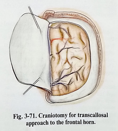
One or two traction sutures are placed through the base of the dural flap just lateral to the sagittal sinus and are suspended over the already reflected dural flap, to facilitate retraction of the dura. About 3 to 4 cm of longitudinal free space between the hemisphere and falx are required for adequate retraction (Fig. 3-71).
The proper direction to the desired part of the corpus callosum has to be established before retracting the hemisphere from the falx. The usual guide is an imaginary line in the midsagit-tal plane drawn from the coronal suture to the line through both external auditory meatuses.
This path will lead toward the midportion of the corpus callosum or slightly anteriorly to it.
The brain hemisphere is gently retracted reaching the inferior margin of the falx. After that the penetration between the hemispheres may occasionally present some difficulties in identifying the midline. Two retractors can be needed for the separation of the hemispheres.
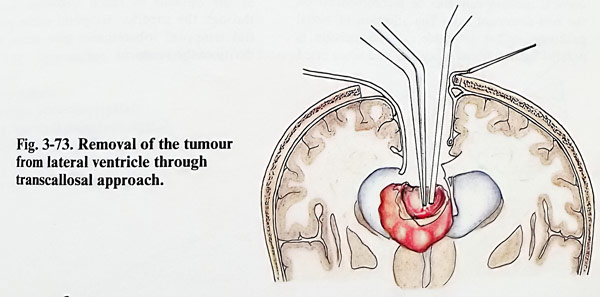
The callosomarginal arteries located above the cingulate gyrus can be mistaken for the perical-losal arteries. The corpus callosum is differentiated from adjacent cortex by its white colour and the two pericallosal arteries situated near each other. The pericallosal arteries are separated (there should not be branches seen crossing between them). We fix a distance of few millimetres between them with cottonoid to obtain their retraction into the more laterally located sulcus of the corpus callosum. Some times, in a case with ventriculomegaly, the pericallosal arteries may not be apparent, but there is no need to search for them if there is adequate corpus callosum surface exposed to open the ventricular system (Fig. 3-72). The corpus callosum is pratically avascular and can be divided with a small blunt dissector, fine bipolar forceps or a small suction tube, and at the expected depth (estimated on the midsagittal MRI image) usually one of the lateral ventricles is penetrated. Penetration is guided also by assessing the asymmetry of dilatation of the ventricles and the shift of the septum pellucidum. In some cases it is the cavum of the septum pellucidum that is penetrated. The approach, dividing the anterior and midpart of the body of the corpus callosum, can be enlarged safely to 4 cm in length. By tilting the spatulas and the microscope anteriorly and posteriorly, a relatively large portion of the ventricular body, anterior horn and even trigonum can be approached (Fig. 3-73).
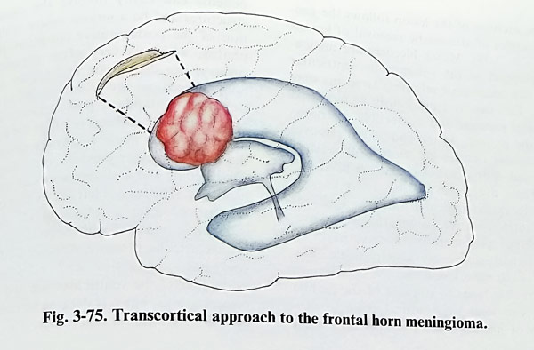
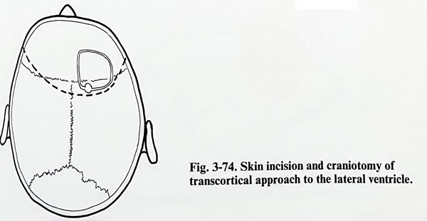
Frontal transcortical approach. The position of the patient in the pin headholder and the coronal incision of the scalp are the same as in a transcallosal approach. The bone flap has a similar location in the anterior-posterior direction but is a little lateral to the midline (Fig. 3-74). Through the superior frontal sulcus the incision penetrates the bulb of the frontal horn and the Monro area (Fig. 3-75). The non-dominant side is of course of less risks for the approach. The convenient exposure of the septum pellucidum permits its fenestration and access to the opposite frontal horn. In some cases, the good exposure of the foramen of Monro allows extension of the exposure to the third ventricle with the transforminal transvelum interpositum approach.
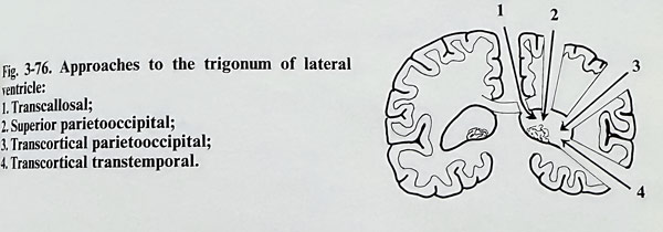
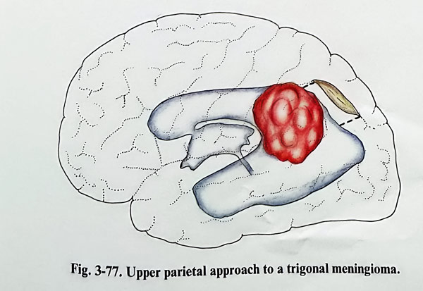
The approaches to the trigonum are important in the removal of meningiomas and papillo-mas. Tumours with this location can be reached in different ways. Several cortical incisions have been proposed: lateral temporoparietal, temporal, transcallosal, and superior parieto-occipital, the last often being performed as a polar resection (Fig. 3-76). The temporoparietal incision, especially on the dominant side, produces significant deficit - dyslexia, agraphia, acalculia and Gerstmann's syndrome have been described after such cortical intervention. However, with a big lesion, this incision on the non-dominant side can provide an access to the trigonal tumour along a relatively short path (Fig. 3-77).
Although the access is the most direct, the vascular supply to the tumour is initially difficult to control, as the supplying branches from the anterior and the posterior choroidal arteries remain hidden from surgeon's view unless a large part of the lesion is removed. Piecemeal removal and debulking of the lesion are the safest method. Even on the non-dominant side, the cortical incision can produce a visual field deficit. The superior temporal sulcus incision, originally designed for hippocampal resection, gives early access to the branches of the anterior choroidal artery and is suitable for the removal of similar lesions. If an additional supply exists from the posterior choroidal arteries, it will remain patent until the final stage of tumour removal. On the dominant side, Wernicke's cortical area can be compromised, but this cortical incision better recommended on the non-dominant side. The affection of visual pathways is less probable, as the incision is parallel to their fibres. They can lead more often to homonymous hemianopia and the visual associative cortex can eventually be damaged. The lesions are approached posteriorly and tumour arterial supply control cannot be an initial step. Another option is the transcallosal route. After the transection in the anterior two-thirds of the corpus callosum body, the lesion is approached anteriorly and close to the midline. As usual, the most difficult part remains the interruption of the feeders and separation from the choroid plexus, naturally after debulking. The superior parietooccipital incision, however, remains as a relatively less damaging cortical penetration. It is approximately 3 cm of length, between the postcentral sulcus and the parietooccipital fissure. There are risks of cortical dysfunction of the posterior parietal lobe (dominant or non-dominant) and they have to be considered before choosing this route.
Approaches to the temporal horn can be used to some extent as an option to reach trigonum. Incisions through the superior temporal sulcus and partial temporal lobectomies give access to the cavity of the ventricle.
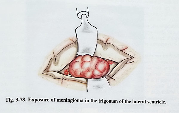
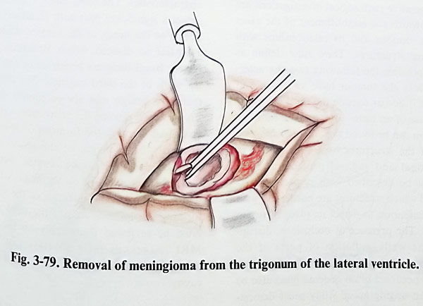
The excision of the lesion follows the general principles of atraumatic removal of lesions (Figs. 3-78; 3-79). Minor bleeders are meticulously stopped as the voluminous ventricular cavities can accumulate relatively large amounts of clots. This can lead insidiously to increased intracranial pressure and irreversible deterioration of the patient. At the end of the removal the ventricular cavities are thoroughly washed of clots and debris particles, and are filled will saline. There are some possible complications peculiar to intraventricular surgery. Risks of massive intraoperative haemorrhage are pre-sent. Delayed postoperative haemorrhages in the ventricular cavity are also possible. The removal of the mass lesion and the fenestration of the ventricle leads to collapse of the mantle and this can lead to subdural collections - hygromas or haematomas. Postoperative brain oedema can easily involve the diencephalic structures and be a serious source of deterioration of the postoperative condition. Hydrocephalus, which is very often present before surgery, can deteriorate and require shunting.
Some procedures are recommended as an addition to the standard postoperative care. An intraventricular catheter is left at the end of the intraventricular work. It permits monitoring of the intraventricular pressure, early diagnosis of any bleeding and withdrawal of CSF amounts if necessary. The tube is left for 48 h or more and is removed after a follow-up CT scan. The catheter is removed with normalisation of the intraventricular pressure. Another CT follow-up study has to be carried out 2 or 3 days later for evaluation of the ventricular size and to detect complications, but it is done as an emergency if clinical deterioration supervenes.