
METASTASES
Only a small number of those patients with intracranial metastases come to the attention of the neurosurgeon. The most common sources of metastases of the brain are tumours of the lung, breast, kidney, and malignant melanomas. Other sources are relatively infrequent, such as tumours of the gastrointestinal tract, thyroid, uterus, ovary, pancreas, prostate, and sarcomas. About the half of the patients have neurological symptoms or signs as the first manifestation of malignant disease. The primary origin of the cancer is usually discovered later, but occasionally the primary tumour cannot be identified, even at autopsy. More than the half the patients in whom the primary tumour is unknown initially are subsequently found to have bronchial carcinoma. There is some predilection towards the finding a metastasis in the middle cerebral artery territory and the cerebellar hemisphere.
A metastasis is discovered either accidentally as being the cause of detected neurological symptoms and signs or as part of the total diagnostic work up of the patient with established malignancy. In the first case, there are some additional goals of surgery.
The differences are as follows:
Accidentally discovered -
Need of a biopsy to obtain histological proof of the lesion.
Symptoms/signs usually due to the brain lesion. Expected relief if removed.
Expected survival cannot be estimated before biopsy; removal preferred.
As the effect of other treatment modalities is unknown, preference is given to surgical removal.
In an established malignant disease -
Diagnosis established.
Morbidity due not only to brain lesion and its relative importance has to be weighted. "How will the patient do without his morbidity due to the cerebral lesion?
Survival prognosis known in general terms.
Expected morbidity and benefit from surgical removal to be compared to the natural course of the disease, and the benefits to be established.
The effect of surgery to be compared to those expected with optimal protocols, including the application of other treatment modalities (as radio-and chemotherapy).
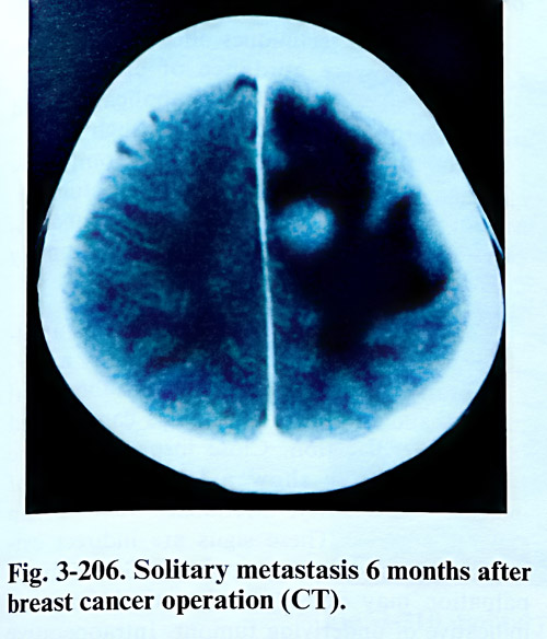
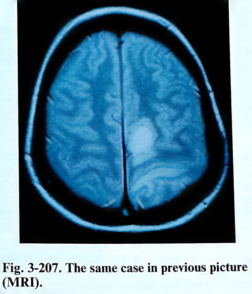
The examination of the patient suspected of having brain metastasis should assess his neurological status and determine the degree of systemic cancer involvement. This cannot exceed the reasonable time for general assessment, especially when the patient is deteriorating neurologically. Neuroimaging studies are essential and must include high-resolution cerebral CT, supplemented by MRI to detect lesions that might go unnoticed by a CT and evaluation of the degree of brain oedema. Incidental, circumscribed nodular lesion with central necrosis can be supposed to be a metastasis. Contrast-enhanced MRI is the best study to detect small metastases and to confirm whether a metastatic lesion is truly solitary (Figs. 3-206; 3-207).
Selection of patients for surgery. The objectives in the treatment of metastases are achieved following the main rules for the management of an intracranial lesion. Modifications to these rules can be imposed as a result of the multiplicity of the lesion, its rapid and fatal evolution, and when at some stage the general condition of the patient is seriously affected.
There is consensus among neurosurgeons that surgical excision should be restricted to patients with a solitary intracerebral metastasis whose general medical status is satisfactory. The opinions are controversial concerning excision of an incapacitating metastasis even when it is known that the tumour is not solitary, or for the
metastases that are multiple, but surgically accessible. In those metastases located deep within the dominant hemisphere and the brain stem the removal can provoke very serious deficit and as a rule they are considered surgically intractable.
External stereotactic radiosurgery can be advised for patients with deeply located and multiple metastases, who are otherwise in good general condition and with good life expectancy according to the type of disease.
The radio- and chemosensitivity of the tumour may play an important role in the decision regarding surgery. Patients with a radioresistant tumour, such as renal cancer or melanomas are more likely than other patients to be offered surgical excision with or without postoperative radiotherapy. Surgery may also be favoured in patients with slow-growing tumours, as suggested by the natural history of the disease or by long latency because that signifies good host defence mechanisms against the primary tumour.
Surgical technique. The surgical technique depends on the aim of the treatment: removal of samples for biopsy or attempt to excise the metastatic tumour. Biopsy alone can establish a histological diagnosis without therapeutic benefit. Only if the therapy after histological diagnosis is radiation and chemotherapy, biopsy can be a satisfactory initial procedure.
Biopsy. Currently, in properly selected patients, image guided stereotactic biopsy is an accurate, highly diagnostic, and low morbidity method to obtain histological confirmation of the tumour type.
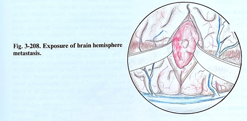
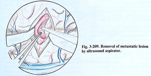
Craniotomy and excision. The surgical excision of a metastatic tumour is performed according the standard neurosurgical technical principles, and metastatic tumours rarely present serious difficulties (Figs. 3-208; 3-209).
Accurate tumour localisation is one of the most important aspects of craniotomy planning for brain metastasis surgery. Dural metastases of tumours that extend to the pial surface are uncommon and most metastases are not visible after dural opening. A detailed review of the axial CT and MRI (tridimentional) images aids tumour localisation. As sometimes there are no external references for the exact placement of the craniotomy (rather from the skull base), frameless stereotaxy and neuronavigation can be a useful technique for precise approach. Stereotactic techniques offer also an alternative for more precise guidance of craniotomy. Stereotactic guidance is best for small subcortical tumours that otherwise are occasionally impossible to find during craniotomy. Although stereotactic frame application and intraoperative CT scanning may slightly prolong the time of surgery, it can completely eliminate the problem of finding the tumour.
When localising techniques are not available, the neurosurgeon is confronted by a normal brain surface and a hidden subcortical tumour, and several practical tips can be used for tumour detection. Close inspection of the pial surface may show a localised area of increased vascularity, arterialized veins, or an expanded gyrus. These signs are indirect evidence of an underlying neoplasm. Gentle finger palpation may detect a subcortical firmness, indicative of underlying tumour. Intraoperative ultrasonography can be very useful to localise some subcortical lesions. Many lesions can be found by ultrasonography, but smaller tumours may evade even this method of detection. After localising the tumour, penetration into the brain tissue is through a small cortical incision. We prefer penetration through the depth of a sulcus. The excision is guided by the identification of the line between oedematous brain tissue and the tumour margin. The metastatic brain tumour often appears grossly separated and can be easily dissected from the surrounding brain.
Smaller tumours can be removed in a single piece, and that can be attempted for large tumours to reduce dissemination. Bleeding is usually minimal once the entire tumour is removed. Careful attention to haemostasis in the tumour bed will reduce the likelihood of haematoma formation in the operative site.
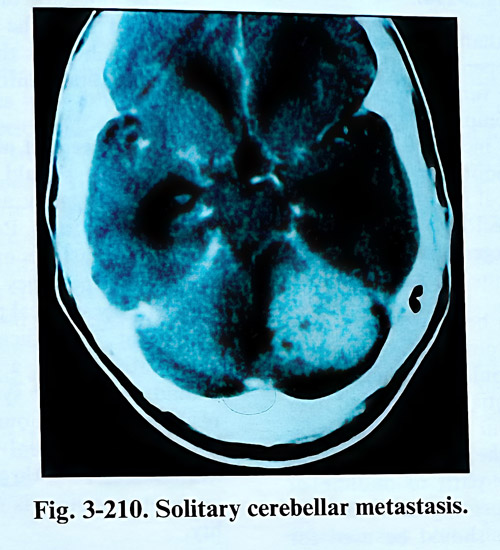
Operations for posterior fossa metastatic tumours have the tendency for greater morbidity and mortality compared to most supratentorial locations because of the tumour's proximity to the brain stem and the tendency to develop hydrocephalus. Tumours of the brain stem are rarely candidates for surgical excision and may be more amenable to radiosurgery. Cerebellar tumours can be located in the hemisphere or vermis and can grow to involve the fourth ventricle, cerebellar peduncle, or the brain stem. Such tumours are usually intraparenchymal and do not often grow primarily in the subarachnoid cisterns or extend through the tentorium (Fig. 3-210).
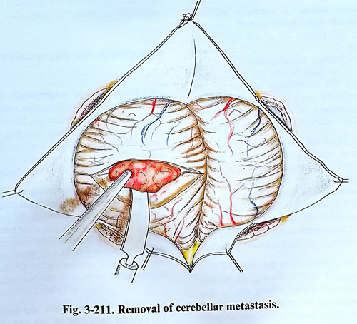
A generous, rather than a limited suboccipital craniectomy (with opening of the foramen magnum) aids in midline tumour exposure and in providing tonsilar decompression in the event of tumour recurrence or increased intracranial pressure. Because of the poor long-term survival and the probability of local recurrence, craniotomy (as opposed to craniectomy) has little place in metastatic tumour surgery in the posterior fossa (Fig. 3-211).
Lateral tumours can be approached via a lateral suboccipital or retromastoid exposure. A straight, direct route should be taken to the tumour to minimise potential neurological deficits associated with tumour excision. Coagulation and incision into the cerebellar vermis provide a low morbidity path to midline or paramedian lesions. A direct approach through the cerebellar hemisphere should be used for
more lateral tumours. The methods of resection further on are the routine ones.
The presence of hydrocephalus should be recognised from the preoperative imaging studies. In such cases the craniectomy is preceded by an occipital ventricular puncture through a burr hole. The placement of a shunt (usually ventriculoperitoneal should be indicated considering the life expectancy and the possible benefit of relief of symptoms. Patients who have advanced systemic metastases or multiple brain metastases and a life expectancy of 1 month or less do not warrant any surgical procedure, including shunt placement. The indication for a shunt operation in a patient with a metastatic brain tumour should be to treat symptomatic hydrocephalus resulting from tumour not amenable to surgical excision. Peritoneal seeding from malignant intracranial cells through the ventriculoperitoneal shunt system is only a theoretical possibility.