PINEAL REGION TUMOURS
A wide variety of tumours can occur in the pineal region and approximately 25 - 30% of them are encapsulated; there is therefore justification to attempt their removal. The neoplasm of the pineal region include: germ cell tumours malignant and benign variants - germinomas, embryonal carcinomas, chorioncarcinomas, teratomas, dermoids and epidermoids; tumours from the pineal cells - pineocytomas and pineoblastomas; tumours from supporting cellular components of the pineal body - astrocytomas, meningiomas, and rarely metastatic lesions. Originating from structures in the neighbourhood, meningiomas from the falcotentorial angle and gliomas from the quadrigeminal plate or splenium can also be found.
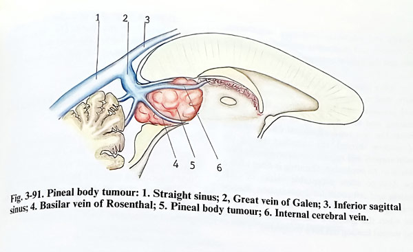
These tumours have central location in the cranial cavity that is equidistant from various cranial points traditionally used as routes of exposure. Such tumours are in intimate contact with the important deep venous system, including the vein of Galen, the precentral cerebellar vein, internal cerebral and basal veins. In most instances the bulk of the tumour, if not all the tumour, lies below the internal cerebral veins and the vein of Galen (Fig. 3-91). Sometimes there may be a firm attachment to these structures, including the tela chorioidea. Therefore, the preservation of the deep veins is one of the main tasks of every pineal tumour removal.
Because of that, it is always preferable to avoid a view in which the veins are obstructing the access to the tumour. The choice of the approach is dictated by the relationship of the tumour to the tributaries of the deep venous system. In the rare cases of the tumour being situated dorsally to the big veins, with or without splenial involvement, an interhemispherical view must also be obtained, and the splenium retracted if needed. The blood supply of the tumours in the pineal region is from the small branches of posterior choroidal arteries and branches of the quadrigeminal arteries.
The main tasks for the management of every tumour, which is a space occupying lesion, are specifically highlighted with this intracranial location, due to the high risk related to the direct approach to this region.
Histopathological identification of the lesion is essential for establishing the degree of malig-nancy, prognosis, and radiosensitivity of the lesion and has to be definen as early as possible. In principle, a stereotactic biopsy is definitely not the procedure of choice, as the surrounding veins are a source of complications and the non-homogeneous structure of some lesions (like teratomas) is misleading concerning the degree of malignancy with such small tissue samples. CSF cytological studies are of some value, but they are obtained usually after shunting. Tumour marker studies can also be helpful to indicate some specific types of lesions.
Intracranial hypertension, caused by obstructive hydrocephalus, must be treated when established in consideration of the future chances of restoring the normal outflow through the aqueduct. If direct surgery is not considered, permanent derivation with a shunt is advisable. However, if direct surgery is possible, the CSF derivation has to precede it, taking care not to collapse the cerebral mantle in extremely enlarged lateral ventricles.
When possible, the intracranial tumour should be removed as totally as this can be achieved without additional harm, especially in the cases when its radiosensitivity is low. Occlusion of any of the main inflowing veins to the straight sinus is not acceptable, unless its role for the drainage is not important and the respective area is well collateralised, for instance with tiny precentral or basal veins. Even then, if possible, such a sacrifice has to be strongly avoided. If the histopathological type of the tumour is not known before the operation, a frozen section study during surgery is mandatory. However, highly malignant tumours must be submitted to surgery very cautiously and selectively, preferring radiotherapy if effective, and thoroughly measuring the benefits, and the expected natural outcome of the tumour type.
After completing the main surgical procedure, final management of the elevated intracranial pressure must be decided on, if it is preexisting and there is also any need of adjuvant treatment. The non-restored natural CSF pathways of outflow through the aqueduct during the main surgery will require permanent shunt derivation to be implanted, if third ventriculostomy has not been done. Shunting is done after a few days to allow acceptable values of CSF protein and cell count for implantation. Radio-or chemotherapy is started as soon as possible, but when the general condition has recovered and wound healing has been completed.
Considering these principles of management, preoperative investigations aim to determine the exact position of the tumour border and its relationship to surrounding structures.
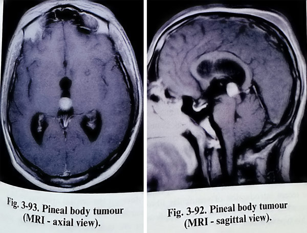
Three dimensional imaging of the area by CT and MRI is the basic investigational approach (Figs. 3-92; 3-93). However angiography is also necessary to establish the degree of vascularity and the pattern of the deep venous outflow. Special attention is given to the position and relationship of the lesion to the deep cerebral veins, the vein of Galen, the Rosenthal (basal) veins and the precentral cerebellar veins.
There are indications for surgical removal of those tumours, which on the basis of investigations, have a particularly high likelihood of being benign, and those patients previously treated with CSF derivation and radiation, without a tissue diagnosis, who have a progressive neurological deficit in the presence of a functioning shunt. Patients in whom cytological examination of CSF shows malignant cells, patients with evidence of either spinal or extraneural metastases, and patients in whom both the anterior and posterior third ventricle tumours are occupied by tumor may not require direct operation. The recent introduction of ventricular fibroendoscopy is currently gaining wider application in this type of lesion.
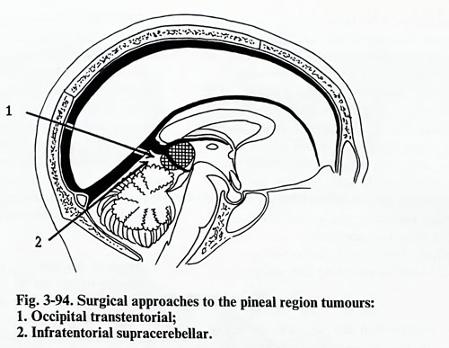
Different surgical approaches to the pineal region have been proposed. The infratentorial supracerebellar approach and the occipital transtentorial approach are the methods of choice (Fig. 3-94)
INFRATENTORIAL SUPRACEREBELLAR APPROACH
The advantages of the infratentorial supra-cerebellar approach are that the surgical corridor is in the midline and reaches the tumour that is centrally seated in the pineal region. The tumours are exposed inferior to the deep venous system. There is no morbidity related to the parietal or occipital lobes as is experienced with supratentorial exposures.
The patient is positioned on the operating table in a sitting or prone position with elevated head and upper thorax - the so called "Concord" position. His head must be strongly flexed so that the best exposure of the tentorial notch can be achieved with the greatest comfort to the surgeon.
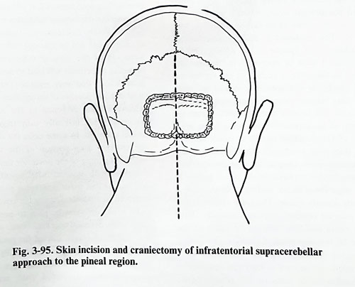
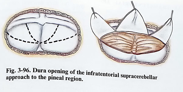
A long midline incision is used, starting from the C4 level and extending into the occipital region up to the lambdoid suture. The muscle attachments and pericranium are elevated on each side, and retracted. A standard craniectomy is performed, which includes exposure of the lateral sinuses and torcular in all instances, without extension to the foramen magnum (Fig. 3-95). Craniotomy of the posterior fossa also can be an option and in such a case an additional nibbling of the superior border is added to expose the lateral sinuses and torcular. If the dura is found tense and nonpulsatile, then the lateral ventricle must be punctured, or dehydrating agent must be applied. The dura can be opened in several different ways, but in any of them the superior surface of the cerebellum should be well exposed. The tentorium should not obstruct the midline view and the dura on the central area has to be retracted upwards to the greatest extent. Secondary incisions are then carried out laterally toward the lateral sinuses. After the completion of the dural incisions, there should be three flaps, a central one and two laterals that reflect upward and expose the superior surface of the cerebellum (Fig. 3-96). Inferiorly the dura mater is left to cover the cerebellar hemispheres during the retraction. The superior border of the cerebellum is gently separated from the lateral sinuses, already under the magnification of the microscope sacrificing the veins entering the sinuses and dissecting the adhesions. Then the superior cerebellar surface is gently retracted, and all bridging veins over the superior surface of the cerebellum (vermis and hemispheres) can be sacrificed until approaching the tentorial incisura.
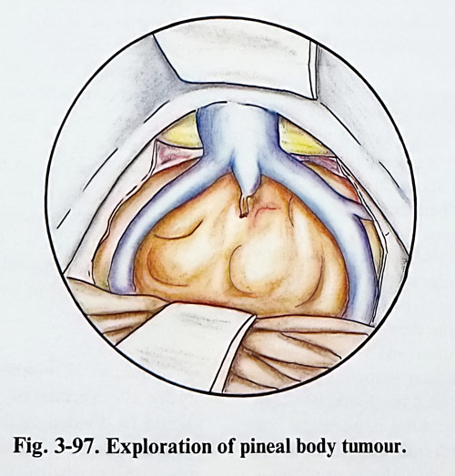
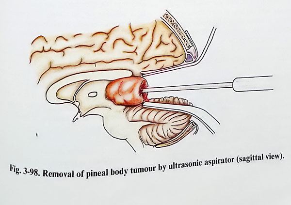
By gravity, with the patient in the sitting position, the cerebellum will drop and provide 1 to 1,5 cm of space between its surface and the tentorium. A self-retaining retractor is placed on the tentorium to the notch lifting torcular and the straight sinus. Another retractor depresses slightly the vermis of the cerebellum (Figs. 3-97; 3-98). The arachnoid of this region is usually thickened and opaque in the presence of the tumour. It must be opened to the midline and close to the anterior surface of the vermis and the cerebellar hemispheres avoiding injury to the deep venous system. The precentral cerebellar vein will be seen coming directly from the region of the superior vermis towards the great vein of Galen, which usually is visible through the thickened arachnoid. This cerebellar vein sometimes needs coagulation and division, permitting further exposure of the pineal region. In this stage the posterior surface of the tumour is usually already visible. The great vein of Galen and internal cerebral veins are generally above the tumour and they may not be visible at this stage of the operation. Laterally, the medial surface of the temporal lobes and the veins of Rosenthal can be seen as they run upwards to the confluence of veins in this region. More retraction of the anterior portion of cerebellum is needed by the inferior self-retaining retractor, exposing the larger posterior surface of the tumour.
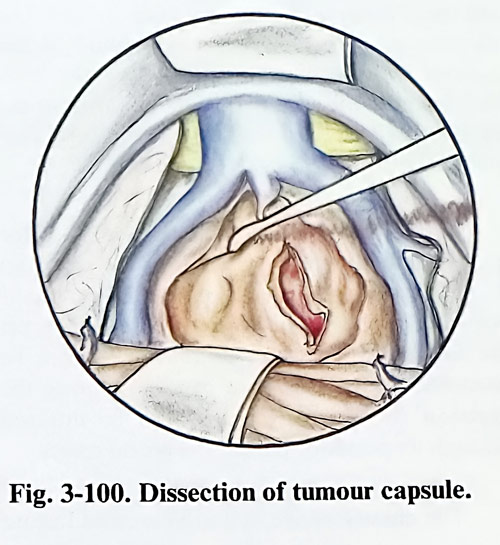
Then the volume of the tumour may be reduced by debulking. In the case of tumours that are benign and encapsulated, it is possible to dissect the capsule of the tumour first laterally, then superiorly, and inferiorly after enucleating its interior. Many tumours are attached anteriorly to the velum interpositum and sometimes laterally to the medial portion of the pulvinar. Attachment inferiorly to the quadrigeminal plate varies according to the nature of the tumour but generally it is modest in encapsulated benign tumours.
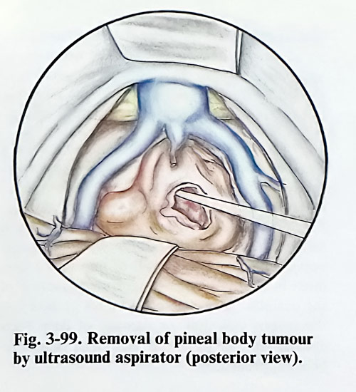
Some of the tumours are extremely soft and may be sucked out by a fine suction tube. Other firmer tumours such as some astrocytomas and teratomas need a laser or cavitron suction aspirator for their debulking. Once the larger central portion of the tumour has been removed, it will become apparent whether the tumour infiltrates or is encapsulated. With infiltrative tumours, it is recommended not to attempt radical resection. Removal of the tumour infiltrating the surrounding structures will lead to severe neurological deficits that may remain permanent. The most difficult portion of encapsulated tumour to remove is that attached to the midbrain or which extends laterally into trigone of the lateral ventricle. In some cases the dissection of the anterior deep portion of the tumour can result in opening of the posterior third ventricle.
Tumours with minimal attachment will often roll out at this point and large pieces may be removed as a whole. During the removal, particular attention must be paid to protecting the deep veins (internal cerebral veins, vein of Galen, and basal veins). Except for packing of the bleeding points, there is practically no other haemostatic method to be used. An attempt to coagulate these distended veins may lead to larger rupture. Even with non-resectable tumours, benefit may be achieved through the performance of internal decompression only (Figs. 3-99; 3-100).
OCCIPITAL TRANSTENTORIAL APPROACH
The advantages of this approach over the infratentorial supracerebellar approach are the greater ability to mobilise the tumour and to visualise major parts of the pineal region. Usually there are no large veins that cross from the occipital lobe into the superior sagittal sinus.
Therefore, no risk of infarction occurs by this approach unless the surgeon makes the mistake of coagulating some big cerebral vein, which drains the occipital lobe into the transverse sinus. Such a vein can be located more laterally and should be outside the operative exposure.
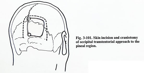
The occipital transtentorial approach is traditionally done with the patient in the sitting position with all precautions against air embolism. More recent and better alternative is the semiprone position with the head rotated about 20 degrees to the side of the operation ("park bench" position). A right occipital craniotomy is made, near to the midline exposing the border of the sagittal sinus and with the lower border of the craniotomy to the transverse sinus (Fig. 3-101). After the bone flap has been turned, the dura is opened parallel to the superior sagittal sinus and the transverse sinus producing a triangular shaped flap that is reflected laterally. Enough dura should be left to the sinuses for an easy dural closure and then the edges are turned out with traction sutures. Some cortical veins entering into the sagittal sinus should be sacrificed, if they are not very big.
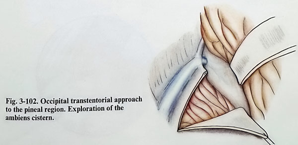
Next, already with the help of the microscope, a self-retaining retractor should be gently applied to the occipital lobe, retracting it laterally and superiorly. Direct compression of the calcarine cortex should be avoided, keeping the retractor more to the occipital pole than deep into the interhemispheric fissure. The tentorium is then opened, making an incision parallel to straight sinus and approximately 1 to 1,5 cm from it, starting at 1 - 2 cm anterior to the transverse sinus and torcular and carried out anteriorly towards the incisura (Fig. 3-102). The tentorium is vascular and the control of bleeding is possible by bipolar coagulation, or occasionally by clips. Traction sutures are placed to hold the tentorium open.
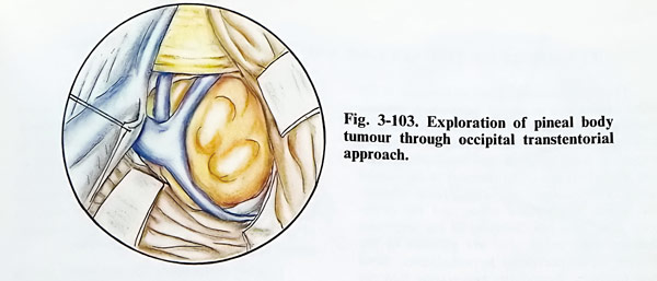
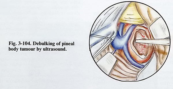
The arachnoid of the ambiens cistern overlying the deep cerebral veins (usually secn through it) must be divided, taking care not to damage the veins. Once the arachnoid is separated from the veins, the relationship of the tumour can be identified and the surgeon can deal with any lesion of the pineal gland, superior vermis, quadrigeminal plate, posterior third ventricle or splenium. The relationship of the deep veins should be identified before initiating the tumour's removal (Fig. 3-103). If there is a pineal tumour or glioma arising from the quadrigeminal plate, these vessels will be displaced superiorly and posteriorly and will be draping over the mass. If the lesion dealt with is a typical pineal tumour, it should be entered by sharp dissection and a biopsy should be obtained. A frozen section will help with the decision on how to proceed. If the tumour proves to be of embryonal origin, such as a teratoma or dermoid, piecemeal or 'en bloc' resection should be done (Fig. 3-104). In such a case an attempt should be made to remove the tumour completely. If the tumour is a germinoma with infiltra-tive growth the removal should be carried out as completely as possible, but without insisting on radicalism.
A pineal tumour may extend downward under the vermis of the cerebellum, which should be divided so that the lower pole of the tumour will be accessible and more easily removed. Similarly, the tumour may extend superiorly into the splenium of the corpus callosum. By tumour debulking and delivering its capsule downward into the operative field, division of the splenium can be avoided. If the tumour arises from the sple-nium of the corpus callosum, the veins usually are displaced inferiorly. In these cases an inter-hemispherical approach is more useful. If the tumour is a glioma, a specimen should be obtained and a modest decompression done.
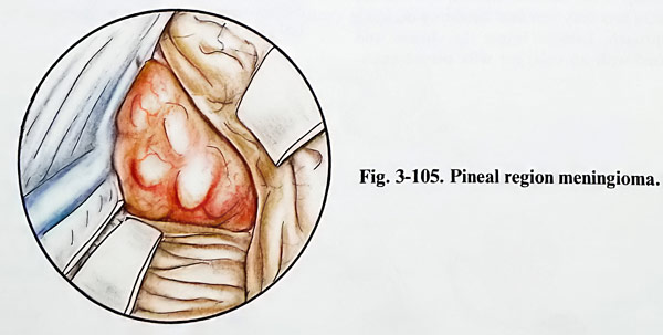
When the tumour grows from the free edge of the tentorium or from the junction of the falx and the tentorium, the deep veins will be displaced anteriorly, creating a problem in identifying them through the tumour (Fig. 3-105). The surgeon must first reduce the volume of the tumour with the ultrasonic aspirator and after that separate the surface of the tumour from the deep veins and surrounding anatomical structures.
Intraaxial tumours, such as gliomas of the quadrigeminal plate or gliomas of the splenium of the corpus callosum displace the deep veins posteriorly, upward or downward depending on the site of origin of the tumour. They can be identified by the lack of a capsule. During the excision the third ventricle can be entered through its posterior part in the pineal recess.
The closure of the operative wound has no special particularities. After complete haemostasis, the wound is irrigated, and the tentorium is approximated with some sutures, to avoid herniation of the occipital lobe into the posterior fossa. The rest of the closure is a routine one.
The complications of this approach are the common ones of any intracranial surgical intervention. Infection, osteomyelitis, and intracranial haemorrhages of various types are possible.
Specific complications when using this approach include the production of homonymous hemianopia. This is a direct result of improper placement of the retractor or vigorous retraction of the occipital lobe. The possibilities of injury to the corpus callosum, or damage of deep veins are the same as in the infratentorial supracerebellar approach.
TUMOURS OF THE SELLAR AND PARASELLAR REGIONS
The most common types of tumours in the sellar region are pituitary adenomas, cranio-pharyngiomas and meningiomas. According to different statistical data, pituitary adenomas constitute from 6 - 8 % and craniopharyngiomas from 2 - 4,5% of all tumours in the cranial cavity. The meningiomas arising from tuberculum sellae, diaphragma sellae, and the cavernous sinus area are discussed in the respective chapter. Less often seen are gliomas of the chiasm, optic nerves or hypothalamus. Skull base tumours, secondarily penetrating into the cranial cavity, as for instance chordomas, carcinomatous metastases and epipharyngeal tumours do, also present with the features of sellar or parasellar lesion.
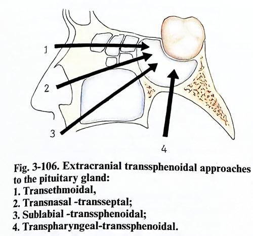
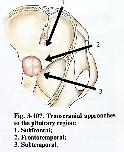
Two main routes of access to the sellar area have been developed through the decades of neurosurgical progress: the extracranial and the transcranial. The extracranial approaches reach the area through the skull base, mainly through the paranasal sinuses, but the transcranial route requires a craniotomy. Extracranial approaches as a rule have a narrow corridor and therefore a limited angle of view, specific for every technique (Figs. 3-106; 3-107).
However, to decide the approach, several common characteristics have to be established in these types of lesion. Although not always possible, the first is to define the structure from which the lesion is thought to derive its origins.
Secondly, because it is important to preserve visual function to its maximum, the relationship of the lesion to chiasm, optic nerves and tracts is a very essential factor in deciding the approach. Lesions below the chiasm and associated with an enlarged sella permit extracranial access.
Lesions in the sellar and parasellar region are closely related to the internal carotid arteries and the circle of Willis. The finding of any compromised important arteries and the main supplying branches is crucial for the safe choice of approach and manipulation during surgery. The unexpected discovery of an intrasellar aneurysmal dilation of an artery during a transsphenoidal approach can lead to a disaster.
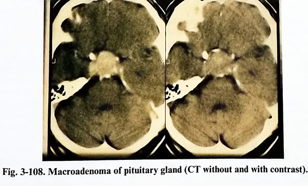
Lesions in the sellar and parasellar area are investigated with all routine imaging modalities as CT and MRI, but the classical skull radiography still gives a general idea of the condition of sellar and parasellar bone structures (Fig. 3- 108). In addition, the lateral X-ray film should always be displayed on the screen during extracranial approaches for comparison with the image on the intraoperative fluoroscopic screen. Angiography is always recommended when vascular pathology (aneurysm, AVM) or very vascularized lesions are suspected.
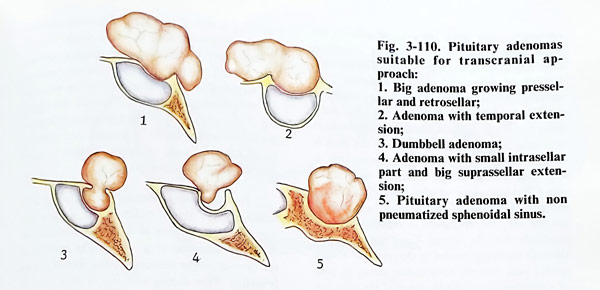
Surgical planning is based on the location of the lesion. Sellar or predominantly sellar lesions, especially those with an enlarged sella, are suitable for a transsphenoidal approach in general as the important structures will be behind the mass along the surgeon's view. Parasellar lesions are not suitable for the same approach, as the carotid artery and the cavernous sinus obstruct the access. In such cases, as well as in any lesion over the chiasm, the only option is transcranial. However, the particular locations of the lesion indicate the different approaches that can be appropriate, as is illustrated in figs. 3-109 and 3-110.
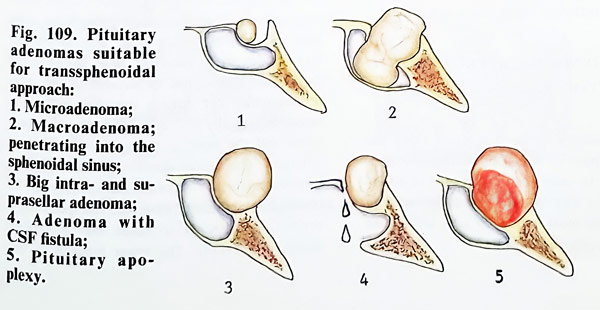
PITUITARY ADENOMAS
Indications and contraindications for surgical treatment. The indications for surgical treatment of pituitary adenomas differ according to the endocrinological activity of the tumour, whether it is non-secreting or hypersecreting.
There are unconditional indications for surgical treatment when the clinical manifestations of the suprasellar growth present affection of the chiasm and optic nerves (disturbances in the visual acuity and fields, and optic atrophy). The same is referred to the lesion of other cranial nerves.
The manifestation of the mass lesion with hydrocephalus due to compression of the third ventricle or increased intracranial pressure provoked by extensive growth in the frontal or temporal direction also requires surgical treatment.
The surgical treatment of an accidentally discovered pituitary adenoma without clinical manifestations should be postponed till its growth is proved by follow up CT scan. Pituitary apoplexy is an indication for urgent operation. Despite the spontaneous improvement of the visual disturbances in some cases, the risk of waiting on spontaneous improvement is larger than that of the urgent operation; this is equally true for elderly patients.
Spontaneous CSF rhinorrhoea in pituitary adenomas is also an indication for surgical treatment. The adenomas presenting with rhinorrhoea usually have aggressive growth and lead to destruction of the dura and the sella.
TRANSSPHENOIDAL APPROACHES TO THE PITUITARY REGION
The pituitary region is reached along the extracranial path by first approaching the sphenoidal sinus. Its cavity can be exposed in different ways by the preceding steps of the operation. The most frequently used techniques to expose the sphenoidal sinus are:
Transsphenoidal sublabial approach. The patient is placed on the operating table in a supine position with the head slightly lifted upward, so that the venous outflow is not obstructed. One of the thighs is prepared and draped also. In case of any need for sellar and sphenoidal packing, an incision can be done on the lateral side of the thigh and the needed fat, muscle pads and fascia easily obtained. The X ray tube with the image intensifier screen is so adjusted that during the whole time of the surgery, the approach, and the intracranial and intrasellar manipulations are efficiently controlled.
After preparing the skin of the face with antiseptic solutions, the gingival mucosa and the upper lip are infiltrated with local anaesthetic at a distance 3 cm from the midline on both sides).
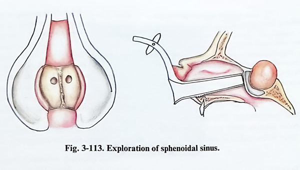
Every nostril is inspected with a speculum and local anaesthetic with adrenalin is injected under the septal mucosa and the floor of the nasal meatus (Fig. 3-111). The upper lip is retracted and an incision on the mucosa of 5 - 6 cm length 1 cm above the teeth is made in the vestibular (mucolabial) fold. The incision is completed to the bone of the alveolar processes and the mucosa with periosteum is stripped upwards until it exposes the lower edge of the anterior nasal apertures. The nasal spine of the maxilla, together with the lower edge of the bony nasal entrances are nibbled (Fig. 3-112). The mucosa is separated subperiosteally from the nasal septum and the floor of the nasal cavity. Deepening the separation, gradually the entire nasal septum is stripped on one side, but on the other only its base. Then the nasal septum is cut at its base and retracted on the side where the mucosa was not detached. The fracture of vomer in the depth that follows has to be close to the sphenoidal sinus. The already detached mucosa is retracted laterally with a specially designed speculum and the view to the sphenoidal sinus is enlarged. The proper direction of the operative approach to the sphenoidal sinus may be checked on the X-ray monitor. The speculum is repositioned deeper to reach the sphenoidal sinus (Fig. 3-113). The lower borders of this retractor, which touch the external edge of the nasal meatus are toothed, and by indenting them any slipping out is prevented. The anterior wall of the sphenoidal sinus is removed. This wall is thin and its perforation is easy with any sharp instrument. The cavity of the sphenoidal sinus and the bulging of the floor of the sella is inspected. The mucous membrane of the sinus is stripped out.
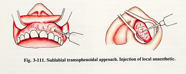
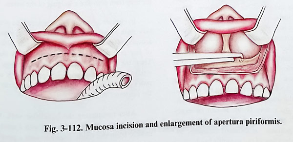
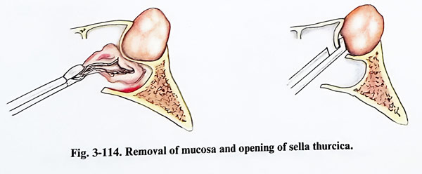
The exposed sellar floor area is identified on the image intensifier as are the other hidden parts of the sphenoidal sinus cavity. The area of sellar floor opening is defined and the speculum is repositioned accordingly. On the lateral X-ray view the line through the upper border of the retractor blades should pass over the tuberculum sellae. In case of a pituitary adenoma the floor of the sella can be thin and there can be a defect, so its opening is very easy. Rarely in microadenomas the sellar floor is hard and a drill has to be used for opening. The opening of the sella is enlarged with a small bone punch to a window of at least 1 cm square, and in an enlarged sella this opening may be even much larger (Fig. 3-114). The sellar dura often has increased vascularity, and bleeding is controlled with bipolar coagulation.
A puncture of the sellar dura has to be done before opening, in order to exclude an aneurysm, empty sella or a cystic tumour, and all this has to be controlled on the X-ray screen for the exact position of the needle.
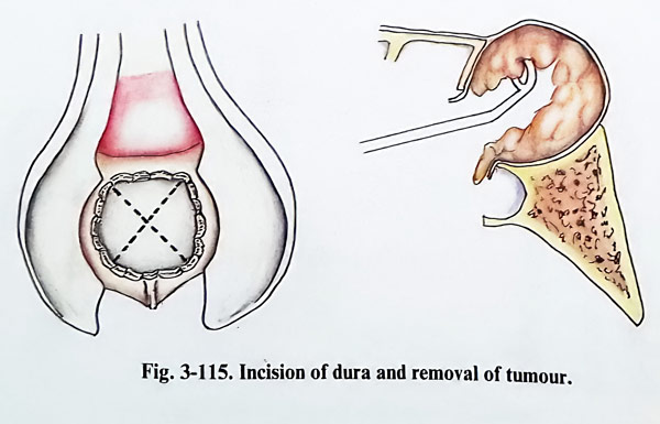
The dura is opened with a crosswise incision, the limit of the incision reaching the edges of the bone defect of the sellar floor. When making the incisions of the dura care should be taken not to injure the internal carotid arteries, which in certain anatomical variants may be situated rather medially. After initial central opening a blunt probe (hook) is inserted under the dura, detecting the presence of a carotid or intracavernous sinus communications in the thickness of the wall. When making the incision, bleeding of the intercavernous anastomoses situated in the dura may occur. It can be easily stopped with bipolar coagulation, packing with cottonoids, gelfoam, or oxidised cellulose, together with elevation of the patient's head.
If it is soft, the removal of tumour tissue is most conveniently done by suction, but when it is hard only by curettage or tumour forceps (Fig. 3-115). It is very important in this removal not to injure the arachnoid and not to provoke a leak of cerebrospinal fluid. Through this opening of the sellar floor it is not possible to inspect the entire tumour cavity. A helpful tool can be a small dentist's type of mirror, which can be inserted inside the tumour cavity.
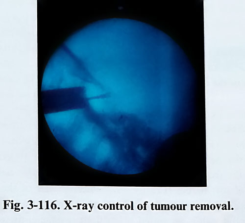
The suprasellar extension of the tumour is removed with curved curettes and suction tip, controlling from time to time the position on the image intensifier (Fig. 3-116). The borders of the tumour on the imaging studies should not be breached. First comes removal of that part of the tumour that is close to the sellar floor, followed by the lateral parts. The suprasellar extension usually drops into the residual cavity and the surgeon can see it pulsating. If this does not occur, the intracranial pressure can be raised moderately by the anaesthetist increasing the positive intrathoracic pressure or positioning the head lower.
In case of a microadenoma, the greyish soft tumour tissue can be seen within the yellowish-brown normal tissue of the pituitary gland. It can be removed easily with dissectors, suction, small pituitary forceps or curettes. If the micro-adenoma is not immediately seen after opening the dura, a horizontal incision of the pituitary gland is made; pressing the gland with a dissector may then provoke a bulging of the lesion.
In elective adenohypophysectomy the pituitary stalk must be exposed initially. The vessels around it, branching out on the anterior surface of the pituitary, are coagulated and divided.
Then the yellowish anterior pituitary tissue is separated from the posterior part, which appears greyish in colour and remains adherent to the posterior sellar wall.
After completing the intrasellar work, the residual cavity, if big, is filled in with a piece of muscle from the thigh and covered with a piece of fascia lata.
If CSF leak should appear during the operation, then, after packing the tumour cavity with muscle, the opening of the sellar floor is closed with bone fragment from the nasal sep-tum. This fragment should be a little longer in one of its dimensions, so that, when introduced inside the sella in a certain way, it can then be rotated to lock inside the sellar opening (Fig. 3-117).
The operation ends only with suture of the vestibular mucosa. Both nostrils are packed with gauze strips, soaked in antibiotic endonasal application ointment.
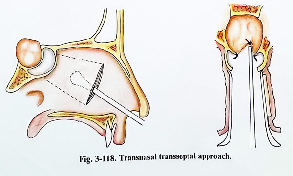
Transnasal transseptal approach. This method greatly simplifies the dissection of the septal mucosa through one nostril. The microscope is in use from the beginning of the operation. A vertical incision is done on the septal mucosa inside the nasal meatus 2 cm behind the nasal entrance. The mucosa is separated from the bone septum until reaching the anterior wall of the sphenoidal sinus. After that the septum is cut and retracted in the same way as in the previous approach, exposing the anterior wall of the sphenoidal sinus bilaterally. Under X-ray control, the speculum is inserted, positioning its blades near to the anterior wall of the sinus and its orifices. Next steps of the approach are the same as in the previous method (Fig. 3-118).
This approach provides a narrower view than the previous, but the distance to the sella is shorter by about 2 cm, and it is suitable for removal of microadenomas.

Transethmoidal approach. The patient is placed in a supine position on the operative table with the head slightly lifted above the heart level. The surgeon stands on the patient's right side. The nasal cavity is loosely packed with cottonoid strips, soaked in 10% cocaine hydrochloride and 1% adrenaline, in order to provoke vasoconstriction of the nasal mucosa.
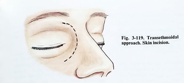
After the application of local anaesthesia a four cm long arch-like incision is made medially to the internal angle of the eyelids (Fig. 3-119).
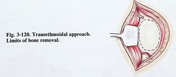
The skin, together with the periosteum is dissected successively from the frontal, and maxillary bones, as a block of soft tissues (Fig. 3-
120). The trochlea is detached and the periosteum from the medial orbital wall is separated, until the anterior ethmoidal artery is reached. A special self-retaining retractor is inserted and the anterior orbital content is displaced laterally to allow an opening between the retractor blades of 2 to 3 cm. The lamina papiracea of the ethmoid is opened, which is easily attained. Only in cases of acromegaly this lamina is thickened and for this opening a fine drill or chisseling can be needed. The bone defect is widened upward, opening the orbital extension of the frontal sinus and the posterior part of the lacrymal fossa. The thin bone plate of the posterior part of the medial orbital wall is preserved and it serves as anatomical landmark for the next stage of the operation. After that the surgeon resorts to the help of the surgical microscope.
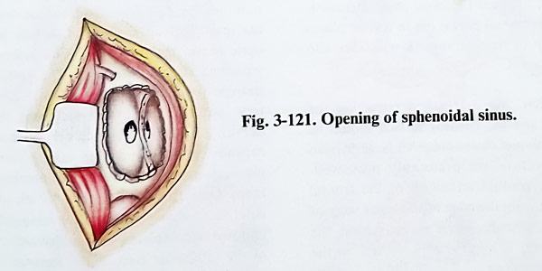
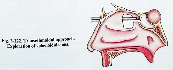
Next is penetrated into the uppermost recess of the right nasal cavity, in which a straight surgical instrument has previously been placed through the right nostril. After the removal of the posterior ethmoidal cells from the right side, the anterior wall of the sphenoidal sinus is reached on the same side (Fig. 3-121). The anterior wall of the sinus is opened and a greater part of it is removed. The midline septum of the sinus cavity is identified, which is not always situated strictly on the midline (Fig, 3-122). It is also removed together with part of the nasal septum and the neighbouring sections of the anterior wall of the sphenoidal sinus from the other side. After this operative stage the sellar floor is sufficiently well exposed.
The opening of the sellar floor and all further steps are carried out as described in the other trassphenoidal methods.
The packs are removed on the third postoperative day. Rarely a pack will slip into the oropharynx. In such a case the pack should be removed and the nostril should be repacked.
A postoperative CSF leak is treated conservatively by lumbar drain for 5 days or repeated lumbar punctures. A leak that visibly drips or does not resolve within 10 days requires surgical revision and closure.
TRANSCRANIAL APPROACHES TO PITUITARY ADENOMAS
The patient is placed in supine position on the operative table, with slight turning of the head to the side opposite to the craniotomy. To decrease the intracranial pressure and facilitate the approach to the tumor almost regularly osmotic diuretics are applied.
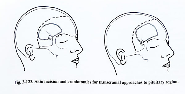
Pituitary adenomas are removed as a rule through a subfrontal or frontotemporal (pterional) craniotomy on the non dominant side, and the skin incision is bitemporal behind the hairline (Fig. 3-123). In very big adenomas with significant subfrontal growth a bifrontal craniotomy can be the most suitable. If the extension is subtemporal, then a temporal craniotomy may be used. As a rule the sellar area is reached following the edge of the lesser sphenoidal wing. In a subfrontal paramedian approach, the retraction is directed to the optic nerves and chiasm.
The dura is opened with an curved incision made at 1 - 2 cm from the basal edge of the craniotomy. The peripheral part is cut in several places and is lifted by traction sutures.
The sellar area is reached retracting the frontal lobe and the cortical surface should be protected with wet cottonoids. All large Sylvian venous collectors are preferably preserved. Through the gradual retraction of the frontal lobe and following the edge of the lesser wing of the sphenoid under magnification, the chiasmal cistern is reached and opened on the side of the craniotomy. The suction of CSF allows the operative approach with less retraction. The optic nerve is followed for exposure of the chiasm and the contralateral optic nerve. Usually both optic nerves are distended and the chiasm is lifted by the tumour.
Before beginning the removal, the tumour is punctured and any possible fluid contents aspirated through a thick needle, in order to exclude an aneurysm or a very vascular tumour. On the other hand, the puncture may reveal a cyst, which appears in 20 percent of all chromophobe adenomas. Cyst evacuation makes the tumour removal easier.
The removal of the tumour is done following certain certain steps in order. Initially the capsule of the tumour (containing diaphragma sellae) is opened by a crosswise or circular incision.
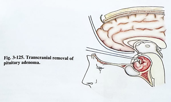
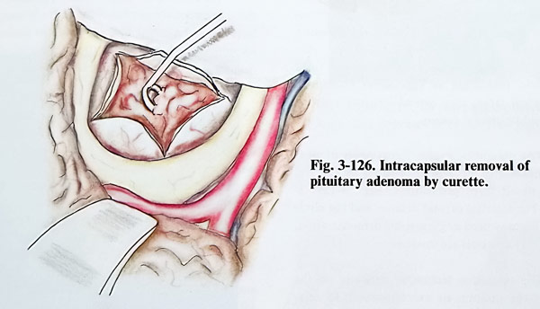
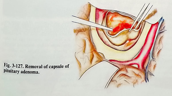
This incision is usually done on the tumour surface between the two optic nerves and chiasm. In some cases with asymmetrical growth of the adenoma, the incision can have another place as is shown in fig. 3-124. This is preceded by coagulation of any large blood vessels at the site of incision. The tumour tissue is removed subcapsularly until a large cavity is produced with the suction, the ultrasonic aspirator and curettes (Figs. 3-125; 3-126). The intra-capsular debulking continues, until the tumour capsule collapses and the compression on the optic nerves and the chiasm is relieved. Then the tumour capsule is dissected from the chiasm and the ipsilateral optic nerve. Laterally from the optic nerve, the internal carotid artery is also separated from the tumour. The opticocarotid triangle (with borders the internal carotid artery, anterior cerebral artery and the optic nerve) is used as a space for dissecting the capsule, at the same time taking care of the pituitary stalk, the IIIrd cranial nerve and the arterial branches located there. Only the blood vessels, which obviously enter the tumour tissue can be coagulated. The removal is completed with additional curettage of the intrasellar part of the tumour. We may consider the removal of an adenoma complete, when the entire suprassellar part is removed including the capsule and the bottom of the sella cavity is curetted, until the surgeon feels the hardness of the sellar floor (Fig. 3-127).
When the adenoma is composed of hard tissue and is strongly adherent to the third ventricle, the complete removal of the tumour often proves impossible. The same applies for large polylobulated tumours. The floor of the sella in some cases is so thin that at the time of removing the deep tumour portions, the floor can be perforated. If so, the bottom of the sella is packed with a piece of muscle.
The removal of an adenoma through a bifrontal craniotomy is very similar to the removal of a meningioma of tuberculum and diaphragma sellae. After the retraction of both frontal lobes, the chiasmatic region is widely exposed. Both internal carotid arteries, and their bifurcations are also exposed in such a way, that larger branches become accessible for the surgeon and they can be separated from the tumour surface with minimal injury.
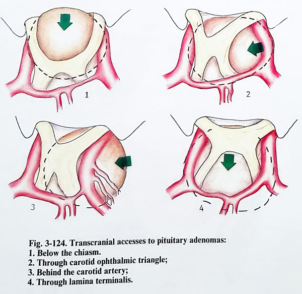
Pituitary adenomas with big temporal fossa extension require temporal or pterional cranioromy. After removal of the temporal extension of the tumour, the remaining part is reached through the opticocarotid triangle and behind the internal carotid artery. Last to be removed is the intrasellar part of the tumour.
After surgery the patient is kept in an intensive care unit overnight with standard monitoring and visual fields frequently evaluated. He is encouraged to be out of bed the next day. Sutures are removed on the seventh postoperative day. The rest of the care is the same as after a transsphenoidal operation.
CRANIOPHARYNGIOMAS
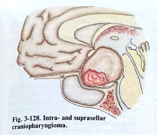
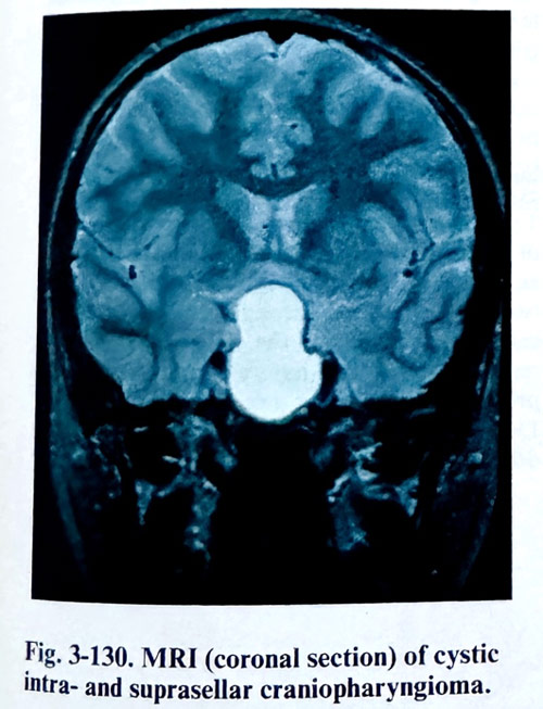
These dysontogenetic tumours, seen mainly in children and in the early decades of life, originate from residual cell nests of the hypophyseal duct in close relation to the diencephalic floor. According to the exact position and later development of these ectopic cellular groups, the craniopharyngiomas can be subpial (hypothalamic and third ventricle), extrapial - subarachnoid with different locations regarding the infundibulum, or extra-arachnoidal. Partially or totally extra-arachnoid craniopharyngiomas can occupy the sella turcica and enlarge it. These tumours often have cystic parts full of yellow to brown fluid and a solid part with calcifications in the tissue (Fig. 3-128). As their formation indicated, they can be found inside the sella, along the infundibulum and inside the third ventricle, displacing all suprasellar structures, and when large, they extend into the anterior and middle cranial fossae.
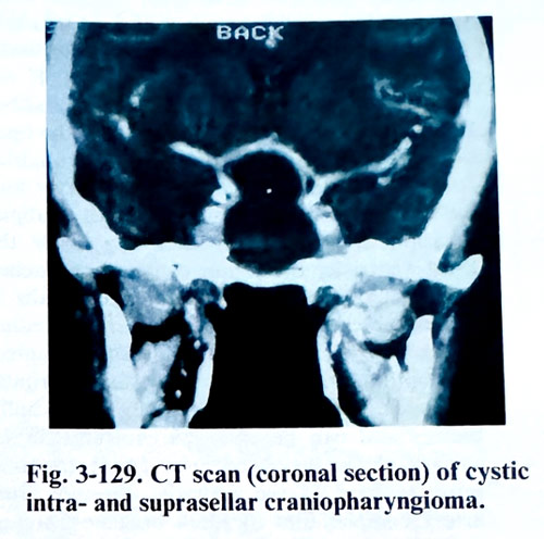
The diagnostic evaluation consists of clear visualisation of all tumour portions by CT and MRI in all three projections (Figs. 3-129: 3-130). The internal carotid arteries and the circle of Willis may need angiographic demonstration, when major vessels are involved.
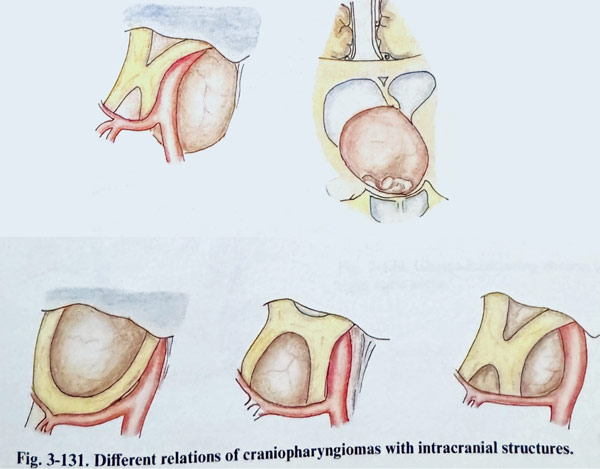
The operative technique depends on the size of the tumour, its exact location, and its relations to the arachnoid of the suprasellar cistern and pia in the area of infundibulum. Further on, the size of the intrasellar part with the subsequent sellar enlargement, as well as the exact locations of the vascular and neural structures regarding the tumour determine additional details of the final decision for the approach and the technique. Craniopharyngiomas with a predominantly suprasellar and intraarachnoid location, associated with a non-enlarged sella or significant extrasellar (subtemporal or subfrontal) extensions, are approached transcranially. Intrasellar lesions, especially those enlarging the sella, with moderate suprasellar extension accessible from the sellar compartment are suitable for a transsphenoidal approach (Fig. 3-131). Cyst evacuations, if one of the main goals of surgery, are done more easily through the trans. sphenoidal route. Those tumours that have a subpial or third-ventricular extension, require exposure of the lamina terminalis and CSF derivation immediately before or during the surgery, by inserting a ventricular drainage. 1f the CSF circulation at the level of the third ventricle is not normalised, the removal of the ventricular drainage is followed by a permanent ventricular shunting.
The main initial steps of the approaches to these lesions - transsphenoidal or transcranial . have been described already with the pituitary adenomas, and only the particular details referring to craniopharyngiomas are mentioned below.
Regardless of the approach, once the tumour is exposed, the first step is always its puncture, aspiration of the tumoural fluid and excision of part of tumoural wall for biopsy. In a transsphenoidal approach, the puncture is done before the dural opening. The dura is opened in the common way and as widely as possible. The tumour is detached from the internal dural surface. As the lesion is collapsed after the evacuation, the wall can be dissected from the wall of the sella, taking care not to damage any intrasellar invagination of arachnoid. Pituitary and infundibulum can be seen posteriorly as an atrophic strip due to the long-lasting compression; they must be preserved as much as possible. As intrasellar tumours can be only partially extraarachnoid, the dissection of the upper portions of the tumour can disrupt the arachnoid.
If no structures protrude and no suprasellar adhesions are seen, the removal can be completed. A diaphragm widely destroyed through suprasellar growth often limits the safety of manipulation due to depth and poor visibility.
Solid tumoural parts create more difficulties than the cystic ones. Any vascular damage will be controlled with considerable difficulty because of the limited transsphenoidal and transsellar view. Transcranial approaches expose the chiasmatic and adjacent cisterns, and the arachnoid is dissected over the tumoural wall. The evacuated cystic part collapses the lesion or part of it.
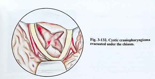
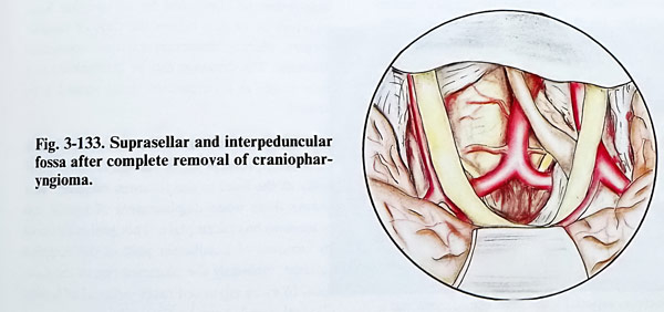
Dissection is performed inside the cystern. The tumour is removed in pieces, dissecting it from the optic pathway structures, infundibulum (identified with difficulty in big tumours) and the big arteries, carefully sparing the small arterial branches (Figs. 3-132; 3-133). If the tumour is located posterior to the infundibulum, it has to be approached through the opticocarotid triangle and the interpeduncular fossa. The third and fourth cranial nerves also have to be preserved. In tumours with a subpial location or secondary penetrating inside the third ventricle, the lesion can be approached with a small incision of the lamina terminalis. It is a suitable access when this structure is either distended by the tumour or is already disrupted.
The optimal view is achieved by the subfrontal approach. The incision is initially a few milli-metres and can be enlarged carefully to the surface of the distended area. Great attention must be paid to the anterior communicating artery complex and its small branches, preserving them as much as possible. Tumour removal follows in a manner requiring minimal manipulation evacuating the cystic part and excising it in small pieces. After evacuation of the cyst, the posterior cerebral and basilar arteries can be identified.
Care should be taken not to injure the III cranial nerve. Preservation of the pituitary stalk is a difficult task for the surgeon. In many cases is impossible to identify the stalk during the dissection of the tumour.
Visual improvement after surgery is obtained in 50% according to most of the statistics. Mortality depends on the size of the lesion and the extent of diencephalic involvement.
GLIOMAS OF THE CHIASM AND OPTIC NERVES
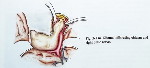
Gliomas of the anterior visual pathways are of two types regarding the primary location of their growth. The lesion arising from the intraorbital part of the optic nerve secondaryly grows intracranially involving the chasm and other suprasellar structures. The management of such type of tumour will be discussed in the respective chapter for orbital and cranio-orbital surgery. The other group of gliomas initiating their growth from the chiasm usually have an infiltrative growth, penetrate into the third ventricle and the orbits, widening the optic canal on one or both sides. The complete removal of this tumour is an exception. The tumour is approached through a frontal craniotomy penetrating toward the pituitary region and after exposing the tumour, a piece is taken for biopsy.
To prevent deterioration of the vision the piece of biopsy should be very small and taken with an incision along the optic pathways. If the tumour penetrates through a widened optic canal into the orbit, the optic canal roof is removed, in order to have a decompression and preserve the vision for a certain period of time (Fig. 3-134). When both optic canals are involved, this decompression is bilateral.