CLIVAL AND PETROCLIVAL MENINGIOMAS
Clival and petroclival meningiomas arise from the upper two-thirds of the clivus and from the petroclival junction medial to the trigeminal nerve. These tumours grow anteriorly or antero-laterally to the brain stem, compressing or involving the basilar artery and its branches. The Vth - VIIIth cranial nerves are usually displaced dorsally. Big clival and petroclival meningiomas may grow superiorly through the tentorial notch, compressing the midbrain.
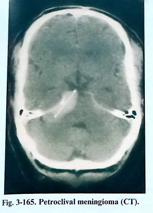
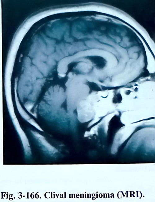
The different modalities of diagnostic imaging (CT, MRI) should demonstrate the size and the location of the tumour, as well as its relationship with the cranial nerves, blood vessels and the brain stem. The indication of the place of attachment by the studies is of essential importance (Figs. 3-165; 3-166). Angiography is needed to show the main feeders of the tumour, the displacement, and the involvement of big vessels in the posterior fossa. In the majority of cases, feeders originate from the meningohypophiseal and marginal tentorial arteries seen enlarged on a subtraction of the angiograms.
Angiography should also indicate any encasement of major basilar branches or the basilar artery itself. Analysis of this information has also the goal of identifying those cases suitable for preoperative embolisation.
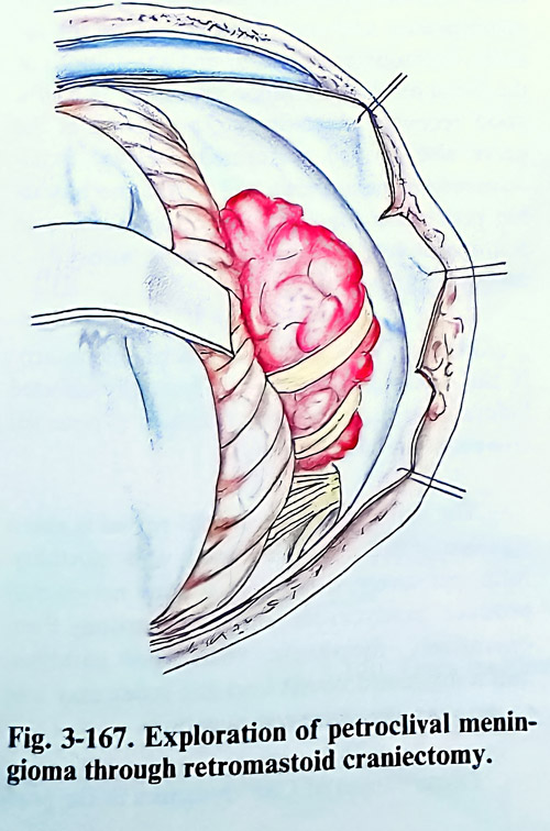
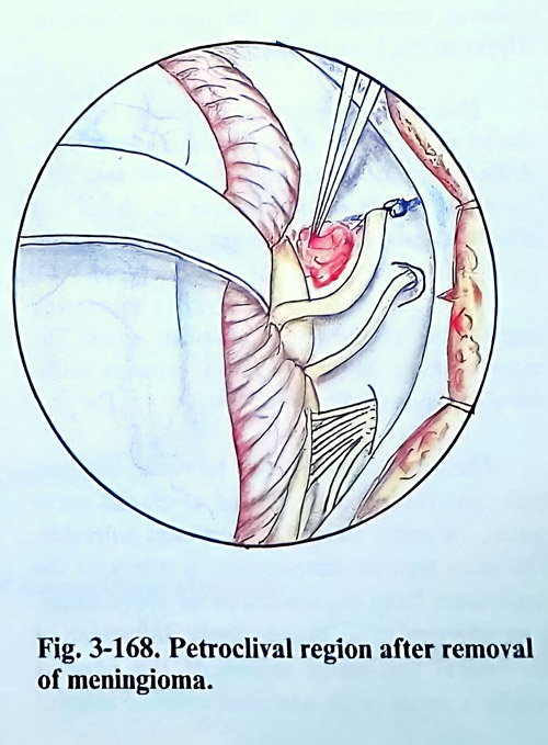
Tumours with this location can be subdivided according to their place of attachment: pure clival, petroclival, petroclival tentorial. The tumours with tentorial involvement may be more supra- or infratentorial. Routes of approach can also be specified as supratentorial, infratentorial or combined. The supratentorial route is convenient for tumours with upper clival and supratentorial attachment. Manipulation on the tumour is performed through the space between the IIIrd and IVth, and the IVth and Vth cranial nerves. An infratentorial attachment place is reached by a lateral suboccipital approach, manipulating the tumour between the Vth and VIIth - VIIIth nerves or below them (Figs. 3-167; 3-168). Petrous attachment is managed more easily than is the clival and extensive radical removals are easier. Transtentorial approaches, retro, and presigmoid transpetrosal approaches, are preferable for those lesions attached to a broader base, supra- and infratentorially, with extensive involvement of arteries and cranial nerves.
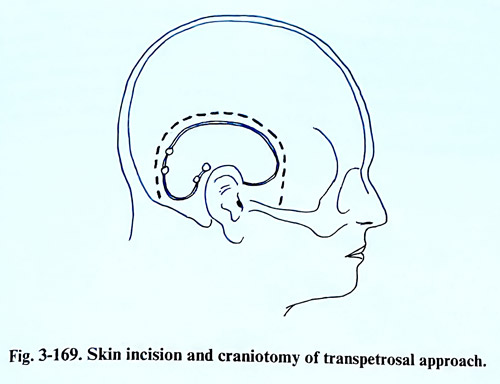
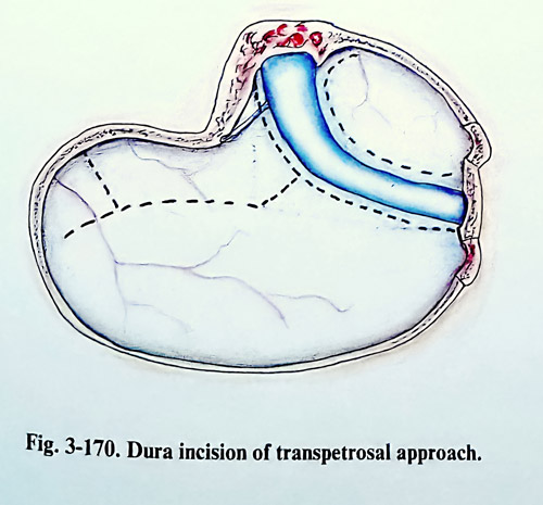
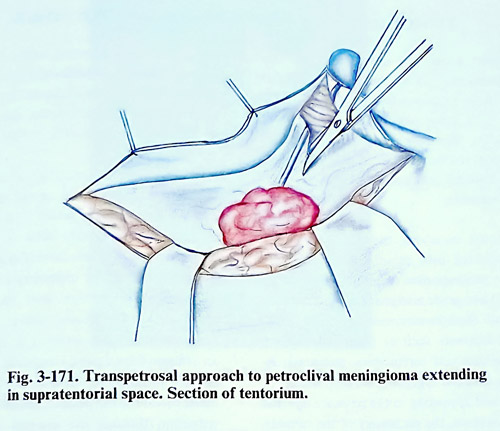
The transpetrosal approach is more appropriate for big tumours extending up through the tentorial notch. The technique of this approach is described in the first chapter of the book (Fig.3-169.The dura anterior to the sigmoid sinus is opened along the anterior margin of the sinus, The incision is then extended toward the supra-tentorial space, parallel to the floor of the temporal fossa. Another incision of the dura is made in the posterior fossa if needed (Fig. 3-170). The temporal lobe is gently retracted, preserving the vein of Labbe. The superior petrosal sinus is clipped or coagulated and transected. The incision is continued on the tentorium, parallel to the edge of the petrous bone and extended through its free edge. Care should be taken to preserve the trochlear nerve, which goes parallel to the tentorial edge. The opening of the tentorium allows excellent exposure of the upper pole of the tumour and the anterior and lateral aspects of the brain stem (Fig. 3-171). Trigeminal nerve roots are frequently stretched and spread by the tumour.
A self-retaining retractor is usually needed to hold the cerebellum medially with the transected edge of the tentorium. With any of the approaches, the initial steps of tumour removal are debulking of the volume and transection of its attachment. The tumour is separated from the tentorial surface, coagulating and cutting the feeding vessels; the same is done from the posterior petrous surface, and clivus. When the tumour is of small or medium size, the VIIth and VIIIth cranial nerves are usually stretched dorsally and they are easily identified. Big tumours may encase these nerves. The arachnoid is dissected from the tumour surface and the tumour is debulked using suction, ultrasonic aspirator or laser with extreme caution since the VIlth, VIth cranial nerves, as well as the posterior inferior cerebellar artery may be encased by the tumour. Then the tumour capsule is dissected from the adjacent structures.
The dissection should be maintained within the arachnoid planes to preserve the neural and vascular structures, often adherent to the tumour surface. Cranial nerves, the basilar artery and its branches may, however, demand meticulous and tedious dissection. The lower cranial nerves are usually easily dissected from the inferior pole of the tumour. Vagus nerve manipulation may provoke hypotension and bradycardia that should be avoided. The sixth nerve is stretched usually anteriorly and inferiorly, and it is also dissected away from the tumour following it distally. Alternating the visualisation of the surgical field between supra-and infratentorial routes allows the tumour capsule to be dissected more easily and more safely from the brain stem, the basilar artery and its branches. Once the tumour has been excised, the area of tumour attachment is preferably vaporised extensively with the laser. If there is extension of the tumour inside the internal auditory meatus, its wall is drilled and the tumour removed. A similar technique is applied for the removal of tumour extension into the jugular foramen. Hyperostotic bone is also removed by drillling.
During the closure, the periosteal flap produced at the initial stage of surgery covers the drilled petrosal bone to avoid CSF leak; the temporal muscle is rotated over the defect and attached to the sternomastoid muscle.
The rate of total removal has increased impressively in the latest published series. The main obstacle for total removal remains extradural invasion of the bone.
The complication rates, however, remain high, and is commonly related to cranial nerve deficit, in many cases transient and tolerable. The most serious complication is injury to the brain stem from manipulation or more likely, from affection of its blood supply. Infarction of the lateral tegmental region of the pons is usually a result of an occluded anterior inferior cerebellar artery, occasionally, the appearance of deficit may be delayed during the postoperative period. Temporal lobe swelling or haemorrhagic infarction can be seen with the subtemporal approach, particularly important on the dominant hemisphere. It is precipitated by coagulation or tearing of the vein of Labbe, or the basilar occipital veins. The cranial nerves from IIIrd to XIIth are also at risk during surgery. Because of the trochlear nerve's close relation to the tentorial border, its injury is a frequent hazard during tentorial splitting. Morbidity resulting from its paralysis, however, is minimal compared to paralysis of other cranial nerves. Morbidity from the trigeminal nerve, however, is more serious, because of the resulting corneal anaesthesia and subsequent keratitis, particularly if the facial nerve is also affected. In these cases immediate tarsorrhaphy should be performed followed by reconstructive surgery for the facial nerve function. A trigeminal nerve lesion may result in facial pain, anaesthesia dolorosa, and trigeminal neuralgia.
The size of the tumour is the most decisive factor for preserving the facial nerve. The facial nerve is usually displaced dorsally in petroclival meningiomas and may be involved by the tumour. Intraoperative end-to-end anastomosis of the facial nerve is an option for repair with 80% good recovery. Intraoperative grafting of the nerve also can be performed. Delayed facial-accessorius anastomosis will be the more suitable procedure for those affections discovered postoperatively and not recovering within 3 - 5 months from surgery.Hearing loss usually exists preoperatively. If the hearing is normal or partially affected before operation, its loss can be also a potential complication of surgery too. The deficit of lower cranial nerves is also a significant cause of morbidity and mortality. Intraoperatively, dissection of these nerves may produce bradycardia and hypotension. Post-operatively, dysphagia, vocal cord paralysis, and a depressed cough and gag reflex may lead to serious pulmonary complications.
Disturbances of CSF dynamics in the postoperative period include CSF leaks. hydrocephalus, and CSF collections under the soft tissue layers. Hydrocephalus may present before surgery and it may persist despite total removal of the mass, but it may also develop postoperatively. Acute postoperative hydrocephalus is usually obstructive and is related to mass effect, while delayed hydrocephalus is usually communicating due to poor absorption of CSF or obliteration of the basal cisterns. A CT scan is the most important diagnostic investigation and the treatment often is shunting, There is a risk of CS leak in the transpetrosal approach, occurring via the skin or through the middle ear. The leak is best avoided with watertight suturing of the dura, application of bone wax to the exposed cavities and sealing a periosteal flap over the drilled temporal bone surface. Prophylactic antibiotic coverage is usual in these circumstances.