TUMOURS OF THE CEREBELLOPONTINE ANGLE
Acoustic neurinomas are the most common cerebellopontine angle tumours. There are other kinds of tumours in the same region such as meningiomas, papillomas, epidermoids, etc. The close relation of the tumours with cranial nerves, brain stem and structures in the petrous bone, determines the specific technical approach to the surgical removal of these lesions.
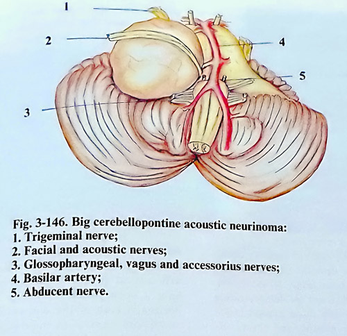
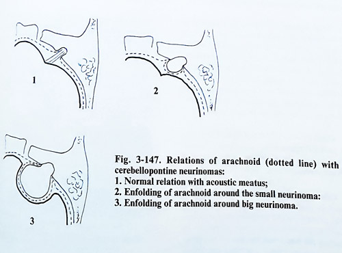
Acoustic neurinomas. These tumours arise usually from the intrameatal portion of the vestibular nerves. They have a round shape, with a smooth or polylobulated surface and variable consistency. When the tumour develops inside the internal auditory canal it leads to canal enlargement early on and later tumor growth continues into the cerebellopontine cistern (Fig. 3-146). According to their size, these tumours are divided into three groups: small size - less than 2 cm in diameter; medium size - from 2 to 4 cm and big - more than 4 cm in diameter. Small tumours are located inside the internal auditory meatus without enlarging it and not adhering to the dura. The facial and acoustic nerves are situated anteriorly to the tumour and are compressed against the anterior wall of the meatus, so they can be dissected easily from the tumour surface. When the tumour reaches 2 cm in diameter, it bulges into the cerebellopontine cistern. The arachnoid remains attached to the petrous bone, and due to the tumour expansion it forld over it as a double layer, containing cisternal elements (vessels and nerves) over the exposed tumour (Fig. 3-14T). The labyrinthine artery is thinner than 1 mm and lies caudally to the tumour and the nerves. Big tumours, already penetrating into the cerebellopontine cistern displace mainly the pons and dorsally they indent on the middle cerebellar peduncle. The medulla oblongata is less displaced. The cerebellar hemisphere in the vicinity of the tumour is also indented by the tumour. With larger displacement, the cerebellar tonsils descend through the foramen magnum and the lower parts of the aqueduct and fourth ventricle can be shifted to the opposite side.
The tumour capsule has different thickness and in many places is adherent to the arachnoid or around the capsule there are enlarged CSF spaces of differing shapes. The base of the tumour is firmly attached to the internal acoustic meatus adhering to the dura. When the tumour is very big, the superior pole may teach the free edge of tentorium. However, it seldom penetrates truly into the supratentorial space, The inferior part of the tumor can reach the foramen magnum.
The relations of neurinomas to the pons are variable. When the neurinoma is growing mainly medially and ventrally, the pons is displaced posteriorly, differing from laterally growing tumours, which displace the pons medially. The relatively dense consistency of these tumours, their slow growth and adherence to the temporal bone cause the compression of cranial nerves inside the cerebellopontine cistern. The vestibular nerve disappears into the tumour capsule still in the region of the internal acoustic meatus. Cochlear and facial nerves are tiny, often seen as transparent ribbons, but in many cases they can be separated from the tumour capsule. In some cases, however, this is impossible to be done, even with the help of higher magnification power of the surgical microscope. The nerves are situated at the anterior surface of the tumour capsule. The trigeminal nerve is displaced ventrally and rostrally from the rostral pole of the tumour, and the trochlear nerve usually is not displaced or compressed by the tumour. The abducent nerve is located ventrally and medially to the tumour mass and usually does not adhere to the tumour. The group of lower cranial nerves (IX. X and XI) are in contact with the tumour at their entrance in the jugular foramen, These nerves are rarely displaced caudally by the lower pole of the tumour.
In a few cases, when the tumour arises from the terminal branches of the vestibular nerve, it may have an intralabyrinthine and intrapyramidal location with destruction of the middle and internal ear.
The blood supply of acoustic neurinomas is carried out from branches of the labyrinthine artery, cortieal branches of the cerebellar arteries and by big branches of anterior and posterior inferior cerebellar arteries. The establishment of the relationships of cerebellar arteries to the acoustic neurinomas is important for tumour removal. The posterior inferior cerebellar artery is situated near the caudal pole of the tumour. It is covered by the tumour, and a big branch of this artery can enter the tumour at this point. The anterior inferior cerebellar artery crosses anteriorly the VIl and VIII cranial nerve, arising from the basilar artery, and is situated deeply on the anteromedial surface of the tumour, between the tumour and the pons.
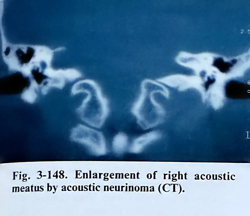
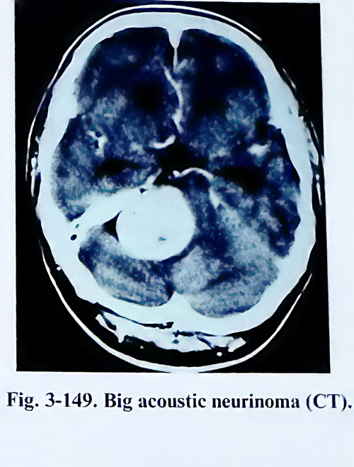
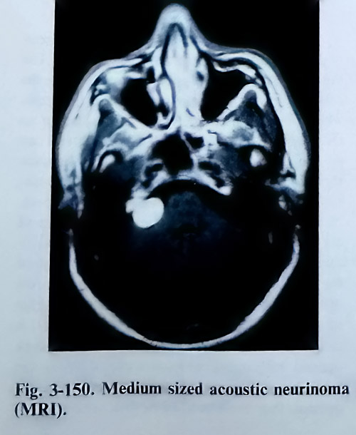
Preoperative diagnostic tests should include high resolution computed tomography (CT) with thin slices through the internal auditory canal. CT is helpful also in demonstrating bone erosion of the petrous bone (Figs. 3-148; 3-149). MRI is a more sensitive test for demonstrating small tumours, especially those located inside the internal acoustic meatus, and shows the relations of larger tumours to the vessels, cranial nerves and the brain stem (Fig. 3-150). Functional studies of the cranial nerves, especially acoustic, vestibular, facial, trigeminal and lower group are also essential preoperatively.
Total surgical removal is the treatment of choice for acoustic neurinomas. In patients with advanced growth of the lesion, causing hydrocephalus and increased intracranial pressure, hydrocephalus treatment has to precede removal by 3 - 4 weeks. In patients over sixty years, the indications for surgery depend on the general conditions. In such a patient partial excision of the tumour can be carried out.
SURGICAL TECHNIQUE.
There are two principal approaches for removal of acoustic neurinomas: retromastoid transmeatal and translabyrinthine.
Retromastoid transmeatal approach. The patient is placed on the operating table in sitting, prone-Concord position, or in a lateral/semisitting position (park-bench position). In the latter, the body and the head must be so inclined to the horizontal plane that the venous outflow from the cranial cavity be facilitated, but at the same time without causing a negative venous pressure, in order to avoid any danger of air embolism. This inclination is approximately between 30 and 45 degrees. The convenience of this position is that when the side of the tumour remains on top, the cerebellar hemisphere has a natural tendency to fall and to separate from the lateral wall of the posterior fossa and this facilitates penetration toward the cerebellopontine angle. The application of diuretics and corticosteriods begins before the operation.
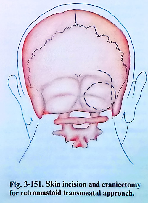
The posterior fossa is opened unilaterally. The skin incision is vertical in the retromastoid area with its upper limit 2 cm above the superior nuchal line and down to the first cervical vertebra. The skin incision may have a hockey-stick form (Fig. 3-151). The horizontal limb of the skin incision is 2 cm above and parallel to the nuchal line. The vertical limb of the incision is made over the mastoid process. The occipital artery and the greater occipital nerve are preserved if possible. A self-retaining retractor is used to maintain exposure. The craniectomy extends up to the edge of the lateral sinus, laterally near the sigmoid sinus and sometimes mastoid cell and emissarium mastoideum may be opened and then sealed with bone wax. Excision of the bone reaches downward near to the foramen magnum but it is not necessary for it to be opened. Medially the bone is excised till the necessary retraction of the cerebellar hemisphere is achieved.
The dura is opened with a curved incision with the flap base directed to the midline. The peripheral remainig part of the dura is lifted off with traction threads. The opening of the dura may be done also in a stellate form with four incisions. If the cerebellar tonsil on that side is under the edge of the foramen magnum, the foramen should be opened, including even the lamina of Cl, if considered appropriate. The arachnoid of the cisterna magna is opened. After sufficiently draining CSF, a self-retaining retractor is gently inserted if needed and the cerebellar hemisphere is retracted.
The size of the neurinoma determines in general the order of steps to follow in its removal. However, every surgery will aim at: 1. Reduction of the size of the lesion by debulking if it is medium or large sized. 2. Dissection of facial and acoustic nerves (if possible) at the internal acoustic meatus after drilling out its posterior rim. 3. Dissection of cranial nerves and the brain stem surface (in bigger tumours) with this completing the total removal.
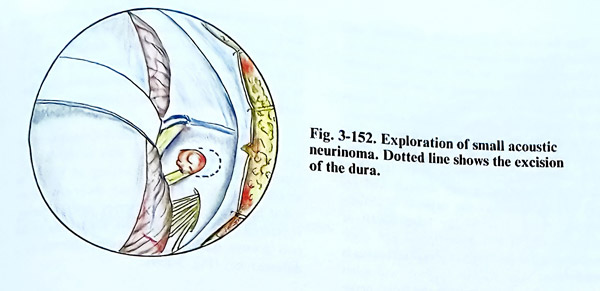
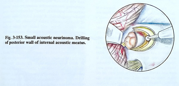
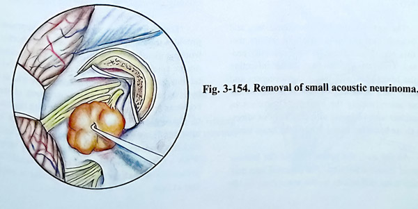
Most cerebellopontine angle neurinomas grow outside the meatus into the subarachnoid space and their further growth is associated with enfolding the arachnoid. A small intrameatal neurinoma, which is less then 2 cm in diameter may be removed totally 'en block'. The posterior wall of the acoustic meatus is drilled out first. This procedure begins with a circular excision of the dura from the petrous bone posterior to the internal auditory meatus (Fig. 3-152). The drilling initially is done using a 4 to 5 mm burr (Fig. 3-153). It is important to know the position of the jugular bulb, (especially if it is high) to avoid damage of its wall. To avoid injury to or opening of the semicircular canal and vestibule, the last part of the bony plate next to the fundus of meatus is left intact at the end of the drilling. The remaining distance to the fundus can be continuously assessed by using an angulated microinstrument (blunt hook) and should not be less than 2 mm. If one of the semicircular canals is fenestrated, suction of the perilymph and endolymph must be avoided, and the fenestration should be closed with fascia, sealed with fibrin glue. After that the meatal dura is opened and the tumour becomes accessible. Using light traction of the tumour out of the meatus the fibres of one of the vestibular nerves can be seen entering the tumour and usually the other is free (Fig. 3- 154). This vestibular nerve, entering into the tumour is divided. The facial and acoustic nerves are located anteriorly to the tumour and can be easily separated with a significant chance of preserving hearing and facial motion. The labyrinthine artery, also located anteriorly to the tumour, should be preserved too. The middle size acoustic neurinomas, which are less than 4 cm of diameter, can be removed easily and in many cases the facial nerve can be saved. Hearing in these cases. however, is usually already lost before surgery. In tumours of this size, there are distinct stages of the surgery, that have been already described above. The stages of debulking and dissection of cranial nerves and brain stem are relatively easy because of the smaller dislocation of structures produced by the lesion.
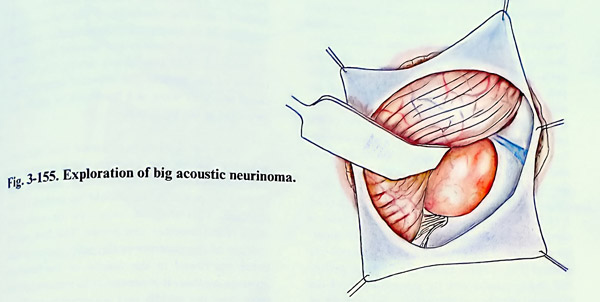
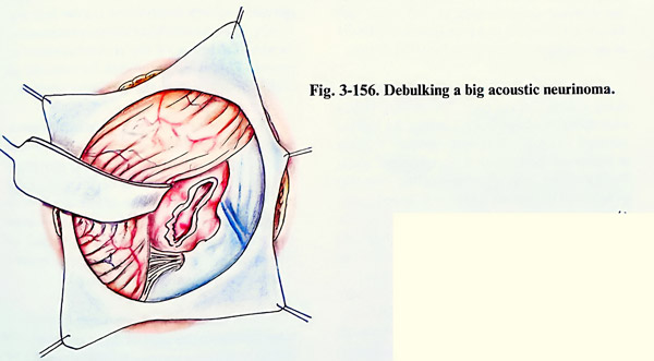
In big tumours (more than 4 cm in dia-meter) the arachnoid should be separated first from the posterior surface of the tumour and the cerebellar hemisphere gently retracted (Fig 3-155). Even with a very large tumour, it is usually not necessary to resect the lateral third of the cerebellar hemisphere to gain exposure. Then the tumour is reduced in size by suction, but better by ultrasonic aspiration if it is soft, or by laser evaporation if it is hard. The bleeding can be stopped by bipolar coagulation. The enucleation of the tumour continues until only thin layers of the tumour tissue remain attached to the capsule and it becomes possible to dissect it from the adjacent brain structures (Fig. 3-156).
The order of manipulations when dissecting the tumour capsule can be variable. The lower pole of the tumour is separated, where the caudal group of cranial nerves are dissected, which, even though they are very close to the tumour, they are not so adherent to it and can be easily detached. Cottonoid is placed between them and the tumour at the intial place of their identification. That prevents also the penetration of blood into the subarachnoid space.
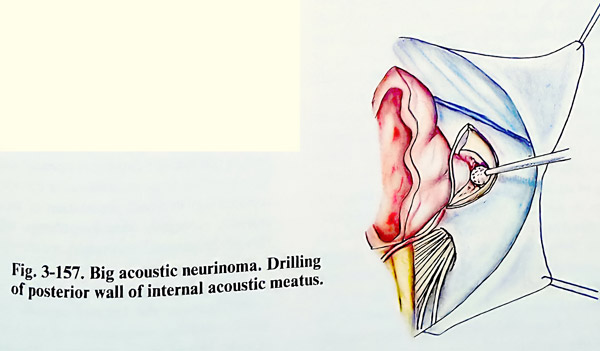
Next is penetrated along the lateral surface of the tumour and the internal auditory canal is reached. The dura along the posterior edge of the canal is coagulated, avoiding injury to the distal part of the sigmoid sinus. The lateral edge of the auditory canal together with its wall is drilled out (Fig. 3-157). The entire canal must be opened sufficiently wide, without opening the labyrinth. If some of the aerated cells are opened, they must be filled in with bone wax.
The facial nerve is located superiorly and anteriorly inside the meatus and is recognised by its yellowish colour. At this stage the mechanical and electrical stimulation of the facial nerve can be monitored intraoperatively. It is necessary to separate the tumour capsule completely from the facial nerve, beginning at the internal acoustic meatus and ending at the brain stem. Sharp dissection is preferred. With large tumours the separation is very difficult and requires a long time, as the capsule is closely adherent to the dura of the lateral wall of posterior fossa. If this dissection is not done immediately after the internal acoustic meatus is cleaned from tumour, the surgeon will meet greater difficulties in preserving the facial nerve at the brain stem.
The most delicate part of the operation is the separation of the tumour capsule from the brain stem. This begins between the tumour's lower pole and the medulla oblongata. The lower cranial nerves, remain distant, already covered with cottonoids. This part of the tumour is retracted laterally from the brain stem and is carefully dissected. Small arterial branches, entering directly into the tumour are coagulated and divided only at their entry point and not away from it. This manipulation is repeated until the tumour is separated from the brain stem. The cottonoids must be used with care and should not be held a long time on the brain stem, as they stick and then cause the tearing of small vessels on its surface. Suction should also be used in this region very cautiously. The anterior inferior cerebellar artery and its branches are the most important arteries encountered in this region and must be very carefully protected. Their coagulation may induce infarction of the brain stem with severe consequences and even a fatal outcome. Several branches of the same artery supply the tumour. They must be identified with certainty and cut, without affecting those branches going toward the brain stem.
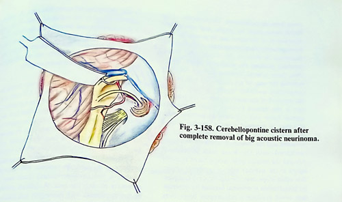
At the end, the upper pole of the tumour capsule is also separated. The petrosal vein may create some difficulties in the separation of the Vth cranial nerve (Fig. 3-158). It is preferable to keep it intact, but its coagulation and cutting may not represent any significant danger, but if it is large, it must be preserved, because it drains a large part of the cerebellar hemisphere and that may provoke congestion and oedema.
Blood transfusion is applied if required to replace the blood loss and the systolic pressure is maintaining through the surgery in a normo-slightly hypotensive range for the patient.
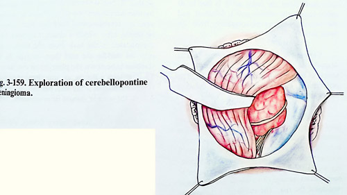
Meningiomas in the cerebellopontine region are approached in the same way as the cerebellopontine neurinomas. According to the dural attachment of these tumours regarding the acoustic meatus they are subdivided into two groups: anterior and posterior. Every one of the two groups displaces the cranial nerves in a different way (Fig. 3-159).
Once the cerebellar hemisphere is retracted and the tumour exposed, it is removed by alternating intratumoural decompression using ultrasonic aspirator, laser, microforceps and bipolar coagulation. A good arachnoidal plane of cleavage usually exists between the tumour and vessels, nerves, and brain. In quite a few instances, the tumour completely surrounds but does not invade nerves and vessels and a careful dissection may preserve neurological function. In tumours “en plaque” it is usually not possible to find this plane and nerve infiltration is evident.
During the tumour removal an attempt should be made to interrupt early the main vascular pedicle of the tumour that often comes from the petrous bone. Generally, the seventh and eighth nerves are well defined and can be separated well. After the tumour is removed, the dural site of attachment should be eliminated and occasionally any abnormal area of the bone drilled.
Translabyrinthine approach. This approach provides the most direct access with the shortest working distance to the internal acoustic meatus in the cerebellopontine region without cerebellar retraction. The disadvantage of the translabyrinthine approach is the destruction of the cochlear and vestibular end organs with the loss of hearing and balance. That disadvantage is relative because in cerebellopontine tumours the cochlear and vestibular functions are lost early. With a normal contralateral ear, the benefit of preserving a malfunctioning inner ear is minimal at best.
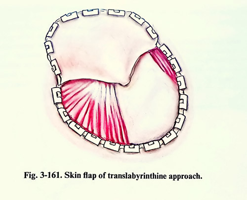
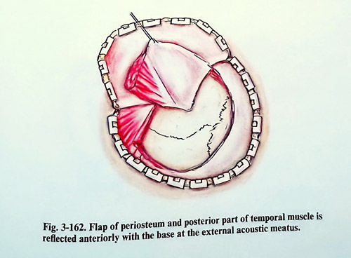
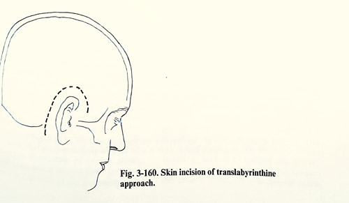
This approach can be indicated in acoustic neurinomas of small and medium size (less than 3,5 cm in diameter), Contraindications include removal of neurinomas that need preserving the hearing or very large tumours (greater than 3.5 cm in diameter). The patient is placed in a supine position with the head turned away from the surgeon. A wide C - shaped retro-auricular incision is made starting just bellow the tip of the mastoid. It runs upwards over the lateral surface of the mastoid to a point 2 cm or so above and behind the tip of the pinna, then curves forward and downward over the temporal muscle to finish 3 cm above the zygomatic arch (Fig. 3-160). A skin flap in the subgaleal plane is elevated forward until the posterior and superior margins of the external auditory meatus are defined (Fig. 3-161). An anteriorly based periosteal flap is incised with the posterior part of the temporal muscle and the pedicle over the area of the external acoustic meatus. All other soft tissues are retracted and preserved for a closure at the conclusion of the procedure (Fig. 3-162).
A complete mastoidectomy is performed, using a large cutting burr. The drilling begins over the mastoid antrum, preserving the tegmen tympani and external acoustic meatus. Drilling is continued posteriorly to the sigmoid sinus, leaving a thin layer of bone residing over that structure. The sigmoid sinus is followed inferiorly to the region of the jugular bulb. The cells of the mastoid tip are completely opened. Further drilling is performed parallel to the stylomastoid foramen until the periosteum of the foramen is identified. The drilling extends closer to the facial nerve, which has to be identified. The facial nerve is then followed superiorly to the region of the horizontal semicircular canal.
Care is taken to visualise the facial nerve throughout its entire vertical portion and the beginning of its horizontal course within the temporal bone to prevent iatrogenic injury. Intraoperative facial nerve elecromyographic monitoring is useful throughout this dissection. The retrofacial mastoid air cells are opened, and bone over the jugular bulb is removed. Drilling then is performed into the labyrinthine portion of the temporal bone. The lateral semicircular canal is carefully drilled with exposure of the secondgenu of the facial nerve in this region. The posterior semicircular canal is identified and opened, and the vestibule of the labyrinth is entered. Next the superior semicircular canal is opened, and the bone in and around this region is removed. The subarcuate artery lies in the centre of the arch of the superior semicircular canal and may serve as a landmark to guide the direction to the petrous tip.
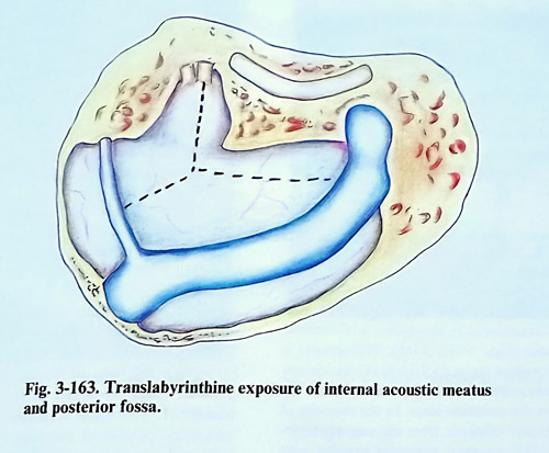
Next the drilling is extended into the petrous tip around the internal acoustic meatus. The bone of the internal acoustic meatus, including its posterior tip should be drilled to an eggshell. This shell of bone can be removed using microdissector and hooks. The free nerve endings of the superior or the inferior vestibular nerve are found in the fundus of the internal acoustic meatus. It is necessary to remove all the bone over the middle and posterior fossa dura, internal auditory meatus and jugular bulb (Fig 3-163).
The transcochlear approach represents a forward extension of the translabyrinthine ex-posure. This is accomplished by removing the cochlea in addition to the vestibular labyrinth.
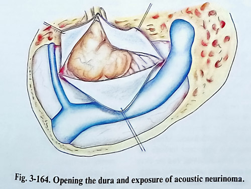
The dura is opened in a stellate manner to expose the cerebellopontine angle and to visua-lise the superior and inferior vestibular nerves. The removal of the tumour begins with identification of the facial nerve at the depth of the meatus (Fig. 3-164). The fibres of the superior and inferior vestibular nerves are interrupted, and just anteriorly, the greyish facial nerve is seen in front of the vertical crest (Bill's bar).
With medium or large sized tumours, the mass fills the entire area of dural exposure of the translabyrinthine approach. The presenting surface of the tumour capsule is opened, and gentle intracapsular tumour removal is carried out and at this point is important to avoid penetration of the tumour capsule, especially anterosuperiorly, where injury of the petrosal vein might occur.
After completion of the internal decompression, the tumour capsule begins to infold, allowing visualisation of the brainstem and IX, X, and XI cranial nerves. The intramental portion of the tumour is carefully dissected away from the facial nerve. The dissection of the facial nerve must be gentle, and mechanical or electric stimulation of the nerve can be monitored with intraoperative elecromyographic recording. All dissection is done in the arachnoidal plane, carefully pushing the arachnoid sheath away.
Only those arterial branches that are definitely entering the tumour are coagulated with bipolar coagulation and then sharply divided. During the removal of the anterior pole of the tumour, care is also needed to avoid injury to the fifth cranial nerve and the superior petrosal vein, that may be stretched over the superior pole of the tumour. This part after debulking can be retracted and the dissection of the seventh nerve continued until the brain stem is reached.
Around the inferior pole, care is taken to avoid injury to the anterior inferior cerebellar artery, that often loops over the eighth nerve complex. Its preservation is essential because its interruption could produce a catastrophic brain stem infarction. After the facial nerve is free in its entire course, the inferior pole is debulked, and the capsule is separated completely from the brain stem.
Closure. A single sheet of fascia lata is draped over the translabyrinthine defect in the temporal bone and filled with strips of fat. The pericranium, subcutaneous layer and the skin are sutured as usual.
Complications. They can occur during surgery and in the postoperative period. With the patient in a sitting position, the most common complication is air embolism. It is usually not a serious problem, but has the potential of becoming a catastrophic one. Careful maintenance of haemostasis during all stages of the operation minimises the risk. The increase of the venous pressure by the anaesthetist from time to time is also a measure to avoid this complication.
Haemorrhage during the operation is usually not a major problem. If the petrosal vein is torn, it can usually be managed with gentle packing with oxidised cellulose. Arterial bleeding is usually due to the tear of a small branch that likewise will respond to packing or bipolar coagulation. During the early postoperative period, monitoring of intracranial pressure aids in the detection of complications. A silastic catheter placed in the subdural space at the and of the operation and brought out accurately monitors posterior fossa pressure. If the posterior fossa pressure remains less then 15 mm Hg for 24 hours, the monitoring is discontinued, and the catheter removed.
Acute hydrocephalus may occur during the early postoperative period and a CT scan may be necessary to differentiate it from a postoperative haemorrhage. Cerebrospinal fluid leak may result from poor wound healing, increased intracranial pressure, wound infection, or opened mastoid cells that have not been sealed well with bone wax. A few simple stitches seldom control a CSF leak unless the increased intracranial pressure is reduced. In patients with hydrocephalus, a ventriculoperitoneal shunt may be necessary to stop the leak. A difficult problem occurs in patients with meningitis and hydrocephalus. At first a ventriculostomy with intraventricular administration of antibiotic is preferred until the cerebrospinal fluid has been shown to be sterile, after which a shunt is inserted. More complicated is the leak from the mastoid cells into the middle ear and then through the Eustachian tube down the pharynx or out the nose. To treat such type of fistula, external lumbar drainage is preferable, but if the leak persists beyond 3 or 4 days, the wound should be re-explored.
When fifth nerve injury occurs, the resultant corneal anaesthesia may lead to corneal ulceration if proper eye care is not provided. The function of the fifth nerve should be evaluated as soon as the patient awakens from anaesthesia. When the corneal reflex is diminished, the eye should be covered with a protective shield and artificial tears applied every 4 hours. If the reflex is absent or if there is an associated facial palsy, it is necessary to do a temporary tarsorrhaphy. Seventh nerve function should also be evaluated in the recovery room. If the face is paralysed, the same care for the eye should be provided as in trigeminus damage. II there is total disruption of the facial nerve during surgery, an operation to recover the paralysed facial muscles is indicated. Generally these procedures can be done in 3 to 4 months after the complete recovery of the patient from the intracranial operation.
The results from the surgical treatment of cerebellopontine neurinomas depend first of all on the size of the tumour and on all other factors determining the indications for surgery.
Small sized tumours (less than 2 cm of diameter) can be removed totally and the facial nerve can be preserved in 100% of patients. Half the patients remain with partial hearing. In 90% of the patients with medium sized tumours (up to 4 cm in diameter) total removal of the tumour is possible with 60% risk of facial nerve damage. Only in 10% of these patients is the facial nerve intact, and in another 10% it is interrupted definitely. In patients with big tumours (over 4 cm in diameter) total removal is achieved in 83% with 40% interruption of the facial nerve. Surgical mortality is 4% with respect to patients with big tumors.