TUMOURS INVOLVING THE CAVERNOUS SINUS
Surgery within the cavernous sinus (CS) has made significant progress through the last decade and now it is possible to remove some tumours that were considered before to be unresectable. The morbidity rate after such operations in the hands of experienced surgeons and with careful selection of patients for surgery is acceptable.
Tumours involving the cavernous sinus may be divided into three groups:1. Benign tumours - meningiomas, angiofibromas, neurinomas; 2. Low-grade malignancies such as chordomas and chondrosarcomas: 3. High grade malignant tumours such as basal cell carcinomas, squamous cell carcinomas, metastatic lesions. The decision regarding surgery should be individualised according to the patient's age and general condition, the pathology of the tumour, and its biological behaviour.
In cases of benign intracavernous sinus tumours, the indication for surgery is the progressive growth of the tumou on the imaging investigations and/or progression of the cranial nerve deficit. Low-grade malignancies such as chordomas and chondosarcomas can be removed from the cavernous sinus whenever complete tumour excision is the goal on a selective basis. This sometimes includes patients with bilateral intracavernous invasion. Indications for removal of high-grade malignancies involving the cavernous sinus are controversial.
Excision in a unilateral involvement of the cavernous sinus is possible in some cases, but bilateral cavernous sinus invasions by high malignancies are not suitable for resection.
CT and MRI with axial and direct CT coronal imaging can demonstrate the cavernous sinus neoplasms, but MRI is more sensitive than CT-scan for visualising small lesions in this area. The relationship of the tumour to the carotid artery and other major vessels in this region can be best delineated by MRI. Angiography provides information on the flow and collateralization and is better associated with a balloon occlusion test (Fig. 3-172).
Operations for removal of tumours invading the cavernous sinus expose the intracavernous carotid artery, which is at high risk of injury. Approximately 80% of patients will tolerate the loss of one carotid artery. However, patients who cannot compensate the reduction of blood flow through their internal carotid artery can suffer massive infarction and sometimes perish if that artery is sacrificed, despite an attempt for an extra - intracranial bypass. To be able to confront these circumstances in the safest mode for the patient, the tolerance to flow reduction through the internal carotid artery has to be tested preoperatively. The most important test is the balloon occlusion test, performed simultaneously with the angiography. An intra-carotid balloon is inflated for up to 15 minutes. If the patient experiences or the physician detects neurological deficit, the test is discontinued. Those patients remaining asymptomatic are submitted to a CBF test with xenon CT. If this test steadily indicates reduction of the flow below 35 ml (100 g) per minute, the patients are also under certain risk, but it is not as high as that of the first group. The remaining patients as a rule tolerate carotid flow interruption (even permanent) without neurological affections.
Safe manipulation of cavernous sinus structures requires the surgeon to be familiar with the morphology of the area beyond the routine requirements. Detailed microsurgical studies have refined those specific areas, which can be selected for penetration into the sinus. They are described conveniently as triangles. Another particular feature of cavernous sinus surgery is the difficulty of haemostasis. Skilful combined application of selective bipolar coagulation and pieces of oxidised cellulose must provide a sufficiently dry' area for proper visualisation and handling of structures.
The cavernous sinus - an anatomical structure built up from dural walls, separates and integrates three types of systems: 1. The internal carotid artery; 2. The cavernous venous plexus system; 3. Cranial nerves traversing it. The venous cavities, because of their extensive anastomosis to adjacent venous collectors, are the only structures which can be partially sacrificed.
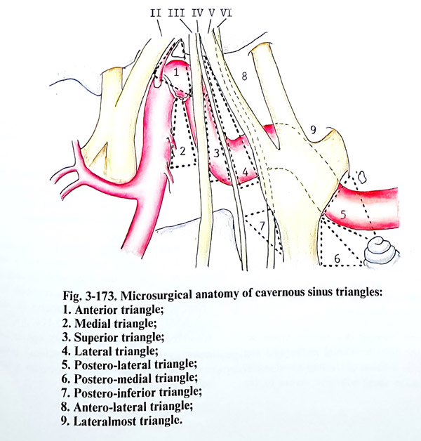
Otherwise, all its arterial or neural structures can be subjected to iatrogenic damage unless special circumstances take place and precautions are not undertaken. The safe approach to all cavernous structures has been standardised through the following triangles (Fig. 3-173):
The anterior triangle is enclosed by the lateral border of the extradural optic nerve, the medial wall of the superior orbital fissure dura and the dural ring, surrounding the internal carotid artery as it penetrates intradurally. The space extradurally contains the genu (C3 segment) of the internal carotid artery.
The medial triangle is produced by locating its tip at the anterior siphon angle (lateral carotid wall) and it must be already exposed, the porus oculomotorius (the intradural exit the third cranial nerve) and the posterior clinoid process. An incision inside this triangle will expose the proximal siphon (C3) and the horizontal intracavernous carotid artery (C4).
The superior triangle has as two borders the third and fourth nerves and posteriorly the dural margin toward the posterior fossa.
Through this triangle are exposed the C4 - C5 segments junction and the origin of the meningohypophiseal trunk.
The lateral triangle is a narrow space between the fourth cranial nerve and the ophthalmic division of the trigeminal nerve, The incision there will give access to the ascending CS segment of the internal carotid artery.
The postero-lateral triangle (Glasscock) is defined by the posterior rim of the foramen ovale, the foramen spinosum, the posterior border of the mandibular division of the trigeminal nerve, and the cochlear apex.
Drilling out the bone in this area will expose the proximal CS segment of the horizontal intrapetrous internal carotid artery. This place is suitable for proximal control or venous bypass graft of the artery.
The postero-medial triangle (Kawase) is confined among the porus trigeminus (extra-dural exit of trigeminal nerve), the posterior border of the mandibular division of the same nerve and the cochlea. This area corresponds to the petrous apex, which can be drilled out to increase the exposure of tri-geminal and brainstem structures.
The Postero-inferior triangle is bounded by the fourth cranial nerve, the posterior clinoid process, and the medial porus trigeminus. An incision in this area will expose the sixth cranial nerve.
The Anterolateral triangle is the arca between the ophthalmic and maxillary division of the fifth cranial nerve. This triangle is used to expose the superior orbital vein and anterolateral extension of tumours within the cavernous sinus.
The Lateralmost triangle is bounded by the second and third divisions of the trigeminal nerve. This is area to expose the lateral extension of cavernous sinus tumours.
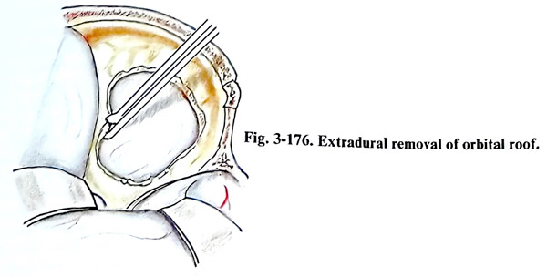
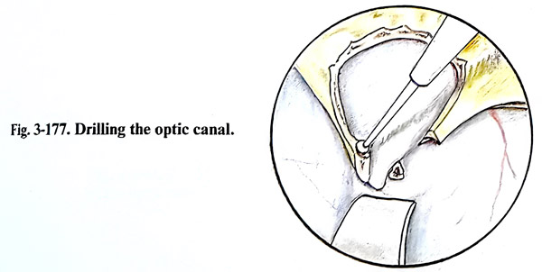
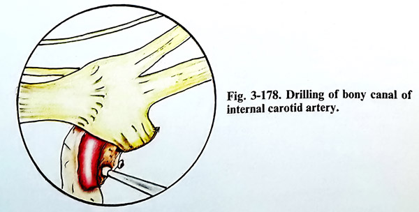
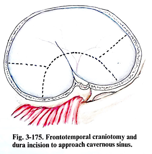
Surgical technique. Surgery of the cavernous sinus requires broad access and a wide range of view for the area. Most specially designed approaches will require a relatively large craniotomy, removing almost always the same cranial bone structures (Figs. 3-174; 3-175). A frontotemporal craniotomy followed in some cases by orbitozygomatic osteotomy permits such a broad approach and provides a better basal view of the area. Extradural removal of the orbital roof including the anterior clinoid process and the optic canal is the next usual step (Figs. 3-176; 3- 177). The dura is elevated from the middle cranial fossa, dissecting the middle meningeal artery, the great petrosal superficial nerve, and the mandibular and maxillary nerves. The greater petrosal superficial nerve and the middle meningeal artery are divided. The horizontal segment of the internal carotid artery is then exposed inside the carotid canal at the petrous apex postero-lateral triangle). The sometimes tiny bony wall of the canal is drilled out with a diamond microdrill head (Fig. 3-178). The technique can be used either for proximal control, as a site for potential bypass, or as a first step for an extra-dural approach following the artery. The Eusta-chian tube is at risk, and is best preserved at this stage by avoiding excessive drilling. An enlarged excision of bones is needed for tumours that have an extension and involvement of the pyramid and the sphenoid.
Extradural approach. The cavernous sinus can be entered extradurally by following the petrous internal carotid artery (inferior approach), especially after the temporary division of the mandibular nerve, if involved in the lesion or between the divisions of trigeminal nerve (anterolateral approach). The space between every two branches is rarely sufficiently broad unless they are distended by the tumour. The cavernous sinus can also be entered by a medial extradural approach if the sphenoid sinus is entered and the optic nerve unroofed after a broad basal frontal exposure of the tumour.
Intradural approach. Large tumours within the cavernous sinus and all meningiomas require an intradural approach. After dural opening, the Sylvian fissure is split and the frontal and temporal lobes are gently separated and retracted. If excessive retraction is necessary, the anterior 2 to 4 cm of the inferior temporal gyrus is resected, sparing the medial temporal lobe structures.
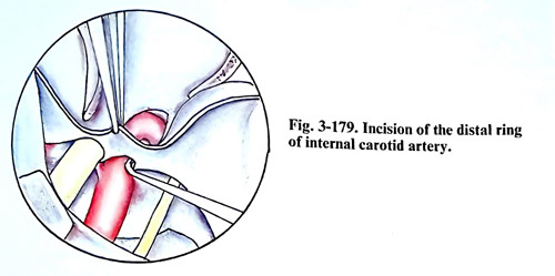
Superior approach. For the superior approach to the cavernous sinus, the optic nerve is unroofed completely and its dural sleeve opened. The anterior clinoid process is removed. The two distal rings around the distal intracavernous internal carotid artery are identified (Fig. 179). The superior wall of the cavernous sinus is opened through the anterior triangle, and the ICA is followed back into the cavernous sinus, exposing consecutively the medial and superior triangles. Intracavernous structures superior to the horizontal intracavernous ICA, the anterior genu, and the vertical segment of the ICA are well exposed by this technique. The sella turcica can also be exposed if necessary.
Lateral approach. This requires opening the lateral wall of the cavernous sinus. It is carried out by working through the Silvian fissure and subtemporally. For meningiomas, the outer dural incision can be crosswise (horizontally below and parallel to the approximate location of cranial nerve IV, and a vertical portion intersecting incision at the most prominent bulge of the tumour). It corresponds to the lateral triangle. For meningiomas, the outer dural layer of the lateral wall is completely peeled away, starting anteriorly in front of the sphenoid sinus, inferiorly near cranial nerve V2 and V3, superiorly from the tentorial edge, and posterorly to the superior petrosal sinus. Cranial nerve III should be identified in the subarachnoid space and followed for 5 mm into the lateral wall of the cavernous sinus. Cranial nerve IV may be found in the lateral wall or followed anteriorly in the subarachnoid space.
Because this last nerve is thin, it is preferable to leave a small cuff of dura around it until the end of the meningioma removal to its porus. Cranial nerve VI may be found in the lateral wall or followed forward from Meckel's cave and the trigeminal ganglion.
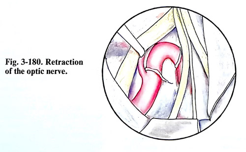
Complications. The most frequent complications are paresis of cranial nerves III to VI. They occur in about 80% of the cases, but most of them improve within the first 3 - 4 months. An important complication is CSF leak due to erosions in the bone caused by the lesion itself. Possible damage of the optic nerve during surgery is due to the opening of the canal by mechanical or thermal damage. This complication can be avoided by careful removal of the optic canal wall (Fig. 3-180). Contusions and/or laceration of the brain, and intracerebral hematomas can be avoided also by exerting only gentle retraction during the operation.
A life-threatening complication is the rupture of the internal carotid artery, which may occur either intra- or postoperatively due to damage of the arterial wall during removal of the tumour. When the tumour is adherent to the wall of the artery, or has even infiltrated it, the preferable decision is to leave part of the tumour around the artery and treat the patient postoperatively by gamma - knife surgery. On the other hand, if a radical operation has been chosen, the artery is then reconstructed with a graft (petrous to intracranial venous bypass).
Another complication can be venous bleeding after complete removal of the tumour. If the bleeding veins during operation are not well packed, fatal haemorrhage may occur post-operatively due to extrusion of the haemostatic pack as a result of straining of the patient. It is therefore advisable, at the end of surgery, after complete removal of the tumour, to place some sutures from one side to the other to prevent this extrusion. Sometimes, however, it is necessary to remove the oxidised cellulose from the cavernous sinus because of pressure exerted on nerves and the carotid. In such a case oxidised cellulose is replaced by loose packing at the same time checking for compression on the internal carotid artery and confirming that venous haemorrhage has been completely stopped.