
Legacy: "Atlast of Neurosurgery" / L.Karaguiosov, A. Ramadan, K.Karaguiosov / Kiwait/ 1998
Read PREVIOUS: 3. Tumors - Chapter 1
Read NEXT: 4. Vascular Diseases of the Brain
TUMOURS OF LATERAL VENTRICLES
Surgery of the lateral ventricular tumours is not very common in current practice, as they account for less than 1% of all intracranial tumours. Very often they are benign or of a low degree of malignancy. Their growth inside the lateral ventricle is usually silent due to the abundance of space and they present themselves at a stage characterised by ventriculomegaly and increased intracranial pressure. The intraventricular mass lesion's growth causes additional displacement and isolation of part or the whole of the lateral ventricle. These features determine the specific surgical management of these lesions. Manipulation of these tumours requires incision of apparently intact brain, as access to them is at a very significant depth. Furthermore, visibility of the tumour border is poor and very often the arterial feeders are not accessible at the beginning of the surgery. As their volume is relatively large, space for manipulation must be gained by tumour removal. Tumours inside the ventricle can have different locations, which therefore require a different approach technique. The most common place for a meningioma is the trigonum. Meningiomas and papillomas can be found in parts of the ventricle containing choroid plexus. Other tumours such as ependymomas, subependymomas, astrocytomas, dermoids and epidermoids are also found elsewhere inside the ventricle, but most often they are near the midline at the foramen of Monro and the septum pellucidum.
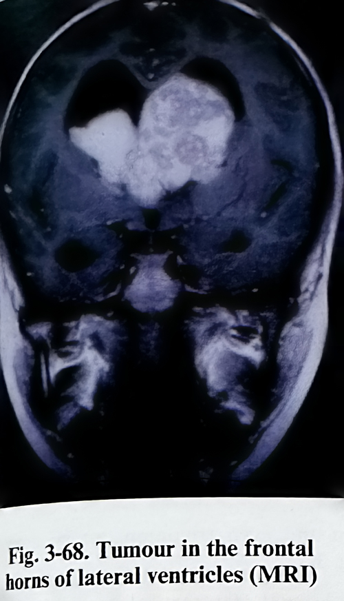
Investigations are based on tridimensional imaging to establish the precise location of the lesion. CT scans - enhanced and non-enhanced in axial, direct coronal and reconstructed images are needed for detailed delineation of the lesion's borders (Fig.3-68). MRI, also Gd enhanced, in at least Tl and T2 weighted images will give additional information on the tumour's texture and location, and on perifocal oedema and the tumour's relationship with important neural and vascular structures. Angiography, studying both the carotid and vertebrobasilar systems, must be used to demonstrate the vascular supply, relationship with choroidal arteries, the major deep venous collectors and their displacement. For the needs of a transcallosal approach, of utmost importance is the knowledge of the position of the big venous cortical parasagittal collectors. Another important area of the investigations is aimed at determining cerebral dominance and predicting the possibility of deficit after cortical incisions in preselected areas. As for the extra requirements of epilepsy surgery, the Wada test must on occasion be considered.
Four main locations inside the lateral ventricle have been specified for clinical surgical needs: frontal horn-Monro area, trigonum, body of the ventricle, and temporal horn. All approaches have been designed to reach the lesion by the shortest path with the largest possible incision of the cerebral mantle, not affecting functionally important (eloquent) cortex and pathways. Cortical incisions are used to reach the lesion, with some locations requiring callosotomies and rarely polar resections.
Every incision or resection gives a specific view and access for manipulation of a limited part of the lateral ventricle. Therefore the approach must be decided after the location of the lesion has been defined in detail. The resections are done following the same rules as in glioma surgery, but corpus callosum divisions require some particular elements in the technique. Isolated parts of the ventricles are dilated and this gives sufficient space for manipulation during the approach to the lesion. Craniotomies are performed over the cortical incision site and are relatively small.
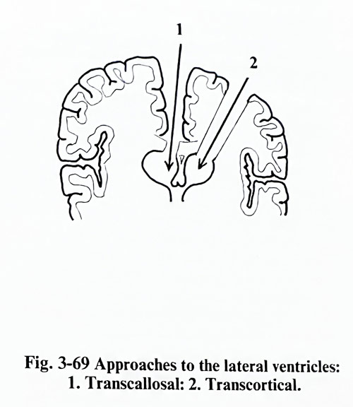
The frontal horn and the ventricular body are approached either by a frontal transcortical approach or by a transcallosal route (Fig. 3-69).
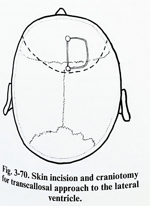
Transcallosal route. The patient is placed in the pin headholder with the head straight and elevated about 20 degrees. A curvilinear incision is made parallel to and about 2 cm behind the coronal suture across the midline (Fig. 3-70).
The incision should be long enough to allow reflection of the skin flap at least 6 cm anterior to the coronal suture. The scalp flap is reflected anteriorly and the sagittal and coronal sutures should be identified on the skull surface.
The bone flap should be made with its medial margin at the midline, using at least two burr holes. One of them is usually made with its medial margin at the midline just behind the junction of the coronal suture with the sagittal suture. The second burr hole should be made with its medial margin at the midline approximately 7 cm anterior to the first burr hole. If the tumour is inside the ventricular body it is helpful to place the bone flap slightly anteriorly. The craniotomy can be preplanned on the midsagittal MR image, properly placing these two burr holes according to the desired angle of view onto the lesion through the callosotomy.
The dural opening follows the outline of the bone flap with its base being hinged at the sagittal sinus, taking care not to avulse any of the cortico-dural veins that may be present. It is preferable to avoid sacrificing any draining vein from the cortex to the sagittal sinus. Small veins anterior to the coronal suture can generally be sacrificed, but if there is a very large draining vein it should be preserved if possible. Craniotomy is also planned considering the position of the major venous cortical tributaries.
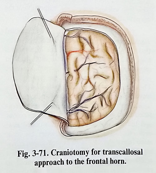
One or two traction sutures are placed through the base of the dural flap just lateral to the sagittal sinus and are suspended over the already reflected dural flap, to facilitate retraction of the dura. About 3 to 4 cm of longitudinal free space between the hemisphere and falx are required for adequate retraction (Fig. 3-71).
The proper direction to the desired part of the corpus callosum has to be established before retracting the hemisphere from the falx. The usual guide is an imaginary line in the midsagit-tal plane drawn from the coronal suture to the line through both external auditory meatuses.
This path will lead toward the midportion of the corpus callosum or slightly anteriorly to it.
The brain hemisphere is gently retracted reaching the inferior margin of the falx. After that the penetration between the hemispheres may occasionally present some difficulties in identifying the midline. Two retractors can be needed for the separation of the hemispheres.
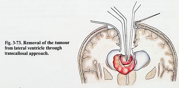
The callosomarginal arteries located above the cingulate gyrus can be mistaken for the perical-losal arteries. The corpus callosum is differentiated from adjacent cortex by its white colour and the two pericallosal arteries situated near each other. The pericallosal arteries are separated (there should not be branches seen crossing between them). We fix a distance of few millimetres between them with cottonoid to obtain their retraction into the more laterally located sulcus of the corpus callosum. Some times, in a case with ventriculomegaly, the pericallosal arteries may not be apparent, but there is no need to search for them if there is adequate corpus callosum surface exposed to open the ventricular system (Fig. 3-72). The corpus callosum is pratically avascular and can be divided with a small blunt dissector, fine bipolar forceps or a small suction tube, and at the expected depth (estimated on the midsagittal MRI image) usually one of the lateral ventricles is penetrated. Penetration is guided also by assessing the asymmetry of dilatation of the ventricles and the shift of the septum pellucidum. In some cases it is the cavum of the septum pellucidum that is penetrated. The approach, dividing the anterior and midpart of the body of the corpus callosum, can be enlarged safely to 4 cm in length. By tilting the spatulas and the microscope anteriorly and posteriorly, a relatively large portion of the ventricular body, anterior horn and even trigonum can be approached (Fig. 3-73).
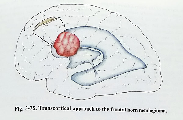
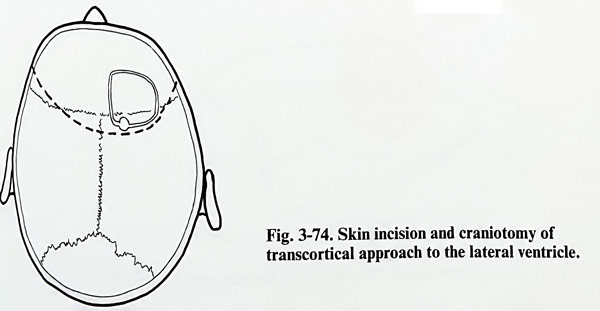
Frontal transcortical approach. The position of the patient in the pin headholder and the coronal incision of the scalp are the same as in a transcallosal approach. The bone flap has a similar location in the anterior-posterior direction but is a little lateral to the midline (Fig. 3-74). Through the superior frontal sulcus the incision penetrates the bulb of the frontal horn and the Monro area (Fig. 3-75). The non-dominant side is of course of less risks for the approach. The convenient exposure of the septum pellucidum permits its fenestration and access to the opposite frontal horn. In some cases, the good exposure of the foramen of Monro allows extension of the exposure to the third ventricle with the transforminal transvelum interpositum approach.
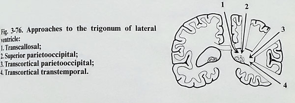
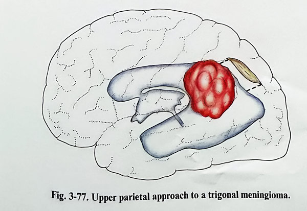
The approaches to the trigonum are important in the removal of meningiomas and papillo-mas. Tumours with this location can be reached in different ways. Several cortical incisions have been proposed: lateral temporoparietal, temporal, transcallosal, and superior parieto-occipital, the last often being performed as a polar resection (Fig. 3-76). The temporoparietal incision, especially on the dominant side, produces significant deficit - dyslexia, agraphia, acalculia and Gerstmann's syndrome have been described after such cortical intervention. However, with a big lesion, this incision on the non-dominant side can provide an access to the trigonal tumour along a relatively short path (Fig. 3-77).
Although the access is the most direct, the vascular supply to the tumour is initially difficult to control, as the supplying branches from the anterior and the posterior choroidal arteries remain hidden from surgeon's view unless a large part of the lesion is removed. Piecemeal removal and debulking of the lesion are the safest method. Even on the non-dominant side, the cortical incision can produce a visual field deficit. The superior temporal sulcus incision, originally designed for hippocampal resection, gives early access to the branches of the anterior choroidal artery and is suitable for the removal of similar lesions. If an additional supply exists from the posterior choroidal arteries, it will remain patent until the final stage of tumour removal. On the dominant side, Wernicke's cortical area can be compromised, but this cortical incision better recommended on the non-dominant side. The affection of visual pathways is less probable, as the incision is parallel to their fibres. They can lead more often to homonymous hemianopia and the visual associative cortex can eventually be damaged. The lesions are approached posteriorly and tumour arterial supply control cannot be an initial step. Another option is the transcallosal route. After the transection in the anterior two-thirds of the corpus callosum body, the lesion is approached anteriorly and close to the midline. As usual, the most difficult part remains the interruption of the feeders and separation from the choroid plexus, naturally after debulking. The superior parietooccipital incision, however, remains as a relatively less damaging cortical penetration. It is approximately 3 cm of length, between the postcentral sulcus and the parietooccipital fissure. There are risks of cortical dysfunction of the posterior parietal lobe (dominant or non-dominant) and they have to be considered before choosing this route.
Approaches to the temporal horn can be used to some extent as an option to reach trigonum. Incisions through the superior temporal sulcus and partial temporal lobectomies give access to the cavity of the ventricle.
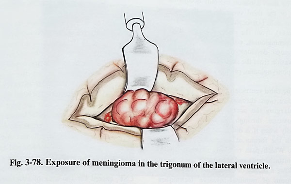
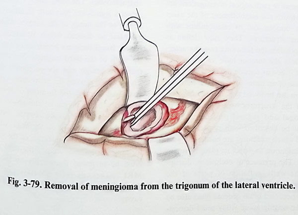
The excision of the lesion follows the general principles of atraumatic removal of lesions (Figs. 3-78; 3-79). Minor bleeders are meticulously stopped as the voluminous ventricular cavities can accumulate relatively large amounts of clots. This can lead insidiously to increased intracranial pressure and irreversible deterioration of the patient. At the end of the removal the ventricular cavities are thoroughly washed of clots and debris particles, and are filled will saline. There are some possible complications peculiar to intraventricular surgery. Risks of massive intraoperative haemorrhage are pre-sent. Delayed postoperative haemorrhages in the ventricular cavity are also possible. The removal of the mass lesion and the fenestration of the ventricle leads to collapse of the mantle and this can lead to subdural collections - hygromas or haematomas. Postoperative brain oedema can easily involve the diencephalic structures and be a serious source of deterioration of the postoperative condition. Hydrocephalus, which is very often present before surgery, can deteriorate and require shunting.
Some procedures are recommended as an addition to the standard postoperative care. An intraventricular catheter is left at the end of the intraventricular work. It permits monitoring of the intraventricular pressure, early diagnosis of any bleeding and withdrawal of CSF amounts if necessary. The tube is left for 48 h or more and is removed after a follow-up CT scan. The catheter is removed with normalisation of the intraventricular pressure. Another CT follow-up study has to be carried out 2 or 3 days later for evaluation of the ventricular size and to detect complications, but it is done as an emergency if clinical deterioration supervenes.
TUMOURS OF THIRD VENTRICLE
The most common tumours occupying the cavity of the third ventricle are those arising from its walls (astrocytomas of all grades, ependymomas), from the choroid plexus (papillomas, meningiomas, colloid cysts), and others with ectopic development or arising as secondary invasion from the skull base as germinomas, craniopharyngiomas, pituitary adenomas.
The third ventricle can be penetrated from three directions: through the foramen of Monro, enlarging the access through the velum interpositum, through the lamina terminalis especially when it is distended or invaded, and between the fornici at an extent of 20 millimetres. Which path to chose depends on the origin (attachment) and the type of the tumour.
For taking a biopsy or evacuation of cysts there are options of 'minimal invasive' techniques. The preference for stereotactic surgery in the past has been recently substituted by the alternative of neuroendoscopy.
Preoperative radiological investigations are directed towards the establishment of the exact size of the tumour and its relationships with surrounding structures. They must define as clearly as possible the following two characteristics: 1. Does the tumour have an origin and attachment inside or outside the third ventricle? 2. Is it a case of an intra- or extraaxial tumour?
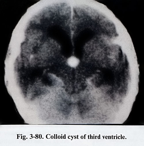
The solution can be found through the complete imaging of the structures by CT and MRI in axial, coronal and sagittal projections. The lesion must be described with respect to size, texture and which structures are bordering, with the added intention to detect its place of origin (Fig. 3-80). The presence of occlusions, disruptions of the walls, isolation of parts of the ventricle (with the dilatation that follows) as well as any perifocal brain oedema must also be detected. An angiographic study must demonstrate the vascularity and blood supply of the lesion, its relationship with the choroid plexus and the deep venous collectors, and for the specific requirements of the approach through the cortex or corpus callosum - the cortical venous drainage to the superior sagittal sinus.
Taking into consideration the characteristics of third ventricle space occupying lesions, with respect to the choice of a corridor for its removal, the lesions can be subdivided into three main categories. The first consists of extra-axial intraventricular lesions, the second – of intraaxial lesions with an intraventricular component, and the third - of basal lesions, with secondary penetration into the third ventricle.
The extraaxial intraventricular lesions are usually benign (colloids cysts, craniopharyngeomas, dermoids, teratomas, papillomas and others). They have well defined borders and are not so adherent to the wall of the ventricle's borders. On imaging studies they are surrounded by CSF, except for the area of attachment. Their margins are usually smooth. Intraaxial lesions with growth inside the third ventricle are most often gliomas, but metastatic tumours – medulloblastomas, germinomas - can have similar characteristics. The MRI and CT investigations detect a significant part with an intraaxial loca-tion. For the needs of the surgical strategy it is essential to discern which structures are affected. Basal tumours penetrate or only elevate the walls of the third ventricle. The majority of them are benign and are potentially excisable.
Those which disrupt the third ventricular wall penetrating from outside can be visualised through the foramen of Monro. The primarily basal origin of the lesion must be detected on the imaging studies. Very often bone changes are seen on the skull X-ray films and CT images. The integrity of the structures forming the inferior wall of the third ventricle must be reviewed thoroughly on the sagittal MRI images, as their affection can indicate penetration of the lesion inside the ventricular cavity.
The indications to the three most accepted paths for entry to the third ventricular cavity can be more precise after the lesion has been classified within one of the three categories mentioned above.
Extraaxial intraventricular tumours require adequate exposure of the ventricle before removing part of the lesion and their nature can very often permit total excision. The widest access to different areas of the ventricular cavity is provided by interforniceal or the transforaminal-transvelum interpositum approaches. Intraaxial lesions with secondary penetration into the ventricular cavity require access not only to the ventricle, but also to the primary site (origin) of the tumour. The involvement of the hypothalamus, thalamus, fornix or other structure will determine which path should be followed, but the surgeon should be able to take a biopsy or partially remove the lesion at least when reaching the ventricular cavity. Any one of the three paths can be chosen. Basal tumours always require a basal approach for the access to the ventricle. Any removal of the basal portion of the tumour preceding the approach to the ventricle provides histopathological proof and better access after debulking. This approach permits better control over the vascular supply of the tumour. If removal through the basal approach and lamina terminalis is not expected to be satisfactory, an additional option can be discussed for the same or another surgical session by one of the superior paths. The craniotomy is preplanned according to these needs.
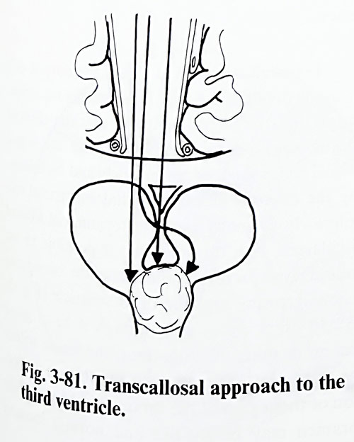
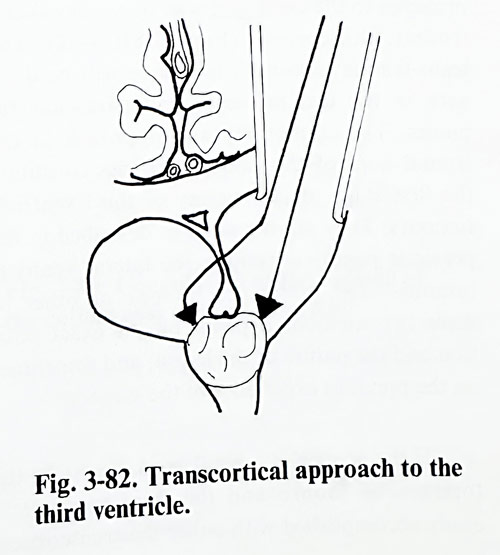
We will present two of the main approaches to the third ventricle: transcallosal and frontal transcortical (Figs. 3-81; 3-82). The trans-lamina terminalis technique will be dealt with in the text on sellar and parasellar tumours. The craniotomy and approach to the frontal horn of the lateral ventricles constitute the first stage of the surgery of third ventricle tumours. They are the same as described in the previous pages concerning the lateral ventricle tumours. The preference for one or other of these two methods depends on the exact position and the nature of the lesion, and sometimes on the previous experience of the surgeon.
If the ventricles are enlarged, access to the foramen of Monro and the third ventricle is easily accomplished with either the transcortical or transcallosal approach. Access to both sides of the third ventricle is obtainable with either exposure, but because of the angle of vision the view of the ipsilateral portion of large lesions can be limited using the transcortical route. The line of vision to the depth of the anterior third ventricle is better with a transcallosal method
Indeed, if the ventricles are small, the transcallosal approach is certainly the better one. The use of the transcortical approach to the ventricle without hydrocephalus requires the disruption of a large amount of cortex and white matter, and the retraction can be difficult. The route also significantly limits mobility if departure from the initial plane of entry into the lateral ventricle is needed. The advantages of the transcallosal approach to the third ventricle are that the anatomy is constant, the distance to the third ventricle is shorter than in the transcortical approach, and there is greater flexibility to explore the anterior-posterior extent of the third ventricle with no disruption of hemisphere tissue, because no cortical incision is necessary. There is excellent unobstructed vision to the depth of the anterior third ventricle, and ventricular size is irrelevant. The advantages of the transcortical exposure are that there is less chance of compromising an essential draining veins going to the sagittal sinus or causing injury to the pericallosal arteries.
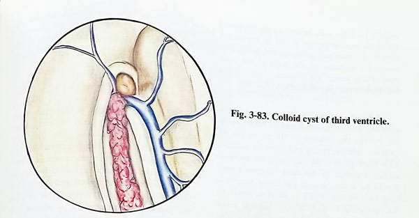
Transcallosal method. Once the corpus callosum is divided, the usual landmarks for orientation into the lateral ventricle are: the choroid plexus, the thalamostriate vein, and the septal vein. The foramen of Monro is found by following the choroid plexus and thalamostriate vein anteriorly. In many cases the foramen of Monro is enlarged and, through it, it is possible to see colloid cysts or other similar space occupying lesions growing into the third ventricle (Fig. 3-83). In other cases, even though the size on imaging investigations was apparently big enough, the lesion within the third ventricle may not be visible on initial inspection of the region of the foramen of Monto. The foramen may be slit-like and normal looking and the mass not seen. In such a case, the closed tip of the bipolar forceps should be introduced into the foramen and gently opened, acting as a temporary retractor on the walls of the foramen. That permits the surgeon to see further into the anterior part of the third ventricle.
When the colloid cyst or tumour is reasonably well seen, an effort should be made, working through the foramen of Monro, to free its anterior, inferior, lateral and posterior surfaces with blunt probes. At this stage the superior surface attached to the third ventricle's roof should be intact because the blood supply is from that surface, which is attached firmly to the ventricular roof. Unless the mass is small and has a tendency to deliver out itself, it is unwise to attempt removal of the lesion intact wothout some volume reduction because the pedicle may be disrupted and bleeding may occur from the roof of the third ventricle, unseen and unsuspected until the third ventricle is filled with blood. The mass is penetrated with a spinal needle and aspiration may then be made.
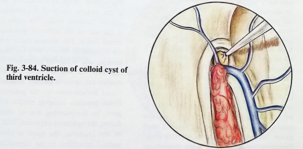
If a cyst content is being aspirated, the capsule shrinks simultaneously with the appearance of fluid in the syringe. If the material is too thick for aspiration through the needle, a larger needle or even a small suction tube may be used (Fig. 3-84). If the cyst content is semisolid, a crosswise incision into the wall allows the use of a small suction tip and small ring curettes to complete the evacuation.
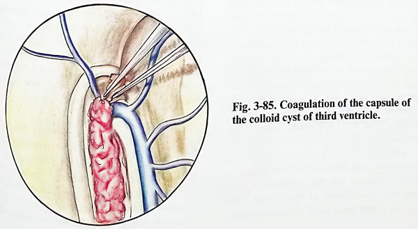
It should never be pushed on the cyst, beacuse of the risk of displacing it posteriorly into the third ventricle and losing sight of it. The complete evacuation of the cyst permits delivery of the capsule by pulling on it gently from various sites around the opening already made through the foramen of Monro. The pedicle will be the last part to be seen as it passes through the foramen of Monro which is then coagulated and divided (Fig. 3-85). If bleeding occurs within the foramen of Monro, coagulation should be used cautiously avoiding obstruction of the thalamostriate and internal cerebral veins. If bleeding continues, one may introduce a soft catheter into the foramen of Monro and then slide down a very small piece of oxidised cellulose between the catheter and the bleeding. point, the catheter serving as a support against the wounded vein. The catheter end is taken outside the wound and removed after 12 to 24 hours.
In many cases the pathological formation may be not visible through the foramen of Monro, or it will be difficult to manage the lesion through the foramen. In such a case the interforniccal approach may be indicated (Figs. 3-86; 3-87). After the clear identification of the anatomical structures in the exposed intraventricular cavity, bipolar coagulation forceps are used to fenestrate the septum pellucidum (septostomy), creating a single ventricular cavity and facilitate a possible shunting procedure in future.
The septum serves as a landmark for guidance to the midline union of the forniceal columns and the forniceal body. The raphe is identified at the site of the septum's attachment on the dorsal fornix. The incision starts at the level of the foramen of Monro with fine tipped bipolar forceps, and is carried posteriorly for 1,5 to 2 cm. The size and shape of the fornices in the midline are variable and are influenced by individual deformation by the pathological mass.
Good preoperative imaging studies help with proper orientation during the operative exposure. With completion of the interforniceal incision, the mass is identified. The normal structures in the diencephalic roof including the tela chorioidea, choroid plexus, internal cerebral veins, and posterior choroidal arteries are often thinner or displaced laterally by the presence of the mass. Retraction at the level of the fornix is often not necessary, or a very narrow retractor with minimum pressure may be used to maintain exposure either initially or after mass decompression is partially completed.
Once internal decompression has been achieved, lateral portions of the mass are separated gently from the third ventricle's wall. With lateral dissection, the internal cerebral veins may be identified and followed anteriorly to the foramen of Monro. The choroid plexus is often intimately adherent to the thalamostriate veins.
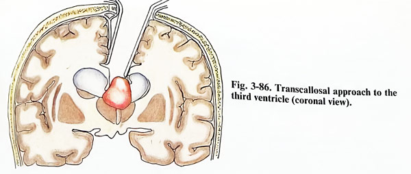
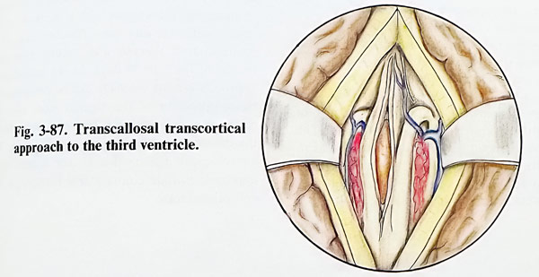
Finally, the posterior component of the mass is approached. For this, sometimes it is necessary to change the position of the microscope. With the mass excision extending from the third ventricle to the dorsum sellae, it is not unusual to visualise the clivus, basilar artery and branches, and prepontine cistern at the completion of lesion excision.
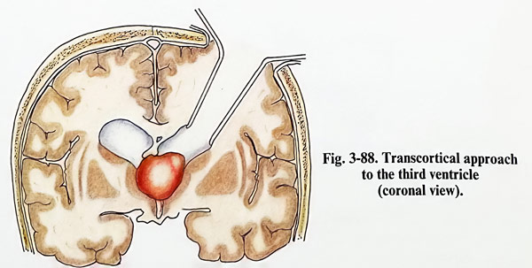
Frontal transcortical approach. The difference from the previous approach is that the foramen of Monro is approached at an angle to the sagittal plane (Fig. 3-88).
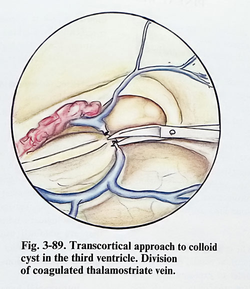
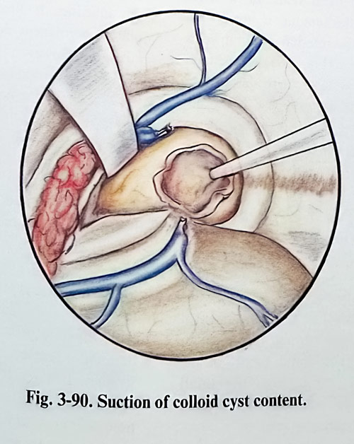
To enlarge the access to the third ventricle cavity, this approach can be expanded with trans-vellum interpositum separation. The choroid plexus near to the foramen of Monro can be elevated slightly and thalmostriate vein coagulated and divided from the internal cerebral vein (Fig. 3-89). The leptomeninges of the velum interpositum are detached from the foramen of Monro posteriorly about 1,5 to 2 cm. The medially inserted retractor is advanced so as to slightly dislocate the fornix with the choroid plexus and internal cerebral vein medially and the lateral retractor slightly depressing the thalamus. The tumour can then be approached between the internal cerebral vein and the thalamus using standard microsurgical instrumentation (Fig. 3-90).
Complications. Most of the complications seen with operation on a lesion expanding into the third ventricle have been related to the location and nature of the primary lesion rather than to the approach. Diabetes insipidus and akinetic mutism can be seen transiently. Aseptic meningitis has occurred in approximately 15% of patients who have undergone intraventricular operations. The signs of aseptic meningitis usually become apparent when corticosteroids are stopped. The CSF shows changes consisting of moderate pleocytosis with a decrease in the glucose content. Patients respond to corticosteroid therapy and require continued treatment for 5 to 30 days. In the transcallosal approach venous drainage can be compromised by compression of the sagittal sinus with the retractor or infarction of the frontal lobe due to the division of a significant draining vein. The neurological deficits due to the transcortical approach include contralateral hemiparesis and epileptic attacks.
PINEAL REGION TUMOURS
A wide variety of tumours can occur in the pineal region and approximately 25 - 30% of them are encapsulated; there is therefore justification to attempt their removal. The neoplasm of the pineal region include: germ cell tumours malignant and benign variants - germinomas, embryonal carcinomas, chorioncarcinomas, teratomas, dermoids and epidermoids; tumours from the pineal cells - pineocytomas and pineoblastomas; tumours from supporting cellular components of the pineal body - astrocytomas, meningiomas, and rarely metastatic lesions. Originating from structures in the neighbourhood, meningiomas from the falcotentorial angle and gliomas from the quadrigeminal plate or splenium can also be found.
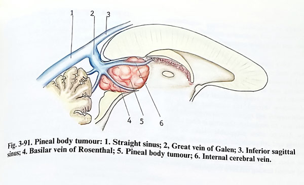
These tumours have central location in the cranial cavity that is equidistant from various cranial points traditionally used as routes of exposure. Such tumours are in intimate contact with the important deep venous system, including the vein of Galen, the precentral cerebellar vein, internal cerebral and basal veins. In most instances the bulk of the tumour, if not all the tumour, lies below the internal cerebral veins and the vein of Galen (Fig. 3-91). Sometimes there may be a firm attachment to these structures, including the tela chorioidea. Therefore, the preservation of the deep veins is one of the main tasks of every pineal tumour removal.
Because of that, it is always preferable to avoid a view in which the veins are obstructing the access to the tumour. The choice of the approach is dictated by the relationship of the tumour to the tributaries of the deep venous system. In the rare cases of the tumour being situated dorsally to the big veins, with or without splenial involvement, an interhemispherical view must also be obtained, and the splenium retracted if needed. The blood supply of the tumours in the pineal region is from the small branches of posterior choroidal arteries and branches of the quadrigeminal arteries.
The main tasks for the management of every tumour, which is a space occupying lesion, are specifically highlighted with this intracranial location, due to the high risk related to the direct approach to this region.
Histopathological identification of the lesion is essential for establishing the degree of malig-nancy, prognosis, and radiosensitivity of the lesion and has to be definen as early as possible. In principle, a stereotactic biopsy is definitely not the procedure of choice, as the surrounding veins are a source of complications and the non-homogeneous structure of some lesions (like teratomas) is misleading concerning the degree of malignancy with such small tissue samples. CSF cytological studies are of some value, but they are obtained usually after shunting. Tumour marker studies can also be helpful to indicate some specific types of lesions.
Intracranial hypertension, caused by obstructive hydrocephalus, must be treated when established in consideration of the future chances of restoring the normal outflow through the aqueduct. If direct surgery is not considered, permanent derivation with a shunt is advisable. However, if direct surgery is possible, the CSF derivation has to precede it, taking care not to collapse the cerebral mantle in extremely enlarged lateral ventricles.
When possible, the intracranial tumour should be removed as totally as this can be achieved without additional harm, especially in the cases when its radiosensitivity is low. Occlusion of any of the main inflowing veins to the straight sinus is not acceptable, unless its role for the drainage is not important and the respective area is well collateralised, for instance with tiny precentral or basal veins. Even then, if possible, such a sacrifice has to be strongly avoided. If the histopathological type of the tumour is not known before the operation, a frozen section study during surgery is mandatory. However, highly malignant tumours must be submitted to surgery very cautiously and selectively, preferring radiotherapy if effective, and thoroughly measuring the benefits, and the expected natural outcome of the tumour type.
After completing the main surgical procedure, final management of the elevated intracranial pressure must be decided on, if it is preexisting and there is also any need of adjuvant treatment. The non-restored natural CSF pathways of outflow through the aqueduct during the main surgery will require permanent shunt derivation to be implanted, if third ventriculostomy has not been done. Shunting is done after a few days to allow acceptable values of CSF protein and cell count for implantation. Radio-or chemotherapy is started as soon as possible, but when the general condition has recovered and wound healing has been completed.
Considering these principles of management, preoperative investigations aim to determine the exact position of the tumour border and its relationship to surrounding structures.
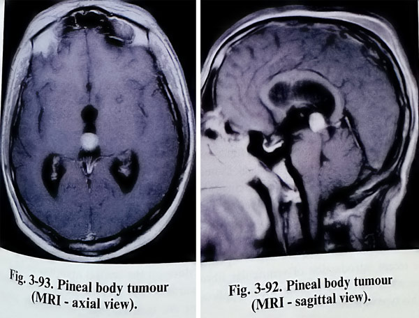
Three dimensional imaging of the area by CT and MRI is the basic investigational approach (Figs. 3-92; 3-93). However angiography is also necessary to establish the degree of vascularity and the pattern of the deep venous outflow. Special attention is given to the position and relationship of the lesion to the deep cerebral veins, the vein of Galen, the Rosenthal (basal) veins and the precentral cerebellar veins.
There are indications for surgical removal of those tumours, which on the basis of investigations, have a particularly high likelihood of being benign, and those patients previously treated with CSF derivation and radiation, without a tissue diagnosis, who have a progressive neurological deficit in the presence of a functioning shunt. Patients in whom cytological examination of CSF shows malignant cells, patients with evidence of either spinal or extraneural metastases, and patients in whom both the anterior and posterior third ventricle tumours are occupied by tumor may not require direct operation. The recent introduction of ventricular fibroendoscopy is currently gaining wider application in this type of lesion.
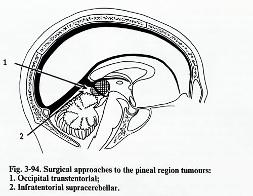
Different surgical approaches to the pineal region have been proposed. The infratentorial supracerebellar approach and the occipital transtentorial approach are the methods of choice (Fig. 3-94)
INFRATENTORIAL SUPRACEREBELLAR APPROACH
The advantages of the infratentorial supra-cerebellar approach are that the surgical corridor is in the midline and reaches the tumour that is centrally seated in the pineal region. The tumours are exposed inferior to the deep venous system. There is no morbidity related to the parietal or occipital lobes as is experienced with supratentorial exposures.
The patient is positioned on the operating table in a sitting or prone position with elevated head and upper thorax - the so called "Concord" position. His head must be strongly flexed so that the best exposure of the tentorial notch can be achieved with the greatest comfort to the surgeon.
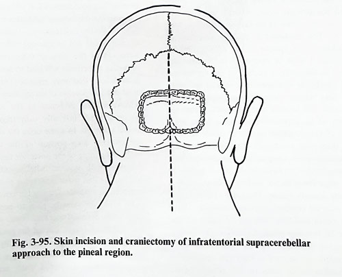
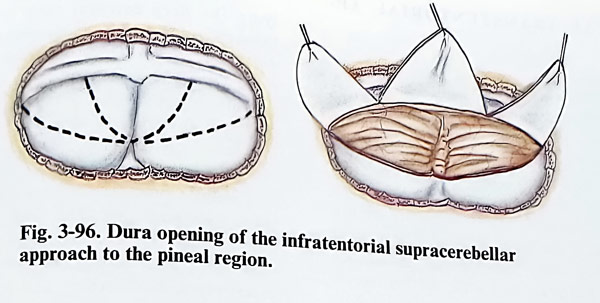
A long midline incision is used, starting from the C4 level and extending into the occipital region up to the lambdoid suture. The muscle attachments and pericranium are elevated on each side, and retracted. A standard craniectomy is performed, which includes exposure of the lateral sinuses and torcular in all instances, without extension to the foramen magnum (Fig. 3-95). Craniotomy of the posterior fossa also can be an option and in such a case an additional nibbling of the superior border is added to expose the lateral sinuses and torcular. If the dura is found tense and nonpulsatile, then the lateral ventricle must be punctured, or dehydrating agent must be applied. The dura can be opened in several different ways, but in any of them the superior surface of the cerebellum should be well exposed. The tentorium should not obstruct the midline view and the dura on the central area has to be retracted upwards to the greatest extent. Secondary incisions are then carried out laterally toward the lateral sinuses. After the completion of the dural incisions, there should be three flaps, a central one and two laterals that reflect upward and expose the superior surface of the cerebellum (Fig. 3-96). Inferiorly the dura mater is left to cover the cerebellar hemispheres during the retraction. The superior border of the cerebellum is gently separated from the lateral sinuses, already under the magnification of the microscope sacrificing the veins entering the sinuses and dissecting the adhesions. Then the superior cerebellar surface is gently retracted, and all bridging veins over the superior surface of the cerebellum (vermis and hemispheres) can be sacrificed until approaching the tentorial incisura.
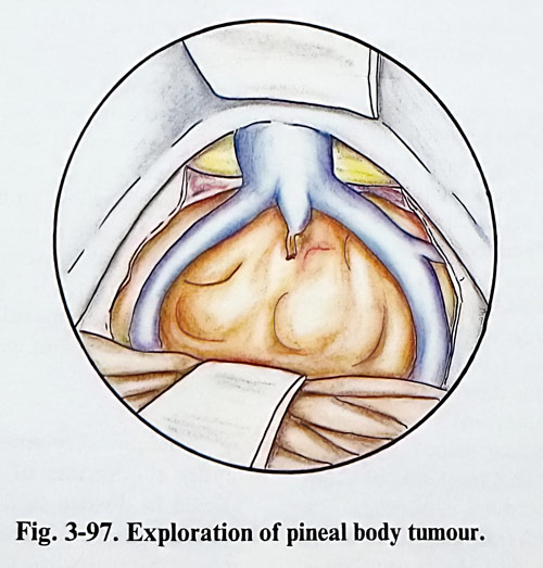
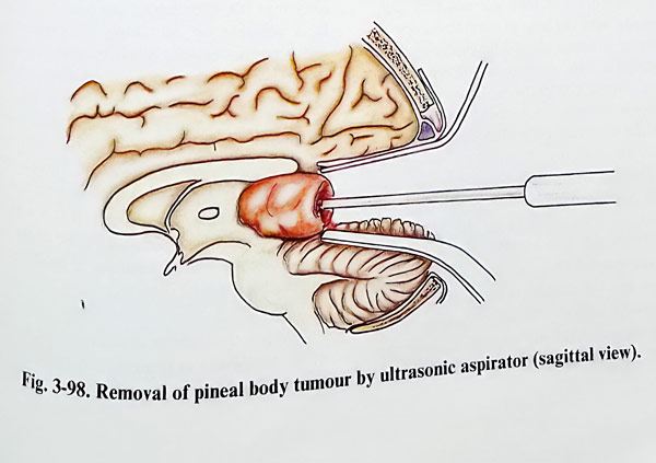
By gravity, with the patient in the sitting position, the cerebellum will drop and provide 1 to 1,5 cm of space between its surface and the tentorium. A self-retaining retractor is placed on the tentorium to the notch lifting torcular and the straight sinus. Another retractor depresses slightly the vermis of the cerebellum (Figs. 3-97; 3-98). The arachnoid of this region is usually thickened and opaque in the presence of the tumour. It must be opened to the midline and close to the anterior surface of the vermis and the cerebellar hemispheres avoiding injury to the deep venous system. The precentral cerebellar vein will be seen coming directly from the region of the superior vermis towards the great vein of Galen, which usually is visible through the thickened arachnoid. This cerebellar vein sometimes needs coagulation and division, permitting further exposure of the pineal region. In this stage the posterior surface of the tumour is usually already visible. The great vein of Galen and internal cerebral veins are generally above the tumour and they may not be visible at this stage of the operation. Laterally, the medial surface of the temporal lobes and the veins of Rosenthal can be seen as they run upwards to the confluence of veins in this region. More retraction of the anterior portion of cerebellum is needed by the inferior self-retaining retractor, exposing the larger posterior surface of the tumour.
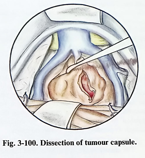
Then the volume of the tumour may be reduced by debulking. In the case of tumours that are benign and encapsulated, it is possible to dissect the capsule of the tumour first laterally, then superiorly, and inferiorly after enucleating its interior. Many tumours are attached anteriorly to the velum interpositum and sometimes laterally to the medial portion of the pulvinar. Attachment inferiorly to the quadrigeminal plate varies according to the nature of the tumour but generally it is modest in encapsulated benign tumours.
Some of the tumours are extremely soft and may be sucked out by a fine suction tube. Other firmer tumours such as some astrocytomas and teratomas need a laser or cavitron suction aspirator for their debulking. Once the larger central portion of the tumour has been removed, it will become apparent whether the tumour infiltrates or is encapsulated. With infiltrative tumours, it is recommended not to attempt radical resection. Removal of the tumour infiltrating the surrounding structures will lead to severe neurological deficits that may remain permanent. The most difficult portion of encapsulated tumour to remove is that attached to the midbrain or which extends laterally into trigone of the lateral ventricle. In some cases the dissection of the anterior deep portion of the tumour can result in opening of the posterior third ventricle.
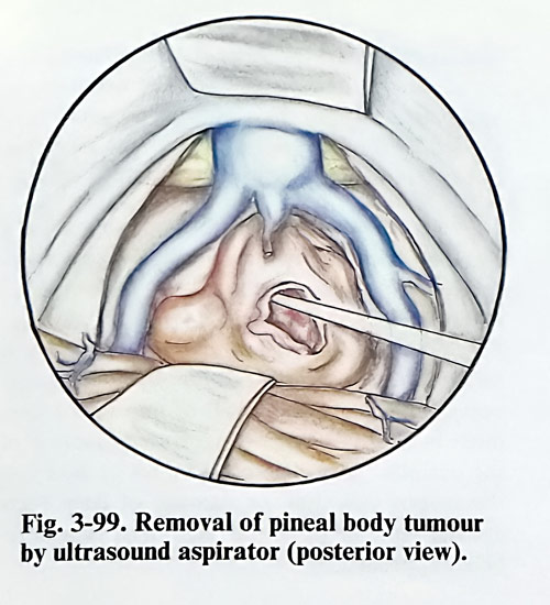
Tumours with minimal attachment will often roll out at this point and large pieces may be removed as a whole. During the removal, particular attention must be paid to protecting the deep veins (internal cerebral veins, vein of Galen, and basal veins). Except for packing of the bleeding points, there is practically no other haemostatic method to be used. An attempt to coagulate these distended veins may lead to larger rupture. Even with non-resectable tumours, benefit may be achieved through the performance of internal decompression only (Figs. 3-99; 3-100).
OCCIPITAL TRANSTENTORIAL APPROACH
The advantages of this approach over the infratentorial supracerebellar approach are the greater ability to mobilise the tumour and to visualise major parts of the pineal region. Usually there are no large veins that cross from the occipital lobe into the superior sagittal sinus.
Therefore, no risk of infarction occurs by this approach unless the surgeon makes the mistake of coagulating some big cerebral vein, which drains the occipital lobe into the transverse sinus. Such a vein can be located more laterally and should be outside the operative exposure.
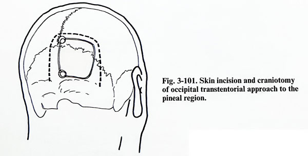
The occipital transtentorial approach is traditionally done with the patient in the sitting position with all precautions against air embolism. More recent and better alternative is the semiprone position with the head rotated about 20 degrees to the side of the operation ("park bench" position). A right occipital craniotomy is made, near to the midline exposing the border of the sagittal sinus and with the lower border of the craniotomy to the transverse sinus (Fig. 3-101). After the bone flap has been turned, the dura is opened parallel to the superior sagittal sinus and the transverse sinus producing a triangular shaped flap that is reflected laterally. Enough dura should be left to the sinuses for an easy dural closure and then the edges are turned out with traction sutures. Some cortical veins entering into the sagittal sinus should be sacrificed, if they are not very big.
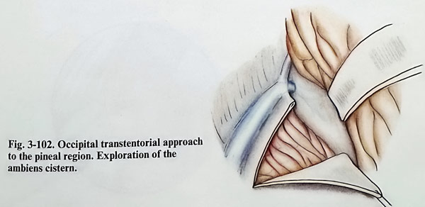
Next, already with the help of the microscope, a self-retaining retractor should be gently applied to the occipital lobe, retracting it laterally and superiorly. Direct compression of the calcarine cortex should be avoided, keeping the retractor more to the occipital pole than deep into the interhemispheric fissure. The tentorium is then opened, making an incision parallel to straight sinus and approximately 1 to 1,5 cm from it, starting at 1 - 2 cm anterior to the transverse sinus and torcular and carried out anteriorly towards the incisura (Fig. 3-102). The tentorium is vascular and the control of bleeding is possible by bipolar coagulation, or occasionally by clips. Traction sutures are placed to hold the tentorium open.
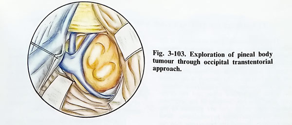
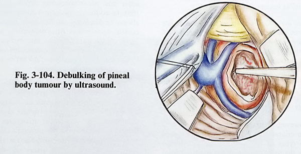
The arachnoid of the ambiens cistern overlying the deep cerebral veins (usually secn through it) must be divided, taking care not to damage the veins. Once the arachnoid is separated from the veins, the relationship of the tumour can be identified and the surgeon can deal with any lesion of the pineal gland, superior vermis, quadrigeminal plate, posterior third ventricle or splenium. The relationship of the deep veins should be identified before initiating the tumour's removal (Fig. 3-103). If there is a pineal tumour or glioma arising from the quadrigeminal plate, these vessels will be displaced superiorly and posteriorly and will be draping over the mass. If the lesion dealt with is a typical pineal tumour, it should be entered by sharp dissection and a biopsy should be obtained. A frozen section will help with the decision on how to proceed. If the tumour proves to be of embryonal origin, such as a teratoma or dermoid, piecemeal or 'en bloc' resection should be done (Fig. 3-104). In such a case an attempt should be made to remove the tumour completely. If the tumour is a germinoma with infiltra-tive growth the removal should be carried out as completely as possible, but without insisting on radicalism.
A pineal tumour may extend downward under the vermis of the cerebellum, which should be divided so that the lower pole of the tumour will be accessible and more easily removed. Similarly, the tumour may extend superiorly into the splenium of the corpus callosum. By tumour debulking and delivering its capsule downward into the operative field, division of the splenium can be avoided. If the tumour arises from the sple-nium of the corpus callosum, the veins usually are displaced inferiorly. In these cases an inter-hemispherical approach is more useful. If the tumour is a glioma, a specimen should be obtained and a modest decompression done.
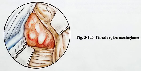
When the tumour grows from the free edge of the tentorium or from the junction of the falx and the tentorium, the deep veins will be displaced anteriorly, creating a problem in identifying them through the tumour (Fig. 3-105). The surgeon must first reduce the volume of the tumour with the ultrasonic aspirator and after that separate the surface of the tumour from the deep veins and surrounding anatomical structures.
Intraaxial tumours, such as gliomas of the quadrigeminal plate or gliomas of the splenium of the corpus callosum displace the deep veins posteriorly, upward or downward depending on the site of origin of the tumour. They can be identified by the lack of a capsule. During the excision the third ventricle can be entered through its posterior part in the pineal recess.
The closure of the operative wound has no special particularities. After complete haemostasis, the wound is irrigated, and the tentorium is approximated with some sutures, to avoid herniation of the occipital lobe into the posterior fossa. The rest of the closure is a routine one.
The complications of this approach are the common ones of any intracranial surgical intervention. Infection, osteomyelitis, and intracranial haemorrhages of various types are possible.
Specific complications when using this approach include the production of homonymous hemianopia. This is a direct result of improper placement of the retractor or vigorous retraction of the occipital lobe. The possibilities of injury to the corpus callosum, or damage of deep veins are the same as in the infratentorial supracerebellar approach.
TUMOURS OF THE SELLAR AND PARASELLAR REGIONS
The most common types of tumours in the sellar region are pituitary adenomas, cranio-pharyngiomas and meningiomas. According to different statistical data, pituitary adenomas constitute from 6 - 8 % and craniopharyngiomas from 2 - 4,5% of all tumours in the cranial cavity. The meningiomas arising from tuberculum sellae, diaphragma sellae, and the cavernous sinus area are discussed in the respective chapter. Less often seen are gliomas of the chiasm, optic nerves or hypothalamus. Skull base tumours, secondarily penetrating into the cranial cavity, as for instance chordomas, carcinomatous metastases and epipharyngeal tumours do, also present with the features of sellar or parasellar lesion.
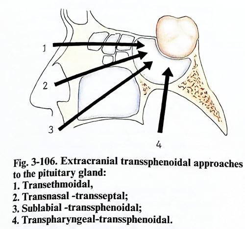
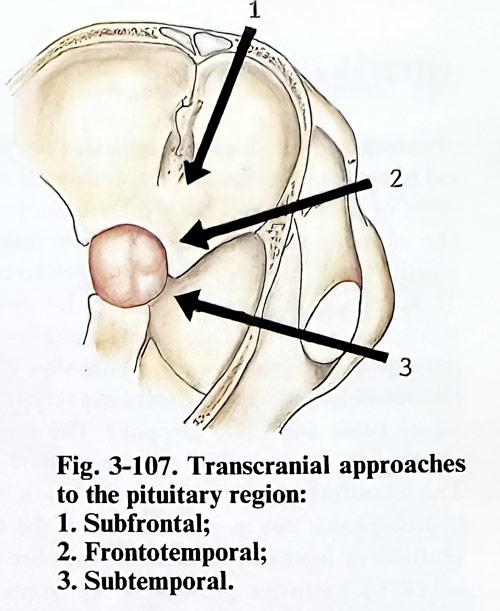
Two main routes of access to the sellar area have been developed through the decades of neurosurgical progress: the extracranial and the transcranial. The extracranial approaches reach the area through the skull base, mainly through the paranasal sinuses, but the transcranial route requires a craniotomy. Extracranial approaches as a rule have a narrow corridor and therefore a limited angle of view, specific for every technique (Figs. 3-106; 3-107).
However, to decide the approach, several common characteristics have to be established in these types of lesion. Although not always possible, the first is to define the structure from which the lesion is thought to derive its origins.
Secondly, because it is important to preserve visual function to its maximum, the relationship of the lesion to chiasm, optic nerves and tracts is a very essential factor in deciding the approach. Lesions below the chiasm and associated with an enlarged sella permit extracranial access.
Lesions in the sellar and parasellar region are closely related to the internal carotid arteries and the circle of Willis. The finding of any compromised important arteries and the main supplying branches is crucial for the safe choice of approach and manipulation during surgery. The unexpected discovery of an intrasellar aneurysmal dilation of an artery during a transsphenoidal approach can lead to a disaster.
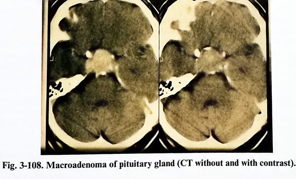
Lesions in the sellar and parasellar area are investigated with all routine imaging modalities as CT and MRI, but the classical skull radiography still gives a general idea of the condition of sellar and parasellar bone structures (Fig. 3- 108). In addition, the lateral X-ray film should always be displayed on the screen during extracranial approaches for comparison with the image on the intraoperative fluoroscopic screen. Angiography is always recommended when vascular pathology (aneurysm, AVM) or very vascularized lesions are suspected.
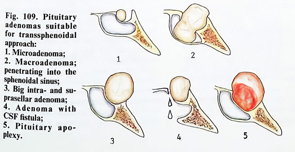
Surgical planning is based on the location of the lesion. Sellar or predominantly sellar lesions, especially those with an enlarged sella, are suitable for a transsphenoidal approach in general as the important structures will be behind the mass along the surgeon's view. Parasellar lesions are not suitable for the same approach, as the carotid artery and the cavernous sinus obstruct the access. In such cases, as well as in any lesion over the chiasm, the only option is transcranial. However, the particular locations of the lesion indicate the different approaches that can be appropriate, as is illustrated in figs. 3-109 and 3-110.
PITUITARY ADENOMAS
Indications and contraindications for surgical treatment. The indications for surgical treatment of pituitary adenomas differ according to the endocrinological activity of the tumour, whether it is non-secreting or hypersecreting.
There are unconditional indications for surgical treatment when the clinical manifestations of the suprasellar growth present affection of the chiasm and optic nerves (disturbances in the visual acuity and fields, and optic atrophy). The same is referred to the lesion of other cranial nerves.
The manifestation of the mass lesion with hydrocephalus due to compression of the third ventricle or increased intracranial pressure provoked by extensive growth in the frontal or temporal direction also requires surgical treatment.
The surgical treatment of an accidentally discovered pituitary adenoma without clinical manifestations should be postponed till its growth is proved by follow up CT scan. Pituitary apoplexy is an indication for urgent operation. Despite the spontaneous improvement of the visual disturbances in some cases, the risk of waiting on spontaneous improvement is larger than that of the urgent operation; this is equally true for elderly patients.
Spontaneous CSF rhinorrhoea in pituitary adenomas is also an indication for surgical treatment. The adenomas presenting with rhinorrhoea usually have aggressive growth and lead to destruction of the dura and the sella.
TRANSSPHENOIDAL APPROACHES TO THE PITUITARY REGION
The pituitary region is reached along the extracranial path by first approaching the sphenoidal sinus. Its cavity can be exposed in different ways by the preceding steps of the operation. The most frequently used techniques to expose the sphenoidal sinus are:
Transsphenoidal sublabial approach. The patient is placed on the operating table in a supine position with the head slightly lifted upward, so that the venous outflow is not obstructed. One of the thighs is prepared and draped also. In case of any need for sellar and sphenoidal packing, an incision can be done on the lateral side of the thigh and the needed fat, muscle pads and fascia easily obtained. The X ray tube with the image intensifier screen is so adjusted that during the whole time of the surgery, the approach, and the intracranial and intrasellar manipulations are efficiently controlled.
After preparing the skin of the face with antiseptic solutions, the gingival mucosa and the upper lip are infiltrated with local anaesthetic at a distance 3 cm from the midline on both sides).
Every nostril is inspected with a speculum and local anaesthetic with adrenalin is injected under the septal mucosa and the floor of the nasal meatus (Fig. 3-111). The upper lip is retracted and an incision on the mucosa of 5 - 6 cm length 1 cm above the teeth is made in the vestibular (mucolabial) fold. The incision is completed to the bone of the alveolar processes and the mucosa with periosteum is stripped upwards until it exposes the lower edge of the anterior nasal apertures. The nasal spine of the maxilla, together with the lower edge of the bony nasal entrances are nibbled (Fig. 3-112). The mucosa is separated subperiosteally from the nasal septum and the floor of the nasal cavity. Deepening the separation, gradually the entire nasal septum is stripped on one side, but on the other only its base. Then the nasal septum is cut at its base and retracted on the side where the mucosa was not detached. The fracture of vomer in the depth that follows has to be close to the sphenoidal sinus. The already detached mucosa is retracted laterally with a specially designed speculum and the view to the sphenoidal sinus is enlarged. The proper direction of the operative approach to the sphenoidal sinus may be checked on the X-ray monitor. The speculum is repositioned deeper to reach the sphenoidal sinus (Fig. 3-113). The lower borders of this retractor, which touch the external edge of the nasal meatus are toothed, and by indenting them any slipping out is prevented. The anterior wall of the sphenoidal sinus is removed. This wall is thin and its perforation is easy with any sharp instrument. The cavity of the sphenoidal sinus and the bulging of the floor of the sella is inspected. The mucous membrane of the sinus is stripped out.
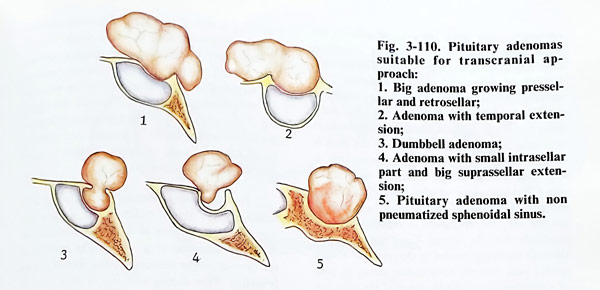
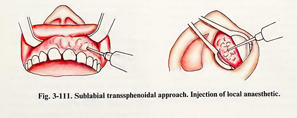
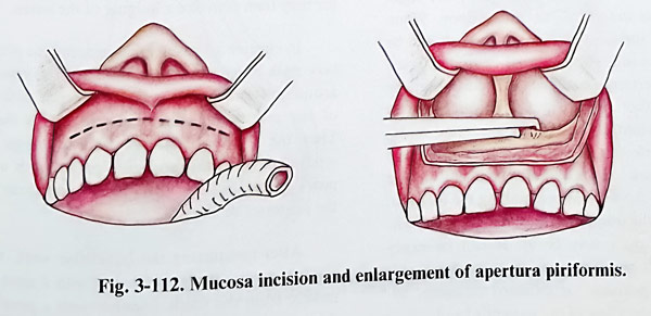
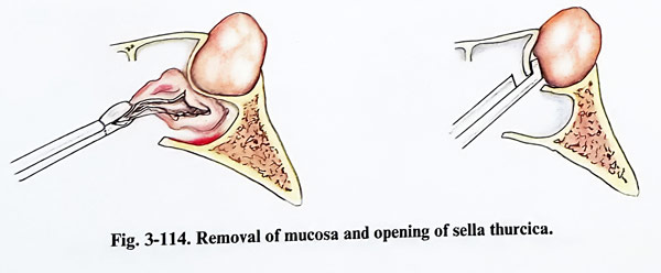
The exposed sellar floor area is identified on the image intensifier as are the other hidden parts of the sphenoidal sinus cavity. The area of sellar floor opening is defined and the speculum is repositioned accordingly. On the lateral X-ray view the line through the upper border of the retractor blades should pass over the tuberculum sellae. In case of a pituitary adenoma the floor of the sella can be thin and there can be a defect, so its opening is very easy. Rarely in microadenomas the sellar floor is hard and a drill has to be used for opening. The opening of the sella is enlarged with a small bone punch to a window of at least 1 cm square, and in an enlarged sella this opening may be even much larger (Fig. 3-114). The sellar dura often has increased vascularity, and bleeding is controlled with bipolar coagulation.
A puncture of the sellar dura has to be done before opening, in order to exclude an aneurysm, empty sella or a cystic tumour, and all this has to be controlled on the X-ray screen for the exact position of the needle.
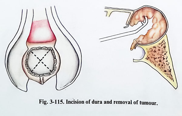
The dura is opened with a crosswise incision, the limit of the incision reaching the edges of the bone defect of the sellar floor. When making the incisions of the dura care should be taken not to injure the internal carotid arteries, which in certain anatomical variants may be situated rather medially. After initial central opening a blunt probe (hook) is inserted under the dura, detecting the presence of a carotid or intracavernous sinus communications in the thickness of the wall. When making the incision, bleeding of the intercavernous anastomoses situated in the dura may occur. It can be easily stopped with bipolar coagulation, packing with cottonoids, gelfoam, or oxidised cellulose, together with elevation of the patient's head.
If it is soft, the removal of tumour tissue is most conveniently done by suction, but when it is hard only by curettage or tumour forceps (Fig. 3-115). It is very important in this removal not to injure the arachnoid and not to provoke a leak of cerebrospinal fluid. Through this opening of the sellar floor it is not possible to inspect the entire tumour cavity. A helpful tool can be a small dentist's type of mirror, which can be inserted inside the tumour cavity.
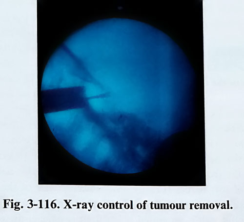
The suprasellar extension of the tumour is removed with curved curettes and suction tip, controlling from time to time the position on the image intensifier (Fig. 3-116). The borders of the tumour on the imaging studies should not be breached. First comes removal of that part of the tumour that is close to the sellar floor, followed by the lateral parts. The suprasellar extension usually drops into the residual cavity and the surgeon can see it pulsating. If this does not occur, the intracranial pressure can be raised moderately by the anaesthetist increasing the positive intrathoracic pressure or positioning the head lower.
In case of a microadenoma, the greyish soft tumour tissue can be seen within the yellowish-brown normal tissue of the pituitary gland. It can be removed easily with dissectors, suction, small pituitary forceps or curettes. If the micro-adenoma is not immediately seen after opening the dura, a horizontal incision of the pituitary gland is made; pressing the gland with a dissector may then provoke a bulging of the lesion.
In elective adenohypophysectomy the pituitary stalk must be exposed initially. The vessels around it, branching out on the anterior surface of the pituitary, are coagulated and divided.
Then the yellowish anterior pituitary tissue is separated from the posterior part, which appears greyish in colour and remains adherent to the posterior sellar wall.
After completing the intrasellar work, the residual cavity, if big, is filled in with a piece of muscle from the thigh and covered with a piece of fascia lata.
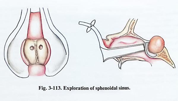
If CSF leak should appear during the operation, then, after packing the tumour cavity with muscle, the opening of the sellar floor is closed with bone fragment from the nasal sep-tum. This fragment should be a little longer in one of its dimensions, so that, when introduced inside the sella in a certain way, it can then be rotated to lock inside the sellar opening (Fig. 3-117).
The operation ends only with suture of the vestibular mucosa. Both nostrils are packed with gauze strips, soaked in antibiotic endonasal application ointment.
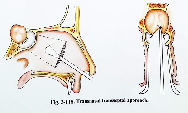
Transnasal transseptal approach. This method greatly simplifies the dissection of the septal mucosa through one nostril. The microscope is in use from the beginning of the operation. A vertical incision is done on the septal mucosa inside the nasal meatus 2 cm behind the nasal entrance. The mucosa is separated from the bone septum until reaching the anterior wall of the sphenoidal sinus. After that the septum is cut and retracted in the same way as in the previous approach, exposing the anterior wall of the sphenoidal sinus bilaterally. Under X-ray control, the speculum is inserted, positioning its blades near to the anterior wall of the sinus and its orifices. Next steps of the approach are the same as in the previous method (Fig. 3-118).
This approach provides a narrower view than the previous, but the distance to the sella is shorter by about 2 cm, and it is suitable for removal of microadenomas.

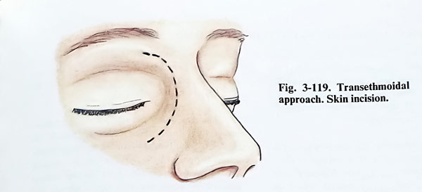
Transethmoidal approach. The patient is placed in a supine position on the operative table with the head slightly lifted above the heart level. The surgeon stands on the patient's right side. The nasal cavity is loosely packed with cottonoid strips, soaked in 10% cocaine hydrochloride and 1% adrenaline, in order to provoke vasoconstriction of the nasal mucosa.
After the application of local anaesthesia a four cm long arch-like incision is made medially to the internal angle of the eyelids (Fig. 3-119).
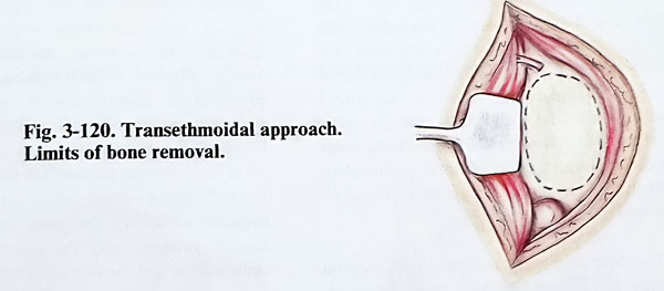
The skin, together with the periosteum is dissected successively from the frontal, and maxillary bones, as a block of soft tissues (Fig. 3-
120). The trochlea is detached and the periosteum from the medial orbital wall is separated, until the anterior ethmoidal artery is reached. A special self-retaining retractor is inserted and the anterior orbital content is displaced laterally to allow an opening between the retractor blades of 2 to 3 cm. The lamina papiracea of the ethmoid is opened, which is easily attained. Only in cases of acromegaly this lamina is thickened and for this opening a fine drill or chisseling can be needed. The bone defect is widened upward, opening the orbital extension of the frontal sinus and the posterior part of the lacrymal fossa. The thin bone plate of the posterior part of the medial orbital wall is preserved and it serves as anatomical landmark for the next stage of the operation. After that the surgeon resorts to the help of the surgical microscope.
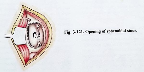
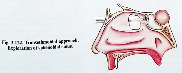
Next is penetrated into the uppermost recess of the right nasal cavity, in which a straight surgical instrument has previously been placed through the right nostril. After the removal of the posterior ethmoidal cells from the right side, the anterior wall of the sphenoidal sinus is reached on the same side (Fig. 3-121). The anterior wall of the sinus is opened and a greater part of it is removed. The midline septum of the sinus cavity is identified, which is not always situated strictly on the midline (Fig, 3-122). It is also removed together with part of the nasal septum and the neighbouring sections of the anterior wall of the sphenoidal sinus from the other side. After this operative stage the sellar floor is sufficiently well exposed.
The opening of the sellar floor and all further steps are carried out as described in the other trassphenoidal methods.
The packs are removed on the third postoperative day. Rarely a pack will slip into the oropharynx. In such a case the pack should be removed and the nostril should be repacked.
A postoperative CSF leak is treated conservatively by lumbar drain for 5 days or repeated lumbar punctures. A leak that visibly drips or does not resolve within 10 days requires surgical revision and closure.
TRANSCRANIAL APPROACHES TO PITUITARY ADENOMAS
The patient is placed in supine position on the operative table, with slight turning of the head to the side opposite to the craniotomy. To decrease the intracranial pressure and facilitate the approach to the tumor almost regularly osmotic diuretics are applied.
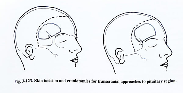
Pituitary adenomas are removed as a rule through a subfrontal or frontotemporal (pterional) craniotomy on the non dominant side, and the skin incision is bitemporal behind the hairline (Fig. 3-123). In very big adenomas with significant subfrontal growth a bifrontal craniotomy can be the most suitable. If the extension is subtemporal, then a temporal craniotomy may be used. As a rule the sellar area is reached following the edge of the lesser sphenoidal wing. In a subfrontal paramedian approach, the retraction is directed to the optic nerves and chiasm.
The dura is opened with an curved incision made at 1 - 2 cm from the basal edge of the craniotomy. The peripheral part is cut in several places and is lifted by traction sutures.
The sellar area is reached retracting the frontal lobe and the cortical surface should be protected with wet cottonoids. All large Sylvian venous collectors are preferably preserved. Through the gradual retraction of the frontal lobe and following the edge of the lesser wing of the sphenoid under magnification, the chiasmal cistern is reached and opened on the side of the craniotomy. The suction of CSF allows the operative approach with less retraction. The optic nerve is followed for exposure of the chiasm and the contralateral optic nerve. Usually both optic nerves are distended and the chiasm is lifted by the tumour.
Before beginning the removal, the tumour is punctured and any possible fluid contents aspirated through a thick needle, in order to exclude an aneurysm or a very vascular tumour. On the other hand, the puncture may reveal a cyst, which appears in 20 percent of all chromophobe adenomas. Cyst evacuation makes the tumour removal easier.
The removal of the tumour is done following certain certain steps in order. Initially the capsule of the tumour (containing diaphragma sellae) is opened by a crosswise or circular incision.
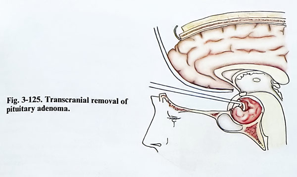
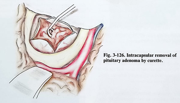
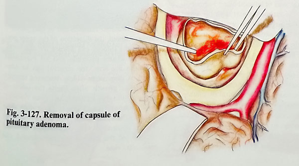
This incision is usually done on the tumour surface between the two optic nerves and chiasm. In some cases with asymmetrical growth of the adenoma, the incision can have another place as is shown in fig. 3-124. This is preceded by coagulation of any large blood vessels at the site of incision. The tumour tissue is removed subcapsularly until a large cavity is produced with the suction, the ultrasonic aspirator and curettes (Figs. 3-125; 3-126). The intra-capsular debulking continues, until the tumour capsule collapses and the compression on the optic nerves and the chiasm is relieved. Then the tumour capsule is dissected from the chiasm and the ipsilateral optic nerve. Laterally from the optic nerve, the internal carotid artery is also separated from the tumour. The opticocarotid triangle (with borders the internal carotid artery, anterior cerebral artery and the optic nerve) is used as a space for dissecting the capsule, at the same time taking care of the pituitary stalk, the IIIrd cranial nerve and the arterial branches located there. Only the blood vessels, which obviously enter the tumour tissue can be coagulated. The removal is completed with additional curettage of the intrasellar part of the tumour. We may consider the removal of an adenoma complete, when the entire suprassellar part is removed including the capsule and the bottom of the sella cavity is curetted, until the surgeon feels the hardness of the sellar floor (Fig. 3-127).
When the adenoma is composed of hard tissue and is strongly adherent to the third ventricle, the complete removal of the tumour often proves impossible. The same applies for large polylobulated tumours. The floor of the sella in some cases is so thin that at the time of removing the deep tumour portions, the floor can be perforated. If so, the bottom of the sella is packed with a piece of muscle.
The removal of an adenoma through a bifrontal craniotomy is very similar to the removal of a meningioma of tuberculum and diaphragma sellae. After the retraction of both frontal lobes, the chiasmatic region is widely exposed. Both internal carotid arteries, and their bifurcations are also exposed in such a way, that larger branches become accessible for the surgeon and they can be separated from the tumour surface with minimal injury.
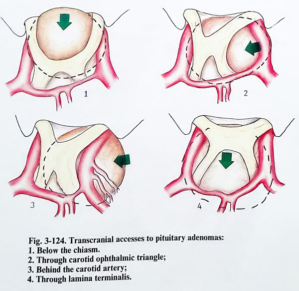
Pituitary adenomas with big temporal fossa extension require temporal or pterional cranioromy. After removal of the temporal extension of the tumour, the remaining part is reached through the opticocarotid triangle and behind the internal carotid artery. Last to be removed is the intrasellar part of the tumour.
After surgery the patient is kept in an intensive care unit overnight with standard monitoring and visual fields frequently evaluated. He is encouraged to be out of bed the next day. Sutures are removed on the seventh postoperative day. The rest of the care is the same as after a transsphenoidal operation.
CRANIOPHARYNGIOMAS
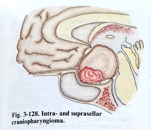
These dysontogenetic tumours, seen mainly in children and in the early decades of life, originate from residual cell nests of the hypophyseal duct in close relation to the diencephalic floor. According to the exact position and later development of these ectopic cellular groups, the craniopharyngiomas can be subpial (hypothalamic and third ventricle), extrapial - subarachnoid with different locations regarding the infundibulum, or extra-arachnoidal. Partially or totally extra-arachnoid craniopharyngiomas can occupy the sella turcica and enlarge it. These tumours often have cystic parts full of yellow to brown fluid and a solid part with calcifications in the tissue (Fig. 3-128). As their formation indicated, they can be found inside the sella, along the infundibulum and inside the third ventricle, displacing all suprasellar structures, and when large, they extend into the anterior and middle cranial fossae.
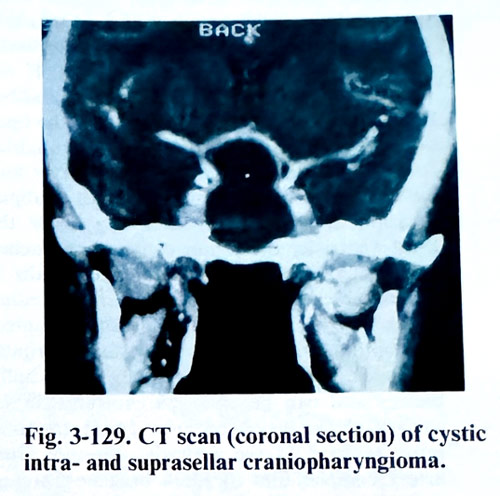
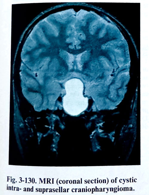
The diagnostic evaluation consists of clear visualisation of all tumour portions by CT and MRI in all three projections (Figs. 3-129: 3-130). The internal carotid arteries and the circle of Willis may need angiographic demonstration, when major vessels are involved.
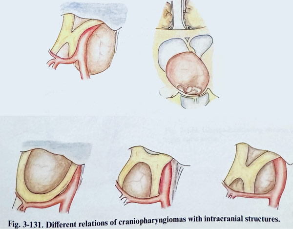
The operative technique depends on the size of the tumour, its exact location, and its relations to the arachnoid of the suprasellar cistern and pia in the area of infundibulum. Further on, the size of the intrasellar part with the subsequent sellar enlargement, as well as the exact locations of the vascular and neural structures regarding the tumour determine additional details of the final decision for the approach and the technique. Craniopharyngiomas with a predominantly suprasellar and intraarachnoid location, associated with a non-enlarged sella or significant extrasellar (subtemporal or subfrontal) extensions, are approached transcranially. Intrasellar lesions, especially those enlarging the sella, with moderate suprasellar extension accessible from the sellar compartment are suitable for a transsphenoidal approach (Fig. 3-131). Cyst evacuations, if one of the main goals of surgery, are done more easily through the trans. sphenoidal route. Those tumours that have a subpial or third-ventricular extension, require exposure of the lamina terminalis and CSF derivation immediately before or during the surgery, by inserting a ventricular drainage. 1f the CSF circulation at the level of the third ventricle is not normalised, the removal of the ventricular drainage is followed by a permanent ventricular shunting.
The main initial steps of the approaches to these lesions - transsphenoidal or transcranial . have been described already with the pituitary adenomas, and only the particular details referring to craniopharyngiomas are mentioned below.
Regardless of the approach, once the tumour is exposed, the first step is always its puncture, aspiration of the tumoural fluid and excision of part of tumoural wall for biopsy. In a transsphenoidal approach, the puncture is done before the dural opening. The dura is opened in the common way and as widely as possible. The tumour is detached from the internal dural surface. As the lesion is collapsed after the evacuation, the wall can be dissected from the wall of the sella, taking care not to damage any intrasellar invagination of arachnoid. Pituitary and infundibulum can be seen posteriorly as an atrophic strip due to the long-lasting compression; they must be preserved as much as possible. As intrasellar tumours can be only partially extraarachnoid, the dissection of the upper portions of the tumour can disrupt the arachnoid.
If no structures protrude and no suprasellar adhesions are seen, the removal can be completed. A diaphragm widely destroyed through suprasellar growth often limits the safety of manipulation due to depth and poor visibility.
Solid tumoural parts create more difficulties than the cystic ones. Any vascular damage will be controlled with considerable difficulty because of the limited transsphenoidal and transsellar view. Transcranial approaches expose the chiasmatic and adjacent cisterns, and the arachnoid is dissected over the tumoural wall. The evacuated cystic part collapses the lesion or part of it.
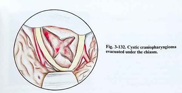
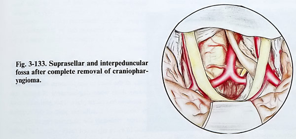
Dissection is performed inside the cystern. The tumour is removed in pieces, dissecting it from the optic pathway structures, infundibulum (identified with difficulty in big tumours) and the big arteries, carefully sparing the small arterial branches (Figs. 3-132; 3-133). If the tumour is located posterior to the infundibulum, it has to be approached through the opticocarotid triangle and the interpeduncular fossa. The third and fourth cranial nerves also have to be preserved. In tumours with a subpial location or secondary penetrating inside the third ventricle, the lesion can be approached with a small incision of the lamina terminalis. It is a suitable access when this structure is either distended by the tumour or is already disrupted.
The optimal view is achieved by the subfrontal approach. The incision is initially a few milli-metres and can be enlarged carefully to the surface of the distended area. Great attention must be paid to the anterior communicating artery complex and its small branches, preserving them as much as possible. Tumour removal follows in a manner requiring minimal manipulation evacuating the cystic part and excising it in small pieces. After evacuation of the cyst, the posterior cerebral and basilar arteries can be identified.
Care should be taken not to injure the III cranial nerve. Preservation of the pituitary stalk is a difficult task for the surgeon. In many cases is impossible to identify the stalk during the dissection of the tumour.
Visual improvement after surgery is obtained in 50% according to most of the statistics. Mortality depends on the size of the lesion and the extent of diencephalic involvement.
GLIOMAS OF THE CHIASM AND OPTIC NERVES
Gliomas of the anterior visual pathways are of two types regarding the primary location of their growth. The lesion arising from the intraorbital part of the optic nerve secondaryly grows intracranially involving the chasm and other suprasellar structures. The management of such type of tumour will be discussed in the respective chapter for orbital and cranio-orbital surgery. The other group of gliomas initiating their growth from the chiasm usually have an infiltrative growth, penetrate into the third ventricle and the orbits, widening the optic canal on one or both sides. The complete removal of this tumour is an exception. The tumour is approached through a frontal craniotomy penetrating toward the pituitary region and after exposing the tumour, a piece is taken for biopsy.
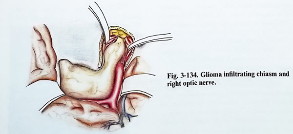
To prevent deterioration of the vision the piece of biopsy should be very small and taken with an incision along the optic pathways. If the tumour penetrates through a widened optic canal into the orbit, the optic canal roof is removed, in order to have a decompression and preserve the vision for a certain period of time (Fig. 3-134). When both optic canals are involved, this decompression is bilateral.
TUMOURS OF CEREBELLUM AND FOURTH VENTRICLE
The tumours of the cerebellum and fourth ventricle have clear age related incidences. Infratentorial tumours in children are three times more frequent than those in the supratentorial space. In adult patients this relation is vice versa. The most common tumours in children are meduloblastomas, astrocytomas, and ependymomas. These types of tumours are rare in the adult patient and at the expense of predominant astrocytomas, metastases and haemangiomas.
There are several diagnostic features to be established before deciding the surgical technique. They are the location of the lesion regarding the midline (vermis) and the IVth ventricle, the type of displacement and involvement of the brain stem, the presence of hydrocephalus, the degree of vascularity, the presence of a cystic portion and finally the histopathological prediction for the lesion.
The non-enhanced and enhanced CT is usually the first investigation to do, and it confirms the existence of a posterior fossa mass lesion, and the displacement and obliteration of the fourth ventricle and cisterns. The density and enhancement of the lesion indicate its possible nature, especially the presence of cysts and a highly vascular nodule (astrocytomas and haemangioblastomas). MRI, however, is superior in its diagnostic value. It is able to demonstrate with better resolution the characteristics of the lesion, the patency of the CSF spaces and grey to white matter margins (Figs. 3-135; 3-136; 3-137). Angiography is reserved for the additional need to demonstrate a vascular pathology or highly vascular tumour.
Surgical planning entails considering the possibilities of current surgical techniques. Sub-cortical cerebellar lesions can be approached either by midline vertical incision of vermis or by a limited incision on the middle or lateral part of the hemispheric surface. Limited incisions are also possible on the lateral and (rarely) on the superior hemispheric surfaces. The lateral part of the hemisphere may be excised. Vermian incisions are possible after midline craniectomies or craniotomies and hemispheric ones - after lateral craniectomies or craniotomies. The choice of bone exposure area depends on the desired surface required for incision and manipulation of the tumour. Hydrocephalus, if clinically symptomatic, must be treated as a preliminary step for improvement of the general condition of the patient. Otherwise it can be controlled by an occipital horn puncture at or just before the time of tumour surgery, placing temporary external ventricular drainage. The drainage can be internalised at a later stage, as a permanent shunt should it be needed.
Another important goal of the approach is the decompression of neural structures, especially at the level of the foramen magnum. It is always done when displacement of tonsils and brain stem has taken place. This goal is achieved by removal of a sufficient part of the occipital squama, including the posterior rim of the foramen. In more advanced cases removal of lamina C1, and even C2 parts can be required.
The burr hole for ventricular drainage is placed 4 cm lateral and 6 cm above the inion on the right side. The lateral ventricle is canulated but rapid ventricular drainage should be avoided particularly if the intracranial pressure is significantly elevated, since it can cause the tearing of bridging cortical veins and create a subdural haematoma.
After dura is opened in a Y - shaped configuration, the cerebellar surface is inspected. When the tumour is located superficially and infiltrates the external cerebellar surface, it is seen immediately after opening of the dura. If it is located subcortically in the cerebellar tissue, exploration is made by first inspecting the cerebellar surface. Special attention is paid to the colour and the configuration of cerebellar sulci and the displacement of blood vessels. The cerebellar hemisphere, in which the tumour is located, seems enlarged in volume and causes a larger protrusion toward the craniotomy, than the opposite. Its sulci are broader and the cerebellar surface is smoother. The cerebellar tonsils are situated usually lower in the cisterna magna or are herniated down under the foramen magnum. The displacement of the vermis to one side also suggests the presence of a tumour in the hemisphere, and when it is bulging, a tumour along the cerebellar midline can be expected (Fig. 3-138). In few cases the cerebellar surface may be carefully palpated to get a tactile feeling about its consistency. The vicinity of the tumour has a consistency that may be more dense or softer than the normal and sometimes in cystic tumours can be felt fluctuating. After this, the cerebellar hemispheres are retracted and the cerebellopontine angles are explored. The exploration may be finalised with exposure of the fourth ventricle.
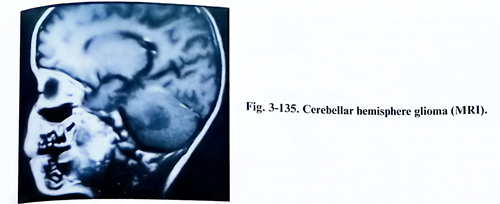
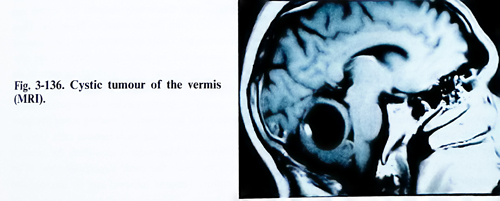
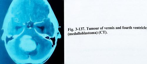
For this purpose the tonsils are separated from one another, until reaching the foramen of Magendie. A fine dissector can be inserted through the opening and a large part of the ventricular cavity is inspected.
Cerebellar astrocytomas may be solid, cystic, and their location in one of the cerebellar hemispheres makes total excision possible (Fig 3-139). The cystic tumour should be punctured first and the mass should be exposed through the cerebellar cortical incision. If the tumour is solid, dissection is carried out around the tumour or it can be debulked with the ultrasonic aspirator. In case of a cystic tumour with a mural nodule, a total excision of the nodule should be carried out, without removal of the wall.
When only one hemisphere is involved by the tumour, a unilateral approach is usually enough for manipulation. The approach was already described, and the size of craniotomy or craniectomy and extent to foramen magnum rim removal decided according to the space required for manipulation and the degree of downward shift of the hemisphere. The dura is usually opened in a stellate fashion, after assuring normalisation of the intracranial pressure.
Direct transdural puncture of a large hemispheric cyst is also acceptable for rapid reduction of the intracranial pressure. After inspection of the hemispheric surface, the craniectomy can be additionally extended after the location of the lesion.
Medulloblastomas are reddish-grey, friable masses that frequently distend the vermis and protrude from the foramen of Magendie. These tumours are easily aspirated, and their removal is carried out by penetrating between the cerebellar tonsils and after vertical incision of the vermis. The subarachnoid space at the foramen magnum is gently packed with cottonoids to reduce the possibility of seeding along the spinal subarachnoid space with tumour cells. Once the foramen of Magendie is identified, the vermis is split to reach the most superficial layer of the tumour. Tumour removal can be accomplished by gentle ordinary or ultrasonic suction and bipolar coagulation or laser evaporation.
Through all debulking, the surgeon should be aware of the position of the dorsal brain stem surface. Initially the obex is found following the dorsal columns, and later - the distorted inferior cerebellar peduncles and the IVth ventricular floor. Having these structures in view and then covered for protection, but not packed with pressure, will provide safe debulking and removal. Tumour removal is continued so that the lateral recesses of the fourth ventricle and aqueduct of Sylvius are visualised. It may not be possible to remove those portions of the tumour which are adherent to the floor of the ventricle. In many cases, the infiltrative growth of these tumours does not permit complete removal of some small portions.
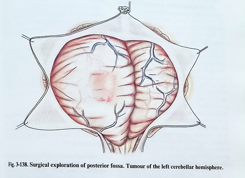
Ependymomas usually grow into the fourth ventricle and bulge out of the foramen Magendie, and extend along the medulla oblongata and upper cervical cord to at least the level of C1 (Fig. 3-140). After opening the cisterna magna, the tumour can be grasped and lifted up to the obex, exposing the floor of the fourth ventricle. At this point is possible to see whether the tumour is adherent to the floor of the fourth ventricle or can be freed from it (Figs. 3-141; 3-142). A cottonoid is slipped between the tumour and the floor of the fourth ventricle. The cerebellar vermis is then split, and with the aid of the ultrasonic aspirator, debulking of the tumour begins. In some cases fourth ventricle ependymomas can go out laterally through one foramen Luschka into the cerebellopontine angle and care is needed to dissect well the involved cranial nerves. The complete removal of these tumours is not usually possible because frequently they arise in the region of the hypoglossal and vagal triangle and are intimately adherent to the underlying tissue of the brain stem.
A variety of cystic masses may be encountered in the midline or near to the midline. They are symptomatic as a result of direct brain stem and cerebellar compression, and obstructive hydrocephalus. Such a cystic lesion should be fully exposed, and the cyst wall carefully examined.
The removal of choroid plexus papillomas from the fourth ventricle is relatively easy, as they are not invasive and can be excised with the bipolar coagulations and the ordinary suction or ultrasonic aspirator. The removal of the tumour attachment in proximity to the foramen of Magendie and from the fourth ventricular floor must be performed very carefully.
Cerebellar haemangiomas/ haemagioblastomas are vascular tumours often located in the cerebellar hemispheres. In 60% of the cases the mass exists as a cystic tumour with a mural nodule. Complete removal of the nodule is curative. The arterial feeders should be easily accessible and be interrupted before an attempt is made to remove tumour tissue. In a case of a very large, solid haemagioblastoma that cannot be removed as a single mass, the surgeon should attempt to interrupt the tumour's arterial supply before trying to remove the lesion.
Dermoid cysts located in the posterior fossa typically arise near the midline. These lesions are present from birth, although they may be not symptomatic until adult life, they grow slowly into a cyst as sebaceous material and desquamated epithelium accumulate inside.
Dermoids and epidermoids should be dissected free of surrounding cerebellum. Care must be taken not to spread the contents of the tumour liquid, because this may provoke irritation and give rise to a chemical meningitis and severe adhesive arachnoiditis.
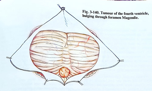
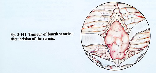
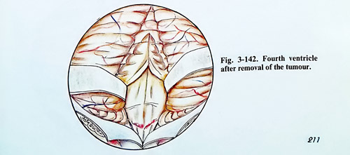
TUMOURS OF BRAIN STEM
Brain stem tumours constitute about 5 to 10% of all intracranial tumours and are most common in childhood and adolescence. Gliomas are the most frequent type of tumour and the ratio of low to high grade gliomas is nearly 50:50. They usually infiltrate the brain stem and cause its enlargement. Some of the gliomas are circumscribed nodülar forms and in such a case an attempt at total removal of the nodule is an option for treatment (Fig. 3-143).
Various surgical approaches are used according to the exact size and location of the lesion. Caudal brain stem tumours are exposed through a midline or retromastoid suboccipital approach to the posterior fossa. The supratentorial occipital approach to the pineal region can be used also for lesions involving the ponto-mesencephalic brain stem.
The microsurgical technique of tumour removal with the use of the ultrasonic aspirator and laser is the widely accepted standard (Figs. 3-144; 3-145). Nodular tumours that have defined borders within the brain tissue should be removed till normal brain tissue is visible. In nodular tumour types surrounded by a wide zone of infiltration, a significant part of the tumour tissue is removed, but the infiltrated zone is left intact. In these cases the postoperative CT and MRI will reveal afterward small parts of residual tumour. In diffuse gliomas, when any border between the tumour and the brain tissue is absent under the microscope, the greater part of the tumour will remain on the follow-up CT and MRI images. The majority of brain stem gliomas are relatively avascular and bipolar coagulation is applied rarely and with extreme caution.
Immediately after surgery a relatively big number of the patients deteriorate in comparison to their preoperative focal deficit. However, follow-up for 2 to 3 months reveals an improvement and it may bring the patients to a better functional level than before surgery.

TUMOURS OF THE CEREBELLOPONTINE ANGLE
Acoustic neurinomas are the most common cerebellopontine angle tumours. There are other kinds of tumours in the same region such as meningiomas, papillomas, epidermoids, etc. The close relation of the tumours with cranial nerves, brain stem and structures in the petrous bone, determines the specific technical approach to the surgical removal of these lesions.
Acoustic neurinomas. These tumours arise usually from the intrameatal portion of the vestibular nerves. They have a round shape, with a smooth or polylobulated surface and variable consistency. When the tumour develops inside the internal auditory canal it leads to canal enlargement early on and later tumor growth continues into the cerebellopontine cistern (Fig. 3-146). According to their size, these tumours are divided into three groups: small size - less than 2 cm in diameter; medium size - from 2 to 4 cm and big - more than 4 cm in diameter. Small tumours are located inside the internal auditory meatus without enlarging it and not adhering to the dura. The facial and acoustic nerves are situated anteriorly to the tumour and are compressed against the anterior wall of the meatus, so they can be dissected easily from the tumour surface. When the tumour reaches 2 cm in diameter, it bulges into the cerebellopontine cistern. The arachnoid remains attached to the petrous bone, and due to the tumour expansion it forld over it as a double layer, containing cisternal elements (vessels and nerves) over the exposed tumour (Fig. 3-14T). The labyrinthine artery is thinner than 1 mm and lies caudally to the tumour and the nerves. Big tumours, already penetrating into the cerebellopontine cistern displace mainly the pons and dorsally they indent on the middle cerebellar peduncle. The medulla oblongata is less displaced. The cerebellar hemisphere in the vicinity of the tumour is also indented by the tumour. With larger displacement, the cerebellar tonsils descend through the foramen magnum and the lower parts of the aqueduct and fourth ventricle can be shifted to the opposite side.
The tumour capsule has different thickness and in many places is adherent to the arachnoid or around the capsule there are enlarged CSF spaces of differing shapes. The base of the tumour is firmly attached to the internal acoustic meatus adhering to the dura. When the tumour is very big, the superior pole may teach the free edge of tentorium. However, it seldom penetrates truly into the supratentorial space, The inferior part of the tumor can reach the foramen magnum.
The relations of neurinomas to the pons are variable. When the neurinoma is growing mainly medially and ventrally, the pons is displaced posteriorly, differing from laterally growing tumours, which displace the pons medially. The relatively dense consistency of these tumours, their slow growth and adherence to the temporal bone cause the compression of cranial nerves inside the cerebellopontine cistern. The vestibular nerve disappears into the tumour capsule still in the region of the internal acoustic meatus. Cochlear and facial nerves are tiny, often seen as transparent ribbons, but in many cases they can be separated from the tumour capsule. In some cases, however, this is impossible to be done, even with the help of higher magnification power of the surgical microscope. The nerves are situated at the anterior surface of the tumour capsule. The trigeminal nerve is displaced ventrally and rostrally from the rostral pole of the tumour, and the trochlear nerve usually is not displaced or compressed by the tumour. The abducent nerve is located ventrally and medially to the tumour mass and usually does not adhere to the tumour. The group of lower cranial nerves (IX. X and XI) are in contact with the tumour at their entrance in the jugular foramen, These nerves are rarely displaced caudally by the lower pole of the tumour.
In a few cases, when the tumour arises from the terminal branches of the vestibular nerve, it may have an intralabyrinthine and intrapyramidal location with destruction of the middle and internal ear.
The blood supply of acoustic neurinomas is carried out from branches of the labyrinthine artery, cortieal branches of the cerebellar arteries and by big branches of anterior and posterior inferior cerebellar arteries. The establishment of the relationships of cerebellar arteries to the acoustic neurinomas is important for tumour removal. The posterior inferior cerebellar artery is situated near the caudal pole of the tumour. It is covered by the tumour, and a big branch of this artery can enter the tumour at this point. The anterior inferior cerebellar artery crosses anteriorly the VIl and VIII cranial nerve, arising from the basilar artery, and is situated deeply on the anteromedial surface of the tumour, between the tumour and the pons.
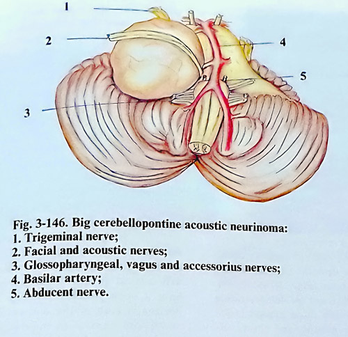
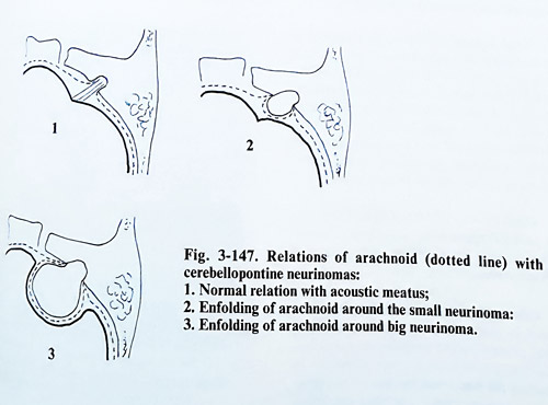
Preoperative diagnostic tests should include high resolution computed tomography (CT) with thin slices through the internal auditory canal. CT is helpful also in demonstrating bone erosion of the petrous bone (Figs. 3-148; 3-149). MRI is a more sensitive test for demonstrating small tumours, especially those located inside the internal acoustic meatus, and shows the relations of larger tumours to the vessels, cranial nerves and the brain stem (Fig. 3-150). Functional studies of the cranial nerves, especially acoustic, vestibular, facial, trigeminal and lower group are also essential preoperatively.
Total surgical removal is the treatment of choice for acoustic neurinomas. In patients with advanced growth of the lesion, causing hydrocephalus and increased intracranial pressure, hydrocephalus treatment has to precede removal by 3 - 4 weeks. In patients over sixty years, the indications for surgery depend on the general conditions. In such a patient partial excision of the tumour can be carried out.
SURGICAL TECHNIQUE.
There are two principal approaches for removal of acoustic neurinomas: retromastoid transmeatal and translabyrinthine.
Retromastoid transmeatal approach. The patient is placed on the operating table in sitting, prone-Concord position, or in a lateral/semisitting position (park-bench position). In the latter, the body and the head must be so inclined to the horizontal plane that the venous outflow from the cranial cavity be facilitated, but at the same time without causing a negative venous pressure, in order to avoid any danger of air embolism. This inclination is approximately between 30 and 45 degrees. The convenience of this position is that when the side of the tumour remains on top, the cerebellar hemisphere has a natural tendency to fall and to separate from the lateral wall of the posterior fossa and this facilitates penetration toward the cerebellopontine angle. The application of diuretics and corticosteriods begins before the operation.
The posterior fossa is opened unilaterally. The skin incision is vertical in the retromastoid area with its upper limit 2 cm above the superior nuchal line and down to the first cervical vertebra. The skin incision may have a hockey-stick form (Fig. 3-151). The horizontal limb of the skin incision is 2 cm above and parallel to the nuchal line. The vertical limb of the incision is made over the mastoid process. The occipital artery and the greater occipital nerve are preserved if possible. A self-retaining retractor is used to maintain exposure. The craniectomy extends up to the edge of the lateral sinus, laterally near the sigmoid sinus and sometimes mastoid cell and emissarium mastoideum may be opened and then sealed with bone wax. Excision of the bone reaches downward near to the foramen magnum but it is not necessary for it to be opened. Medially the bone is excised till the necessary retraction of the cerebellar hemisphere is achieved.
The dura is opened with a curved incision with the flap base directed to the midline. The peripheral remainig part of the dura is lifted off with traction threads. The opening of the dura may be done also in a stellate form with four incisions. If the cerebellar tonsil on that side is under the edge of the foramen magnum, the foramen should be opened, including even the lamina of Cl, if considered appropriate. The arachnoid of the cisterna magna is opened. After sufficiently draining CSF, a self-retaining retractor is gently inserted if needed and the cerebellar hemisphere is retracted.
The size of the neurinoma determines in general the order of steps to follow in its removal. However, every surgery will aim at: 1. Reduction of the size of the lesion by debulking if it is medium or large sized. 2. Dissection of facial and acoustic nerves (if possible) at the internal acoustic meatus after drilling out its posterior rim. 3. Dissection of cranial nerves and the brain stem surface (in bigger tumours) with this completing the total removal.
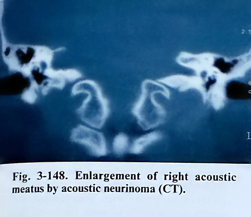
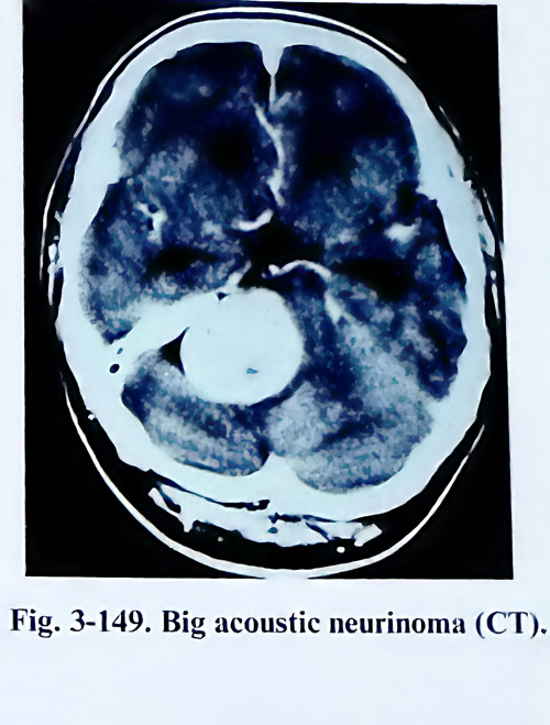
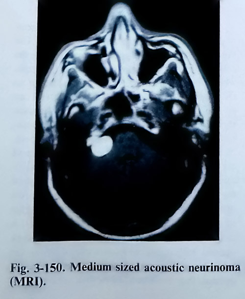
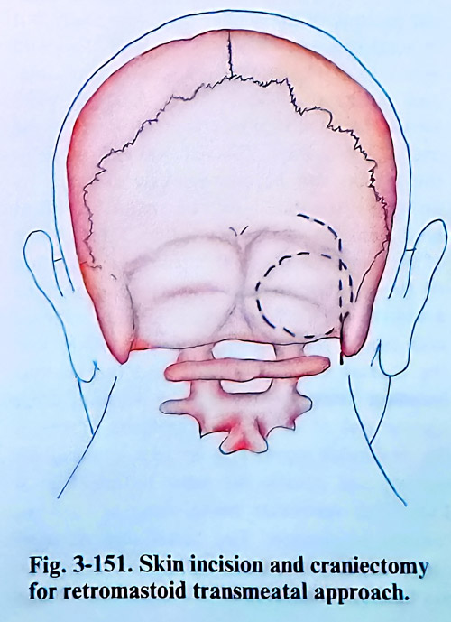
Most cerebellopontine angle neurinomas grow outside the meatus into the subarachnoid space and their further growth is associated with enfolding the arachnoid. A small intrameatal neurinoma, which is less then 2 cm in diameter may be removed totally 'en block'. The posterior wall of the acoustic meatus is drilled out first. This procedure begins with a circular excision of the dura from the petrous bone posterior to the internal auditory meatus (Fig. 3-152). The drilling initially is done using a 4 to 5 mm burr (Fig. 3-153). It is important to know the position of the jugular bulb, (especially if it is high) to avoid damage of its wall. To avoid injury to or opening of the semicircular canal and vestibule, the last part of the bony plate next to the fundus of meatus is left intact at the end of the drilling. The remaining distance to the fundus can be continuously assessed by using an angulated microinstrument (blunt hook) and should not be less than 2 mm. If one of the semicircular canals is fenestrated, suction of the perilymph and endolymph must be avoided, and the fenestration should be closed with fascia, sealed with fibrin glue. After that the meatal dura is opened and the tumour becomes accessible. Using light traction of the tumour out of the meatus the fibres of one of the vestibular nerves can be seen entering the tumour and usually the other is free (Fig. 3- 154). This vestibular nerve, entering into the tumour is divided. The facial and acoustic nerves are located anteriorly to the tumour and can be easily separated with a significant chance of preserving hearing and facial motion. The labyrinthine artery, also located anteriorly to the tumour, should be preserved too. The middle size acoustic neurinomas, which are less than 4 cm of diameter, can be removed easily and in many cases the facial nerve can be saved. Hearing in these cases. however, is usually already lost before surgery. In tumours of this size, there are distinct stages of the surgery, that have been already described above. The stages of debulking and dissection of cranial nerves and brain stem are relatively easy because of the smaller dislocation of structures produced by the lesion.
In big tumours (more than 4 cm in dia-meter) the arachnoid should be separated first from the posterior surface of the tumour and the cerebellar hemisphere gently retracted (Fig 3-155). Even with a very large tumour, it is usually not necessary to resect the lateral third of the cerebellar hemisphere to gain exposure. Then the tumour is reduced in size by suction, but better by ultrasonic aspiration if it is soft, or by laser evaporation if it is hard. The bleeding can be stopped by bipolar coagulation. The enucleation of the tumour continues until only thin layers of the tumour tissue remain attached to the capsule and it becomes possible to dissect it from the adjacent brain structures (Fig. 3-156).
The order of manipulations when dissecting the tumour capsule can be variable. The lower pole of the tumour is separated, where the caudal group of cranial nerves are dissected, which, even though they are very close to the tumour, they are not so adherent to it and can be easily detached. Cottonoid is placed between them and the tumour at the intial place of their identification. That prevents also the penetration of blood into the subarachnoid space.
Next is penetrated along the lateral surface of the tumour and the internal auditory canal is reached. The dura along the posterior edge of the canal is coagulated, avoiding injury to the distal part of the sigmoid sinus. The lateral edge of the auditory canal together with its wall is drilled out (Fig. 3-157). The entire canal must be opened sufficiently wide, without opening the labyrinth. If some of the aerated cells are opened, they must be filled in with bone wax.
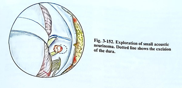
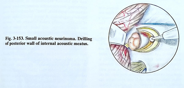
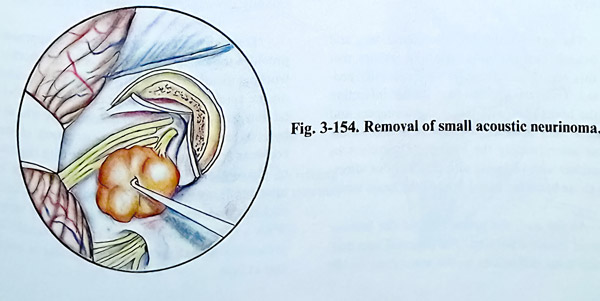
The facial nerve is located superiorly and anteriorly inside the meatus and is recognised by its yellowish colour. At this stage the mechanical and electrical stimulation of the facial nerve can be monitored intraoperatively. It is necessary to separate the tumour capsule completely from the facial nerve, beginning at the internal acoustic meatus and ending at the brain stem. Sharp dissection is preferred. With large tumours the separation is very difficult and requires a long time, as the capsule is closely adherent to the dura of the lateral wall of posterior fossa. If this dissection is not done immediately after the internal acoustic meatus is cleaned from tumour, the surgeon will meet greater difficulties in preserving the facial nerve at the brain stem.
The most delicate part of the operation is the separation of the tumour capsule from the brain stem. This begins between the tumour's lower pole and the medulla oblongata. The lower cranial nerves, remain distant, already covered with cottonoids. This part of the tumour is retracted laterally from the brain stem and is carefully dissected. Small arterial branches, entering directly into the tumour are coagulated and divided only at their entry point and not away from it. This manipulation is repeated until the tumour is separated from the brain stem. The cottonoids must be used with care and should not be held a long time on the brain stem, as they stick and then cause the tearing of small vessels on its surface. Suction should also be used in this region very cautiously. The anterior inferior cerebellar artery and its branches are the most important arteries encountered in this region and must be very carefully protected. Their coagulation may induce infarction of the brain stem with severe consequences and even a fatal outcome. Several branches of the same artery supply the tumour. They must be identified with certainty and cut, without affecting those branches going toward the brain stem.
At the end, the upper pole of the tumour capsule is also separated. The petrosal vein may create some difficulties in the separation of the Vth cranial nerve (Fig. 3-158). It is preferable to keep it intact, but its coagulation and cutting may not represent any significant danger, but if it is large, it must be preserved, because it drains a large part of the cerebellar hemisphere and that may provoke congestion and oedema.
Blood transfusion is applied if required to replace the blood loss and the systolic pressure is maintaining through the surgery in a normo-slightly hypotensive range for the patient.
Meningiomas in the cerebellopontine region are approached in the same way as the cerebellopontine neurinomas. According to the dural attachment of these tumours regarding the acoustic meatus they are subdivided into two groups: anterior and posterior. Every one of the two groups displaces the cranial nerves in a different way (Fig. 3-159).
Once the cerebellar hemisphere is retracted and the tumour exposed, it is removed by alternating intratumoural decompression using ultrasonic aspirator, laser, microforceps and bipolar coagulation. A good arachnoidal plane of cleavage usually exists between the tumour and vessels, nerves, and brain. In quite a few instances, the tumour completely surrounds but does not invade nerves and vessels and a careful dissection may preserve neurological function. In tumours “en plaque” it is usually not possible to find this plane and nerve infiltration is evident.
During the tumour removal an attempt should be made to interrupt early the main vascular pedicle of the tumour that often comes from the petrous bone. Generally, the seventh and eighth nerves are well defined and can be separated well. After the tumour is removed, the dural site of attachment should be eliminated and occasionally any abnormal area of the bone drilled.
Translabyrinthine approach. This approach provides the most direct access with the shortest working distance to the internal acoustic meatus in the cerebellopontine region without cerebellar retraction. The disadvantage of the translabyrinthine approach is the destruction of the cochlear and vestibular end organs with the loss of hearing and balance. That disadvantage is relative because in cerebellopontine tumours the cochlear and vestibular functions are lost early. With a normal contralateral ear, the benefit of preserving a malfunctioning inner ear is minimal at best.
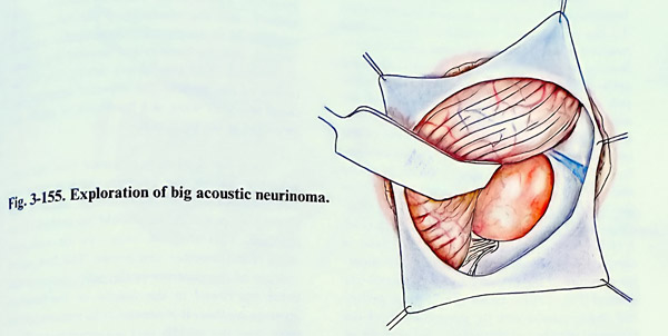
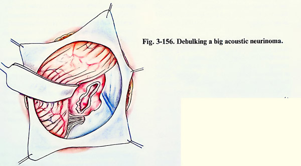
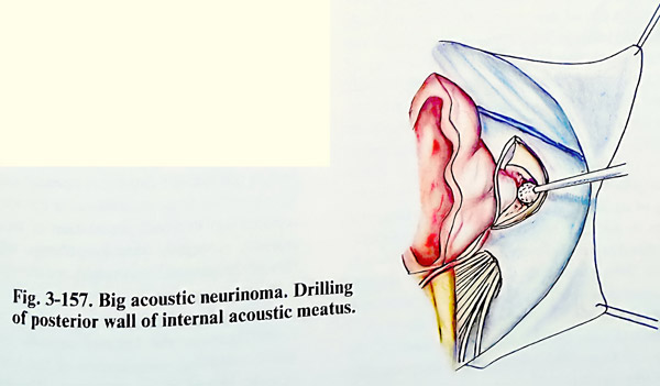
This approach can be indicated in acoustic neurinomas of small and medium size (less than 3,5 cm in diameter), Contraindications include removal of neurinomas that need preserving the hearing or very large tumours (greater than 3.5 cm in diameter). The patient is placed in a supine position with the head turned away from the surgeon. A wide C - shaped retro-auricular incision is made starting just bellow the tip of the mastoid. It runs upwards over the lateral surface of the mastoid to a point 2 cm or so above and behind the tip of the pinna, then curves forward and downward over the temporal muscle to finish 3 cm above the zygomatic arch (Fig. 3-160). A skin flap in the subgaleal plane is elevated forward until the posterior and superior margins of the external auditory meatus are defined (Fig. 3-161). An anteriorly based periosteal flap is incised with the posterior part of the temporal muscle and the pedicle over the area of the external acoustic meatus. All other soft tissues are retracted and preserved for a closure at the conclusion of the procedure (Fig. 3-162).
A complete mastoidectomy is performed, using a large cutting burr. The drilling begins over the mastoid antrum, preserving the tegmen tympani and external acoustic meatus. Drilling is continued posteriorly to the sigmoid sinus, leaving a thin layer of bone residing over that structure. The sigmoid sinus is followed inferiorly to the region of the jugular bulb. The cells of the mastoid tip are completely opened. Further drilling is performed parallel to the stylomastoid foramen until the periosteum of the foramen is identified. The drilling extends closer to the facial nerve, which has to be identified. The facial nerve is then followed superiorly to the region of the horizontal semicircular canal.
Care is taken to visualise the facial nerve throughout its entire vertical portion and the beginning of its horizontal course within the temporal bone to prevent iatrogenic injury. Intraoperative facial nerve elecromyographic monitoring is useful throughout this dissection. The retrofacial mastoid air cells are opened, and bone over the jugular bulb is removed. Drilling then is performed into the labyrinthine portion of the temporal bone. The lateral semicircular canal is carefully drilled with exposure of the secondgenu of the facial nerve in this region. The posterior semicircular canal is identified and opened, and the vestibule of the labyrinth is entered. Next the superior semicircular canal is opened, and the bone in and around this region is removed. The subarcuate artery lies in the centre of the arch of the superior semicircular canal and may serve as a landmark to guide the direction to the petrous tip.
Next the drilling is extended into the petrous tip around the internal acoustic meatus. The bone of the internal acoustic meatus, including its posterior tip should be drilled to an eggshell. This shell of bone can be removed using microdissector and hooks. The free nerve endings of the superior or the inferior vestibular nerve are found in the fundus of the internal acoustic meatus. It is necessary to remove all the bone over the middle and posterior fossa dura, internal auditory meatus and jugular bulb (Fig 3-163).
The transcochlear approach represents a forward extension of the translabyrinthine ex-posure. This is accomplished by removing the cochlea in addition to the vestibular labyrinth.
The dura is opened in a stellate manner to expose the cerebellopontine angle and to visua-lise the superior and inferior vestibular nerves. The removal of the tumour begins with identification of the facial nerve at the depth of the meatus (Fig. 3-164). The fibres of the superior and inferior vestibular nerves are interrupted, and just anteriorly, the greyish facial nerve is seen in front of the vertical crest (Bill's bar).
With medium or large sized tumours, the mass fills the entire area of dural exposure of the translabyrinthine approach. The presenting surface of the tumour capsule is opened, and gentle intracapsular tumour removal is carried out and at this point is important to avoid penetration of the tumour capsule, especially anterosuperiorly, where injury of the petrosal vein might occur.
After completion of the internal decompression, the tumour capsule begins to infold, allowing visualisation of the brainstem and IX, X, and XI cranial nerves. The intramental portion of the tumour is carefully dissected away from the facial nerve. The dissection of the facial nerve must be gentle, and mechanical or electric stimulation of the nerve can be monitored with intraoperative elecromyographic recording. All dissection is done in the arachnoidal plane, carefully pushing the arachnoid sheath away.
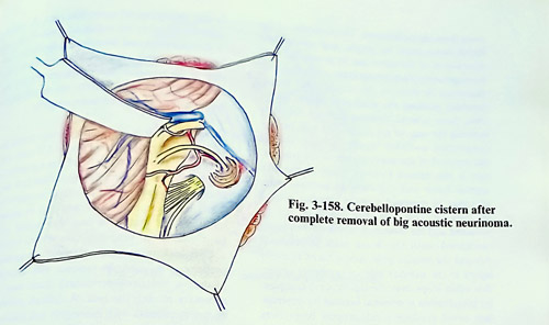
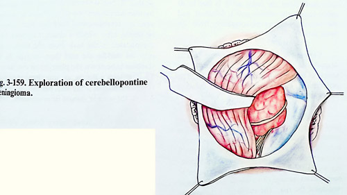
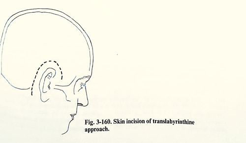
Only those arterial branches that are definitely entering the tumour are coagulated with bipolar coagulation and then sharply divided. During the removal of the anterior pole of the tumour, care is also needed to avoid injury to the fifth cranial nerve and the superior petrosal vein, that may be stretched over the superior pole of the tumour. This part after debulking can be retracted and the dissection of the seventh nerve continued until the brain stem is reached.
Around the inferior pole, care is taken to avoid injury to the anterior inferior cerebellar artery, that often loops over the eighth nerve complex. Its preservation is essential because its interruption could produce a catastrophic brain stem infarction. After the facial nerve is free in its entire course, the inferior pole is debulked, and the capsule is separated completely from the brain stem.
Closure. A single sheet of fascia lata is draped over the translabyrinthine defect in the temporal bone and filled with strips of fat. The pericranium, subcutaneous layer and the skin are sutured as usual.
Complications. They can occur during surgery and in the postoperative period. With the patient in a sitting position, the most common complication is air embolism. It is usually not a serious problem, but has the potential of becoming a catastrophic one. Careful maintenance of haemostasis during all stages of the operation minimises the risk. The increase of the venous pressure by the anaesthetist from time to time is also a measure to avoid this complication.
Haemorrhage during the operation is usually not a major problem. If the petrosal vein is torn, it can usually be managed with gentle packing with oxidised cellulose. Arterial bleeding is usually due to the tear of a small branch that likewise will respond to packing or bipolar coagulation. During the early postoperative period, monitoring of intracranial pressure aids in the detection of complications. A silastic catheter placed in the subdural space at the and of the operation and brought out accurately monitors posterior fossa pressure. If the posterior fossa pressure remains less then 15 mm Hg for 24 hours, the monitoring is discontinued, and the catheter removed.
Acute hydrocephalus may occur during the early postoperative period and a CT scan may be necessary to differentiate it from a postoperative haemorrhage. Cerebrospinal fluid leak may result from poor wound healing, increased intracranial pressure, wound infection, or opened mastoid cells that have not been sealed well with bone wax. A few simple stitches seldom control a CSF leak unless the increased intracranial pressure is reduced. In patients with hydrocephalus, a ventriculoperitoneal shunt may be necessary to stop the leak. A difficult problem occurs in patients with meningitis and hydrocephalus. At first a ventriculostomy with intraventricular administration of antibiotic is preferred until the cerebrospinal fluid has been shown to be sterile, after which a shunt is inserted. More complicated is the leak from the mastoid cells into the middle ear and then through the Eustachian tube down the pharynx or out the nose. To treat such type of fistula, external lumbar drainage is preferable, but if the leak persists beyond 3 or 4 days, the wound should be re-explored.
When fifth nerve injury occurs, the resultant corneal anaesthesia may lead to corneal ulceration if proper eye care is not provided. The function of the fifth nerve should be evaluated as soon as the patient awakens from anaesthesia. When the corneal reflex is diminished, the eye should be covered with a protective shield and artificial tears applied every 4 hours. If the reflex is absent or if there is an associated facial palsy, it is necessary to do a temporary tarsorrhaphy. Seventh nerve function should also be evaluated in the recovery room. If the face is paralysed, the same care for the eye should be provided as in trigeminus damage. II there is total disruption of the facial nerve during surgery, an operation to recover the paralysed facial muscles is indicated. Generally these procedures can be done in 3 to 4 months after the complete recovery of the patient from the intracranial operation.
The results from the surgical treatment of cerebellopontine neurinomas depend first of all on the size of the tumour and on all other factors determining the indications for surgery.
Small sized tumours (less than 2 cm of diameter) can be removed totally and the facial nerve can be preserved in 100% of patients. Half the patients remain with partial hearing. In 90% of the patients with medium sized tumours (up to 4 cm in diameter) total removal of the tumour is possible with 60% risk of facial nerve damage. Only in 10% of these patients is the facial nerve intact, and in another 10% it is interrupted definitely. In patients with big tumours (over 4 cm in diameter) total removal is achieved in 83% with 40% interruption of the facial nerve. Surgical mortality is 4% with respect to patients with big tumors.
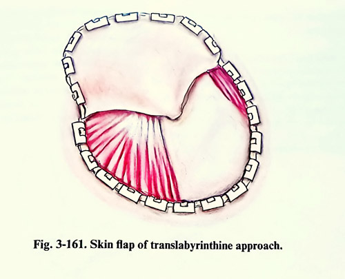
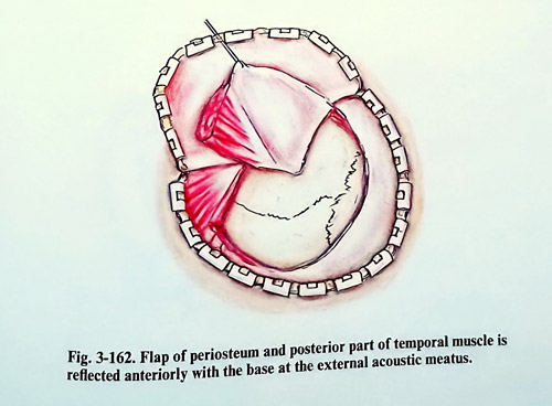
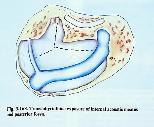
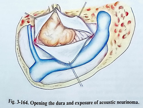
CLIVAL AND PETROCLIVAL MENINGIOMAS
Clival and petroclival meningiomas arise from the upper two-thirds of the clivus and from the petroclival junction medial to the trigeminal nerve. These tumours grow anteriorly or antero-laterally to the brain stem, compressing or involving the basilar artery and its branches. The Vth - VIIIth cranial nerves are usually displaced dorsally. Big clival and petroclival meningiomas may grow superiorly through the tentorial notch, compressing the midbrain.
The different modalities of diagnostic imaging (CT, MRI) should demonstrate the size and the location of the tumour, as well as its relationship with the cranial nerves, blood vessels and the brain stem. The indication of the place of attachment by the studies is of essential importance (Figs. 3-165; 3-166). Angiography is needed to show the main feeders of the tumour, the displacement, and the involvement of big vessels in the posterior fossa. In the majority of cases, feeders originate from the meningohypophiseal and marginal tentorial arteries seen enlarged on a subtraction of the angiograms.
Angiography should also indicate any encasement of major basilar branches or the basilar artery itself. Analysis of this information has also the goal of identifying those cases suitable for preoperative embolisation.
Tumours with this location can be subdivided according to their place of attachment: pure clival, petroclival, petroclival tentorial. The tumours with tentorial involvement may be more supra- or infratentorial. Routes of approach can also be specified as supratentorial, infratentorial or combined. The supratentorial route is convenient for tumours with upper clival and supratentorial attachment. Manipulation on the tumour is performed through the space between the IIIrd and IVth, and the IVth and Vth cranial nerves. An infratentorial attachment place is reached by a lateral suboccipital approach, manipulating the tumour between the Vth and VIIth - VIIIth nerves or below them (Figs. 3-167; 3-168). Petrous attachment is managed more easily than is the clival and extensive radical removals are easier. Transtentorial approaches, retro, and presigmoid transpetrosal approaches, are preferable for those lesions attached to a broader base, supra- and infratentorially, with extensive involvement of arteries and cranial nerves.
The transpetrosal approach is more appropriate for big tumours extending up through the tentorial notch. The technique of this approach is described in the first chapter of the book (Fig.3-169.The dura anterior to the sigmoid sinus is opened along the anterior margin of the sinus, The incision is then extended toward the supra-tentorial space, parallel to the floor of the temporal fossa. Another incision of the dura is made in the posterior fossa if needed (Fig. 3-170). The temporal lobe is gently retracted, preserving the vein of Labbe. The superior petrosal sinus is clipped or coagulated and transected. The incision is continued on the tentorium, parallel to the edge of the petrous bone and extended through its free edge. Care should be taken to preserve the trochlear nerve, which goes parallel to the tentorial edge. The opening of the tentorium allows excellent exposure of the upper pole of the tumour and the anterior and lateral aspects of the brain stem (Fig. 3-171). Trigeminal nerve roots are frequently stretched and spread by the tumour.
A self-retaining retractor is usually needed to hold the cerebellum medially with the transected edge of the tentorium. With any of the approaches, the initial steps of tumour removal are debulking of the volume and transection of its attachment. The tumour is separated from the tentorial surface, coagulating and cutting the feeding vessels; the same is done from the posterior petrous surface, and clivus. When the tumour is of small or medium size, the VIIth and VIIIth cranial nerves are usually stretched dorsally and they are easily identified. Big tumours may encase these nerves. The arachnoid is dissected from the tumour surface and the tumour is debulked using suction, ultrasonic aspirator or laser with extreme caution since the VIlth, VIth cranial nerves, as well as the posterior inferior cerebellar artery may be encased by the tumour. Then the tumour capsule is dissected from the adjacent structures.
The dissection should be maintained within the arachnoid planes to preserve the neural and vascular structures, often adherent to the tumour surface. Cranial nerves, the basilar artery and its branches may, however, demand meticulous and tedious dissection. The lower cranial nerves are usually easily dissected from the inferior pole of the tumour. Vagus nerve manipulation may provoke hypotension and bradycardia that should be avoided. The sixth nerve is stretched usually anteriorly and inferiorly, and it is also dissected away from the tumour following it distally. Alternating the visualisation of the surgical field between supra-and infratentorial routes allows the tumour capsule to be dissected more easily and more safely from the brain stem, the basilar artery and its branches. Once the tumour has been excised, the area of tumour attachment is preferably vaporised extensively with the laser. If there is extension of the tumour inside the internal auditory meatus, its wall is drilled and the tumour removed. A similar technique is applied for the removal of tumour extension into the jugular foramen. Hyperostotic bone is also removed by drillling.
During the closure, the periosteal flap produced at the initial stage of surgery covers the drilled petrosal bone to avoid CSF leak; the temporal muscle is rotated over the defect and attached to the sternomastoid muscle.
The rate of total removal has increased impressively in the latest published series. The main obstacle for total removal remains extradural invasion of the bone.
The complication rates, however, remain high, and is commonly related to cranial nerve deficit, in many cases transient and tolerable. The most serious complication is injury to the brain stem from manipulation or more likely, from affection of its blood supply. Infarction of the lateral tegmental region of the pons is usually a result of an occluded anterior inferior cerebellar artery, occasionally, the appearance of deficit may be delayed during the postoperative period. Temporal lobe swelling or haemorrhagic infarction can be seen with the subtemporal approach, particularly important on the dominant hemisphere. It is precipitated by coagulation or tearing of the vein of Labbe, or the basilar occipital veins. The cranial nerves from IIIrd to XIIth are also at risk during surgery. Because of the trochlear nerve's close relation to the tentorial border, its injury is a frequent hazard during tentorial splitting. Morbidity resulting from its paralysis, however, is minimal compared to paralysis of other cranial nerves. Morbidity from the trigeminal nerve, however, is more serious, because of the resulting corneal anaesthesia and subsequent keratitis, particularly if the facial nerve is also affected. In these cases immediate tarsorrhaphy should be performed followed by reconstructive surgery for the facial nerve function. A trigeminal nerve lesion may result in facial pain, anaesthesia dolorosa, and trigeminal neuralgia.
The size of the tumour is the most decisive factor for preserving the facial nerve. The facial nerve is usually displaced dorsally in petroclival meningiomas and may be involved by the tumour. Intraoperative end-to-end anastomosis of the facial nerve is an option for repair with 80% good recovery. Intraoperative grafting of the nerve also can be performed. Delayed facial-accessorius anastomosis will be the more suitable procedure for those affections discovered postoperatively and not recovering within 3 - 5 months from surgery.Hearing loss usually exists preoperatively. If the hearing is normal or partially affected before operation, its loss can be also a potential complication of surgery too. The deficit of lower cranial nerves is also a significant cause of morbidity and mortality. Intraoperatively, dissection of these nerves may produce bradycardia and hypotension. Post-operatively, dysphagia, vocal cord paralysis, and a depressed cough and gag reflex may lead to serious pulmonary complications.
Disturbances of CSF dynamics in the postoperative period include CSF leaks. hydrocephalus, and CSF collections under the soft tissue layers. Hydrocephalus may present before surgery and it may persist despite total removal of the mass, but it may also develop postoperatively. Acute postoperative hydrocephalus is usually obstructive and is related to mass effect, while delayed hydrocephalus is usually communicating due to poor absorption of CSF or obliteration of the basal cisterns. A CT scan is the most important diagnostic investigation and the treatment often is shunting, There is a risk of CS leak in the transpetrosal approach, occurring via the skin or through the middle ear. The leak is best avoided with watertight suturing of the dura, application of bone wax to the exposed cavities and sealing a periosteal flap over the drilled temporal bone surface. Prophylactic antibiotic coverage is usual in these circumstances.
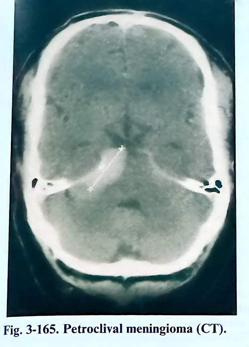
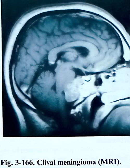
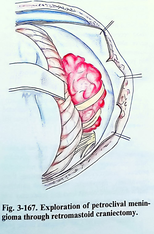
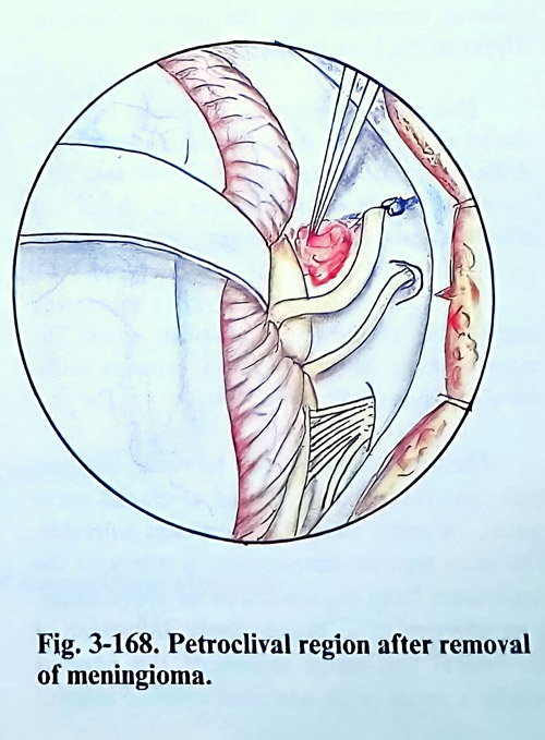
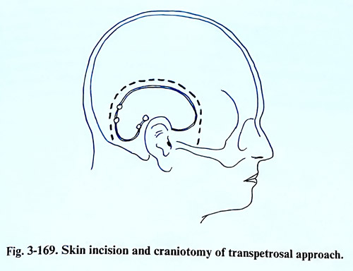
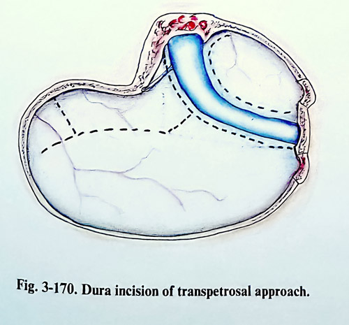
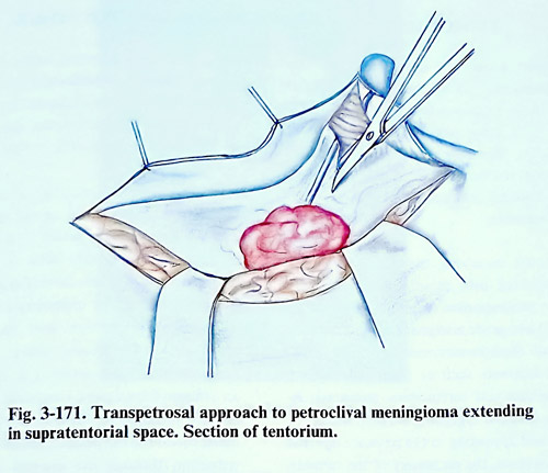
TUMOURS INVOLVING THE CAVERNOUS SINUS
Surgery within the cavernous sinus (CS) has made significant progress through the last decade and now it is possible to remove some tumours that were considered before to be unresectable. The morbidity rate after such operations in the hands of experienced surgeons and with careful selection of patients for surgery is acceptable.
Tumours involving the cavernous sinus may be divided into three groups:1. Benign tumours - meningiomas, angiofibromas, neurinomas; 2. Low-grade malignancies such as chordomas and chondrosarcomas: 3. High grade malignant tumours such as basal cell carcinomas, squamous cell carcinomas, metastatic lesions. The decision regarding surgery should be individualised according to the patient's age and general condition, the pathology of the tumour, and its biological behaviour.
In cases of benign intracavernous sinus tumours, the indication for surgery is the progressive growth of the tumou on the imaging investigations and/or progression of the cranial nerve deficit. Low-grade malignancies such as chordomas and chondosarcomas can be removed from the cavernous sinus whenever complete tumour excision is the goal on a selective basis. This sometimes includes patients with bilateral intracavernous invasion. Indications for removal of high-grade malignancies involving the cavernous sinus are controversial.
Excision in a unilateral involvement of the cavernous sinus is possible in some cases, but bilateral cavernous sinus invasions by high malignancies are not suitable for resection.
CT and MRI with axial and direct CT coronal imaging can demonstrate the cavernous sinus neoplasms, but MRI is more sensitive than CT-scan for visualising small lesions in this area. The relationship of the tumour to the carotid artery and other major vessels in this region can be best delineated by MRI. Angiography provides information on the flow and collateralization and is better associated with a balloon occlusion test (Fig. 3-172).
Operations for removal of tumours invading the cavernous sinus expose the intracavernous carotid artery, which is at high risk of injury. Approximately 80% of patients will tolerate the loss of one carotid artery. However, patients who cannot compensate the reduction of blood flow through their internal carotid artery can suffer massive infarction and sometimes perish if that artery is sacrificed, despite an attempt for an extra - intracranial bypass. To be able to confront these circumstances in the safest mode for the patient, the tolerance to flow reduction through the internal carotid artery has to be tested preoperatively. The most important test is the balloon occlusion test, performed simultaneously with the angiography. An intra-carotid balloon is inflated for up to 15 minutes. If the patient experiences or the physician detects neurological deficit, the test is discontinued. Those patients remaining asymptomatic are submitted to a CBF test with xenon CT. If this test steadily indicates reduction of the flow below 35 ml (100 g) per minute, the patients are also under certain risk, but it is not as high as that of the first group. The remaining patients as a rule tolerate carotid flow interruption (even permanent) without neurological affections.
Safe manipulation of cavernous sinus structures requires the surgeon to be familiar with the morphology of the area beyond the routine requirements. Detailed microsurgical studies have refined those specific areas, which can be selected for penetration into the sinus. They are described conveniently as triangles. Another particular feature of cavernous sinus surgery is the difficulty of haemostasis. Skilful combined application of selective bipolar coagulation and pieces of oxidised cellulose must provide a sufficiently dry' area for proper visualisation and handling of structures.
The cavernous sinus - an anatomical structure built up from dural walls, separates and integrates three types of systems: 1. The internal carotid artery; 2. The cavernous venous plexus system; 3. Cranial nerves traversing it. The venous cavities, because of their extensive anastomosis to adjacent venous collectors, are the only structures which can be partially sacrificed.
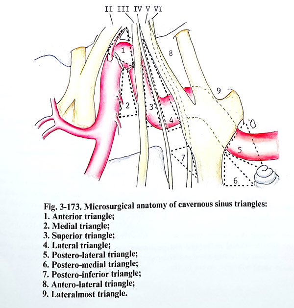
Otherwise, all its arterial or neural structures can be subjected to iatrogenic damage unless special circumstances take place and precautions are not undertaken. The safe approach to all cavernous structures has been standardised through the following triangles (Fig. 3-173):
The anterior triangle is enclosed by the lateral border of the extradural optic nerve, the medial wall of the superior orbital fissure dura and the dural ring, surrounding the internal carotid artery as it penetrates intradurally. The space extradurally contains the genu (C3 segment) of the internal carotid artery.
The medial triangle is produced by locating its tip at the anterior siphon angle (lateral carotid wall) and it must be already exposed, the porus oculomotorius (the intradural exit the third cranial nerve) and the posterior clinoid process. An incision inside this triangle will expose the proximal siphon (C3) and the horizontal intracavernous carotid artery (C4).
The superior triangle has as two borders the third and fourth nerves and posteriorly the dural margin toward the posterior fossa.
Through this triangle are exposed the C4 - C5 segments junction and the origin of the meningohypophiseal trunk.
The lateral triangle is a narrow space between the fourth cranial nerve and the ophthalmic division of the trigeminal nerve, The incision there will give access to the ascending CS segment of the internal carotid artery.
The postero-lateral triangle (Glasscock) is defined by the posterior rim of the foramen ovale, the foramen spinosum, the posterior border of the mandibular division of the trigeminal nerve, and the cochlear apex.
Drilling out the bone in this area will expose the proximal CS segment of the horizontal intrapetrous internal carotid artery. This place is suitable for proximal control or venous bypass graft of the artery.
The postero-medial triangle (Kawase) is confined among the porus trigeminus (extra-dural exit of trigeminal nerve), the posterior border of the mandibular division of the same nerve and the cochlea. This area corresponds to the petrous apex, which can be drilled out to increase the exposure of tri-geminal and brainstem structures.
The Postero-inferior triangle is bounded by the fourth cranial nerve, the posterior clinoid process, and the medial porus trigeminus. An incision in this area will expose the sixth cranial nerve.
The Anterolateral triangle is the arca between the ophthalmic and maxillary division of the fifth cranial nerve. This triangle is used to expose the superior orbital vein and anterolateral extension of tumours within the cavernous sinus.
The Lateralmost triangle is bounded by the second and third divisions of the trigeminal nerve. This is area to expose the lateral extension of cavernous sinus tumours.
Surgical technique. Surgery of the cavernous sinus requires broad access and a wide range of view for the area. Most specially designed approaches will require a relatively large craniotomy, removing almost always the same cranial bone structures (Figs. 3-174; 3-175). A frontotemporal craniotomy followed in some cases by orbitozygomatic osteotomy permits such a broad approach and provides a better basal view of the area. Extradural removal of the orbital roof including the anterior clinoid process and the optic canal is the next usual step (Figs. 3-176; 3- 177). The dura is elevated from the middle cranial fossa, dissecting the middle meningeal artery, the great petrosal superficial nerve, and the mandibular and maxillary nerves. The greater petrosal superficial nerve and the middle meningeal artery are divided. The horizontal segment of the internal carotid artery is then exposed inside the carotid canal at the petrous apex postero-lateral triangle). The sometimes tiny bony wall of the canal is drilled out with a diamond microdrill head (Fig. 3-178). The technique can be used either for proximal control, as a site for potential bypass, or as a first step for an extra-dural approach following the artery. The Eusta-chian tube is at risk, and is best preserved at this stage by avoiding excessive drilling. An enlarged excision of bones is needed for tumours that have an extension and involvement of the pyramid and the sphenoid.
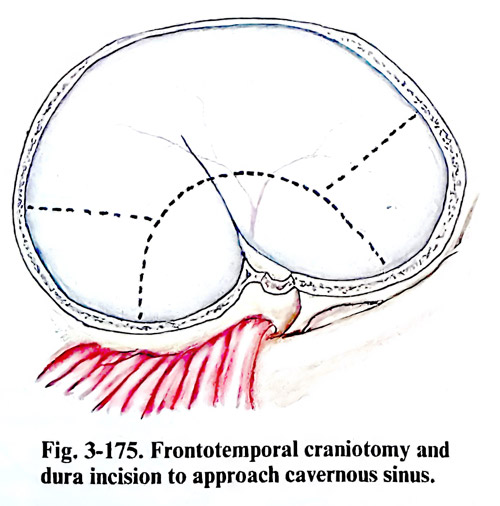
Extradural approach. The cavernous sinus can be entered extradurally by following the petrous internal carotid artery (inferior approach), especially after the temporary division of the mandibular nerve, if involved in the lesion or between the divisions of trigeminal nerve (anterolateral approach). The space between every two branches is rarely sufficiently broad unless they are distended by the tumour. The cavernous sinus can also be entered by a medial extradural approach if the sphenoid sinus is entered and the optic nerve unroofed after a broad basal frontal exposure of the tumour.
Intradural approach. Large tumours within the cavernous sinus and all meningiomas require an intradural approach. After dural opening, the Sylvian fissure is split and the frontal and temporal lobes are gently separated and retracted. If excessive retraction is necessary, the anterior 2 to 4 cm of the inferior temporal gyrus is resected, sparing the medial temporal lobe structures.
Superior approach. For the superior approach to the cavernous sinus, the optic nerve is unroofed completely and its dural sleeve opened. The anterior clinoid process is removed. The two distal rings around the distal intracavernous internal carotid artery are identified (Fig. 179). The superior wall of the cavernous sinus is opened through the anterior triangle, and the ICA is followed back into the cavernous sinus, exposing consecutively the medial and superior triangles. Intracavernous structures superior to the horizontal intracavernous ICA, the anterior genu, and the vertical segment of the ICA are well exposed by this technique. The sella turcica can also be exposed if necessary.
Lateral approach. This requires opening the lateral wall of the cavernous sinus. It is carried out by working through the Silvian fissure and subtemporally. For meningiomas, the outer dural incision can be crosswise (horizontally below and parallel to the approximate location of cranial nerve IV, and a vertical portion intersecting incision at the most prominent bulge of the tumour). It corresponds to the lateral triangle. For meningiomas, the outer dural layer of the lateral wall is completely peeled away, starting anteriorly in front of the sphenoid sinus, inferiorly near cranial nerve V2 and V3, superiorly from the tentorial edge, and posterorly to the superior petrosal sinus. Cranial nerve III should be identified in the subarachnoid space and followed for 5 mm into the lateral wall of the cavernous sinus. Cranial nerve IV may be found in the lateral wall or followed anteriorly in the subarachnoid space.
Because this last nerve is thin, it is preferable to leave a small cuff of dura around it until the end of the meningioma removal to its porus. Cranial nerve VI may be found in the lateral wall or followed forward from Meckel's cave and the trigeminal ganglion.
Complications. The most frequent complications are paresis of cranial nerves III to VI. They occur in about 80% of the cases, but most of them improve within the first 3 - 4 months. An important complication is CSF leak due to erosions in the bone caused by the lesion itself. Possible damage of the optic nerve during surgery is due to the opening of the canal by mechanical or thermal damage. This complication can be avoided by careful removal of the optic canal wall (Fig. 3-180). Contusions and/or laceration of the brain, and intracerebral hematomas can be avoided also by exerting only gentle retraction during the operation.
A life-threatening complication is the rupture of the internal carotid artery, which may occur either intra- or postoperatively due to damage of the arterial wall during removal of the tumour. When the tumour is adherent to the wall of the artery, or has even infiltrated it, the preferable decision is to leave part of the tumour around the artery and treat the patient postoperatively by gamma - knife surgery. On the other hand, if a radical operation has been chosen, the artery is then reconstructed with a graft (petrous to intracranial venous bypass).
Another complication can be venous bleeding after complete removal of the tumour. If the bleeding veins during operation are not well packed, fatal haemorrhage may occur post-operatively due to extrusion of the haemostatic pack as a result of straining of the patient. It is therefore advisable, at the end of surgery, after complete removal of the tumour, to place some sutures from one side to the other to prevent this extrusion. Sometimes, however, it is necessary to remove the oxidised cellulose from the cavernous sinus because of pressure exerted on nerves and the carotid. In such a case oxidised cellulose is replaced by loose packing at the same time checking for compression on the internal carotid artery and confirming that venous haemorrhage has been completely stopped.
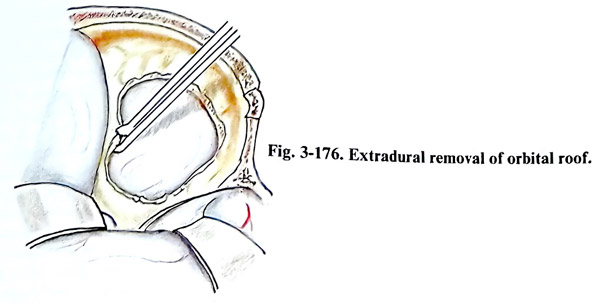
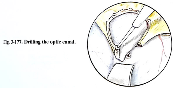
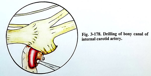
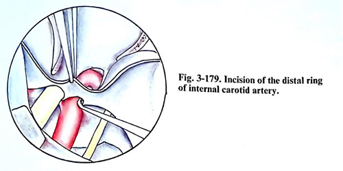
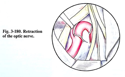
TUMOURS INVOLVING THE CAVERNOUS SINUS
The tumours of the foramen magnum and craniovertebral junction occupy space in the posterior fossa and the spinal canal. Mostly these lesions are extracerebral tumours such as neurinomas, arising from the intracranial part of the cranial nerves and the upper cervical roots or meningiomas, attached to the mid- or lower clivus, foramen magnum or adjacent dura. In addition to these locations, technical difficulties of approach to these lesions are related to the jugular and hypoglossal foramina with the related to them neural and vascular strucrtures, and also the foramen magnum parts located anteriorly and/or laterally to the cord.
Most lesions in this area are investigated by imaging studies to demonstrate the relationship of the tumour to the rim of foramen magnum, the skull base foramina, and the structures passing through them, as well as the major vascular structures: vertebral and basilar arteries, their major branches, and the jugular bulb. Essential techniques of investigation are CT: enhanced, with images demonstrating bone and neural structures alternatively, in axial and coronal projections, and MRI. Bone destruction, erosion, sclerosis, foraminal enlargement and calcification are seen on the CT, but the more precise assessment of neural structures is left for the MRI imaging (Figs. 3-181; 3-182). Angiography should evaluate both carotid and vertebrobasilar circulations and should give the necessary data of collateral supply. It will also indicate the displacement or wall compromise of vascular structures, as well as the degree of vascularity of the lesion.
From the imaging studies the following must be decided upon: 1. If the vertebral or basilar arterial walls are affected by the lesion, immediate arterial reconstruction has to be possible through the surgical approach. 2. If the jugular bulb wall is affected, a venous sinus ligature must be prepared in case of rupture. 3. It should be decided which cranial nerves are encased and will pose a threat of iatrogenic damage or will be required to be sacrificed. The strategy decided on should implicate surgical planning in such a way that the surgical approach provides the necessary access to thenaffected structures and space for performing the manipulations.
When the tumour is located posterior to the brain stem and spinal cord, the posterior fossa is opened in a standard way and the laminae of the upper cervical vertebrae are resected. Meningiomas and neurinomas situated posteriorly are removed comparatively easily, applying the general principles for such removal. Laterally or mainly anteriorly situated tumours need the lower lateral craniocervical approach. For this approach the patient is placed in a semisitting or park-bench position.
The skin incision is similar to that used for the retromastoid craniectomy but is extended caudally to the level of C2, exposing laminae of C1 and C2 unilaterally (Fig. 3-183). After transecting the muscles along the incision to the occipital squama, occipital muscles are detached from the occipital bone predominantly in a lateral direction. The incision of muscles can be performed along the midline with less bleeding, but retraction laterally can be difficult. The central and caudal part of the skin incision can be placed for these reasons also along the midline.
The separation is extended laterally to the tip of the mastoid and transverse processes of C1 - C2 are exposed. The deep muscle layers are detached with care to avoid injury to the vertebral artery. The C1 - C2 joint is identified, and the vertebral artery is exposed along the superior margin of the C1 lamina (the horizontal extracranial segment). If a broader space is required, the C2 spinal nerve can be transected close to its dural entry point. The suboccipital craniectomy is completed and extended as far laterally as the sigmoid sinus, including removal of the lateral rim of the foramen magnum and the lateral half of the Cl and C2 laminae with the posteromedial portion of C1 - C2 joint. If necessary the posterior half of occipital condyle can be removed and that has no effect on stability of the craniocervical junction. It is done with the high-speed diamond drill. It is important to expose the margin of the sigmoid sinus and to follow it caudally to the posterior border of the jugular foramen to the area of the tuberculum jugulare, That increases exposure and prevents unexpected injury. The vertebral artery entry area through the dura is dissected meanwhile, so the dura around it is safely accessible.
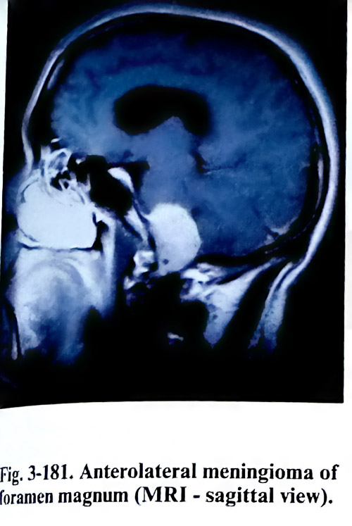
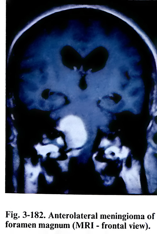
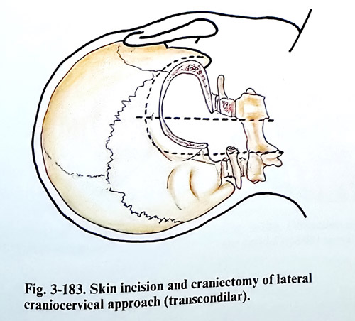
The dura is opened in a Y - shaped incision and the corresponding dural flaps are fixed with traction sutures (Fig, 3-184). Initially the tumor is enucleated by manipulation between the roots of the cranial nerves, cvhoosing the interspaces depending on the location of the tumour. The first denticulate ligament requires division. After the complete debulking follows the separation of the tumour capsule. In cases when the lower cranial nerves are encased by the tumour, neurophysiological monitoring and nerve stimulation can help with their identification and preservation. Dissection of the tumour capsule from the nerves is relatively easier than from the vertebral and basilar arteries and their branches. The preservation of the perforating vessels to the brain stem is extremely important for the outcome of these risky operations. Vascular compromise to neural tissue that occurs in this area can lead to catastrophic complications of lower brain stem dysfunction (Figs, 3-185; 3-186).
Particular difficulties arise in meningiomas attached to the dura at the intracranial entry of the vertebral artery, the jugular foramen and hypoglossal foramina. The vertebral artery can be mobilised opening its dural ring, the C2 transversal foramen and also be temporarily transposed until completing manipulation ventral to it. The parts of the artery immediately proximal and distal to the expected affected segment should be accessible for temporary trapping. The adventitial layer serves as a guide to the safe dissection of the wall. In case of true tumour infiltration of the wall of the vertebral artery, an attempt at total removal should be avoided (actually leaving only that portion, infiltrating the wall on a thin layer). The jugular bulb must be preserved. The hypoglossal nerve is sacrificed only if there is no accessible portion at the entry into the foramen.
The results of surgical therapy depend on the completeness of the resection, the involvement of various anatomical structures by the tumour, and the location and texture of the tumour. Functional recovery is related to disease progress and the promptness of the diagnosis. The earlier the diagnosis is established, the more complete resection is possible and the better results are obtained.
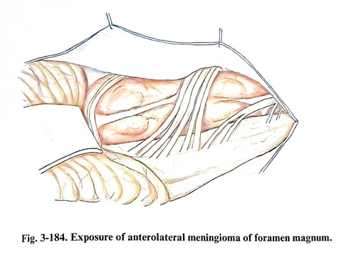
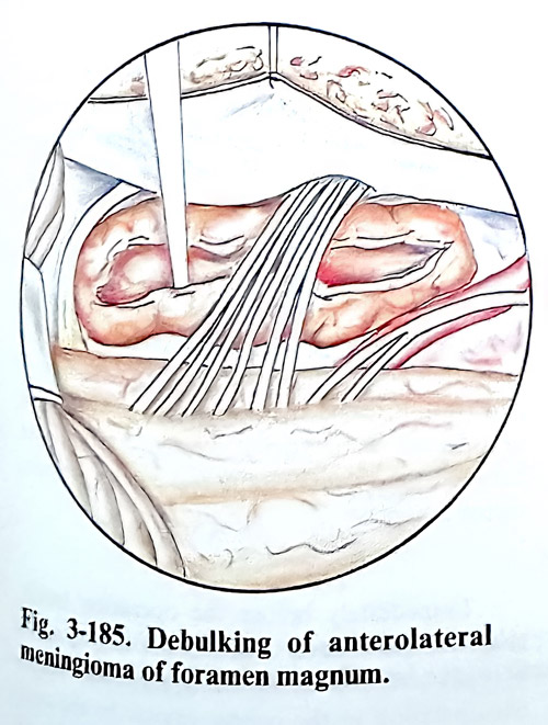
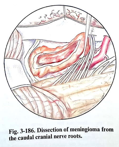
ORBITAL AND CRANIOORBITAL TUMOURS
The great variety of tumours and other mass lesions that occur into the orbit are of interest to several surgical specialities. Ophthalmologists deal with many of these problems by anterior direct approaches. The otolaryngologists manage many conditions arising within the sinuses and involving subsequently the superior, medial and inferior walls of the orbit. Neurosurgeons have access to those tumours, located deeply inside the orbital cavity and/or involving both the intracranial and intraorbital spaces.
When an orbital tumour is suspected clinically because of exophthalmos, visual impairment or limitation of eye movements, the patient needs a detailed examination. With good quality of the X rays of the skull, orbits, and optic canals, calcifications, hyperostosis, gross destructive lesions and sinus diseases can be seen.
An essential task of the imaging studies is to differentiate tumours from intraorbital inflammation. CT and MRI usually reveal the exact location and the size of the tumour, and in many cases the histology of the lesion can be predicted. CT should provide complete imaging of the orbital bony structures and contents in direct coronal and axial planes. Enhancement of the lesion by intravenous contrast is important for the differential diagnosis. MRI provides direct multiplane imaging, which is a very valuable contribution to the topography of the lesion. This modality also adds information about lesions close to the optic nerve and it is superior in visualising them inside the optic canal.
Many different surgical approaches to the orbital cavity have been introduced into practice by ophthalmologists, otolaryngologists, and neurosurgeons. There are controversial opinions on the indications for this large number of approaches. Several factors determine the choice of approach. These factors are related to the position of the tumour and its relationship to surrounding structures. We can specify them as follows:
1. Position of the tumour regarding the muscle cone - inside or outside.
2. Relation of the tumour to the optic nerve - superior, inferior, medial or lateral.
3. Presence of involvement of the optic nerve inside the canal.
4. Defining the lesion as primary of the orbit or secondary - penetrating from adjacent structures and cavities.
Personal preferences of surgeons have strongly influenced the selection and recommendations of approaches to the orbit. Irrespectively of that, we would like to suggest some guidelines. The lesions inside the muscle cone require a broader space for dissection, usually a transcranial approach, providing space for safe manipulation, compared to those mass lesions located outside the cone, which are usually quite accessible under the removed part of the orbital wall. Even more, if they are totally anterior to the equatorial plane of the bulb, they can be removed by an anterior orbitotomy. In those lesions, penetrating into the orbit from an adjacent structure or cavity, the approach should provide access simultaneously to that structure of origin to guarantee as much as possible radical removal (Figs. 3- 187; 3-188).
Tumours in close relation to the nerve have to be approached through broader transcranial orbitotomies and the position of the tumour regarding the optic nerve has a significant importance for the choice. The rationale of choice is that the nerve should not be on the way to the tumour when it is broadly splitting the extraocular muscles. When the tumour has mass effect and the configuration of muscles is grossly or moderately deformed, preferential ways of penetration inside the muscle cone are selected. Such small or intrinsic optic nerve tumours are removed between the medial rectus and levator/superior rectus muscles.
Immediately before the operation tarsorrhaphy is performed to ensure protection of the eyeball and especially of the cornea during manipulation in the orbital cavity. In the postoperative period it is left for a few days, until the oedema of the orbital tissue subsides (Fig. 3-189).
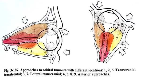
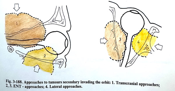
ANTERIOR SURGICAL APPROACHES
The anterior surgical approaches are suitable for tumours, located mainly anterior to the equator of the eye-bull. These tumours usually can be palpated through the orbital entrance (tig. 3-190). The anterior surgical approaches can be transcutaneous or transconjunctival. The transconjunctival approaches usually are performed by ophthalmologists.
ANTERIOR TRANSCUTANEOUS APPROACHES
When the tumour is located superior to the eyeball and under the orbital roof, the skin incision is done through the eyebrow (Fig. 3-191). The orbicular muscle is split along its fibres, reaching the orbital edge of the frontal bone. The supraorbital incisura and foramen containing the frontal nerve and artery are preserved to avoid postoperative anaesthesia of the forehead. The fibrous septum of the upper eyelid is opened 2 - 3 mm away from its attachment to the bone. Its transection is cautious and should avoid injury to the levator palpebrae superioris muscle (Fig. 3-192). This approach is suitable for the removal of haemangiomas, dermoids, and mucoceles. After the bone has been exposed, the periorbit is separated from the bone and the tumour surface is reached. When the tumour is situated medially and superiorly to the eye-ball, the skin incision can be extended medially between medial cantus and the midline at the base of the nose. The deep penetration into the orbital cavity should avoid the damage of trochlea and the lacrimal sac (Fig. 3-193).
The closure of the operative wound is in three layers, suturing the septum of the eye-lid, the orbicular muscle and the skin. In the majority of cases placement of drainage is not necessary.
If the tumour is located inferior to the eyeball, the incision is along the inferior border of the orbital entrance. It should reach the periosteum close to the edge of the orbital entrance and its separation from the edge allows penetration into the orbital cavity. The periorbit is opened according to the pathology expected and found. The inferior rectus muscle and the lacrimal sac are the anatomical structures that need protection.
There are no differences in the closure of the operative wound compared to the previous method.
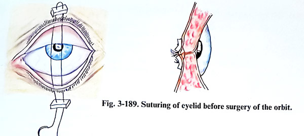
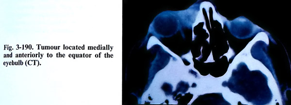
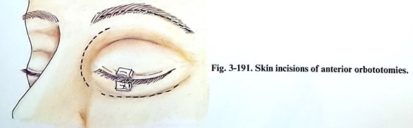
LATERAL EXTRACRANIAL ORBITOTOMY WITH BONE FLAP
This approach is suitable for removing laterally and anteriorly located tumours into the orbital cavity. The skin incision is horizontal and along the superior border of the zygomatic arch. It begins 2 cm from the lateral end of the eye-lid and reaches near to the tragus. There is a second option for the skin incision as is shown in fig. 3-194. The lateral ligament of the eyelids is separated from the bone. The orbicular muscle is detached from the lateral orbital edge and the neighbouring area. The periosteum is cut near to the border of the orbital entrance and after that the periorbit is separated from the bone of the lateral orbital wall and adjacent surfaces of the orbital roof and floor. The lateral rim and the wall of the orbit are cut in two lines: the first a little superior to the zygomatico-frontal suture, and the other at the level of the superior border of the zygomatic arch. The flap is fractured across its base and the bone piece is preserved in saline. The opening of the orbit can be enlarged, nibbling additionally the greater wing of the sphenoid. The periorbit is opened in a way suitable for manipulation inside the orbital cavity. At the end of surgery the bone flap is fixed in its place by wiring or small metallic osteosynthetic plates (Fig. 3-195).
Complications: Extensive orbital oedema can complicate the postoperative period. In such cases corticosteroids are applied. They are continued according to the response to the treatment. Postoperative infection is rare. Visual impairment or loss is also rare and is due to optic nerve ischaemia associated with occlusion of posterior ciliary arteries or the central retinal artery. Ocular movements are affected consequent to the retraction of the intraorbital (extraocular) muscles and almost always this deficit is temporary. Postoperative enophthalmos due to atrophy of orbital fat is common. but usually is not a cosmetic defect of significance.
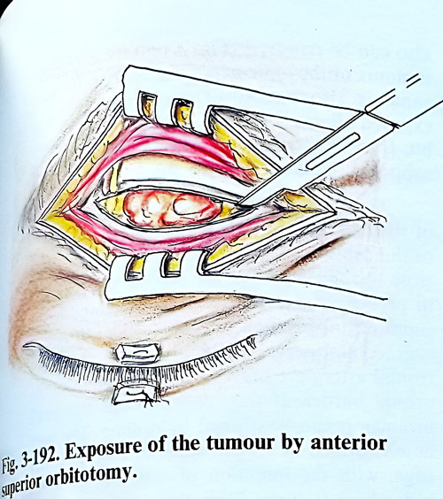
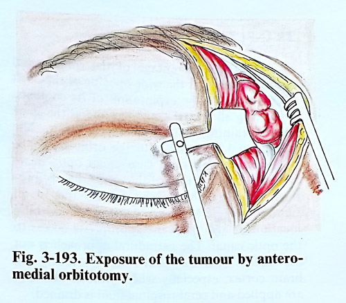
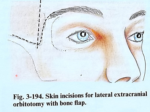
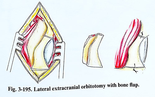
TRANSCRANIAL APPROACHES
The main advantage of the transcranial approaches is that the wide opening of the orbit makes possible the exploration of its contents with preservation of all orbital structures and at the same time obtaining a good cosmetic effect. The transcranial approaches to the orbit are to be preferred, when it is a case of an optic nerve tumour, meningiomas of the orbit, neurofibromas, osteomas or encephalocele, and in all tumours, where there are data of cranioorbital location. It also gives an appropriate access to the optic canal. The extradural approach is not associated with a significant risk of injuring the brain cortex, especially when osmotic diuretics are applied and CSF is drained.
Transfrontal approach. The craniotomy is similar to the described in the approaches to the pituitary region. When it is a case of a tumour that occupies only the orbital cavity, the dura is detached from the orbital roof up to the base of the anterior clinoid process, and is not opened. A burr hole is made in the orbital roof and it is enlarged with a bone nibbler. The excision of the orbital roof anteriorly depends on the size of the frontal sinus. Backward the orbital roof can be resected to the edge of the lesser wing of the sphenoid and the superior orbital fissure can be opened if needed. At the same time a large part of the lateral orbital wall also can be removed (Fig. 3-196). In cases with tumours of the optic nerve, the roof of the optic canal is carefully drilled out. The orbital content is exposed after a Y-shaped incision of the periorbit. If an intracranial extension of the tumour is supposed, the dura is also opened.
The frontal flap can be enlarged with part of the orbital roof, gaining additional exposure to the orbit. Two burr holes drilled close to the skull base are placed very precisely in predefined positions. The medial one has a supraorbital placement, medial to the exit of the supraorbital nerve. The supraorbital nerve and frontal artery are previously separated from the orbital edge. If they pass through a small foramen, it can be opened by drilling. This burr hole is drilled at about 1,5 cm superior to the orbital edge, with the intention of exposing the dura just where it turns from the convexity to the base over the orbital roof. The exposed dura is retracted and the orbital roof - perforated. The orbital edge is divided at that location (Fig. 3-197). The second burr hole is in the most anterior part of the temporal fossa and should coincide with the orbital roof in a way that after being drilled, the anterior cranial fossa and the orbit are opened simultaneously. Through this burr hole the orbital roof is additionally nibbled. The periorbit is separated from the inferior surface of the orbital roof and the flap fractured along a line on the roof, connecting the burr holes.
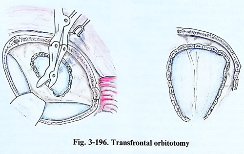
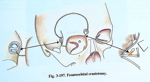
Orbitotomy with two bone flaps. After a craniotomy in the frontotemporal region, a second bone flap from the orbital roof and its lateral wall is lifted, together with the orbital edge. The incision of epicranial tissue is bitemporal. The flap is separated until the lateral and the major part of the superior orbital edge is exposed together with a part of the superior edge of the zygomatic arch. The second bone flap consists of the orbital roof and the lateral wall of the orbit. This is done in the following way: the anterior part of the superior edge of the zygomatic arch - where it forms an angle with the zygomatic process of the maxilla, is dissected through an incision of the periosteum. Next sliding with a fine periosteal elevator on the bone surface of the temporal fossa is reached the inferior orbital fissure. Separating under the periorbit inside the orbital cavity, the inferior orbital fissure is reached through the orbital cavity. With a curved guide needle or hook a thick thread is inserted for passing a Gigly saw through the fissure. With the saw the lateral orbital wall is cut as low as possible, usually at the level of the superior margin of the zygomatic arch. The same bone division can be done easily with a vibrating saw (Fig. 3-198).
Next the frontobasal dura is separated from the orbital roof and is retracted until the lesser wing of the sphenoid is exposed. Behind the lesser wing edge the superior orbital fissure is penetrated. Under the orbital roof, the periorbit is separated, beginning from the orbital edge. A Gigli saw is introduced under the superior orbital edge until it comes out from the superior orbital fissure. The orbital roof is transected together with the orbital edge in the sagittal plane. This incision is best done just lateral to the exit of the supraorbital nerve or medially if the nerve is separated from the bone foramen. This bone incision can be made easily and safer now by the vibrating saw. These two incisions separate a large part of the orbital roof and the whole lateral orbital wall, and they remain fixed to the cranial base only by the greater wing of the sphenoid between the superior and inferior orbital fissures. This part of the bone is nibbled partially beginning at the edge of the craniotomy and reaching in a basal direction as close as possible to the inferior orbital fissure. With a slight effort the second bone flap base is fractured. It consists of the whole lateral wall of the orbit and a large part of the orbital roof, fixed to a part of the temporal muscle. It can be also separated from the muscle as a free bone flap. If necessary, the optic canal is also opened, removing its superior wall. When the work inside the cranial and orbital cavities is completed, both flaps are repositioned and fixed by wiring or metallic osteosynthetic plates.
Comparing it with other methods, this operative approach provides probably the widest operative field, both for the cranial and orbital cavities and does not cause any bone defect. It is suitable for removal of deeply located complex orbital and cranioorbital tumours (Fig.3-199).
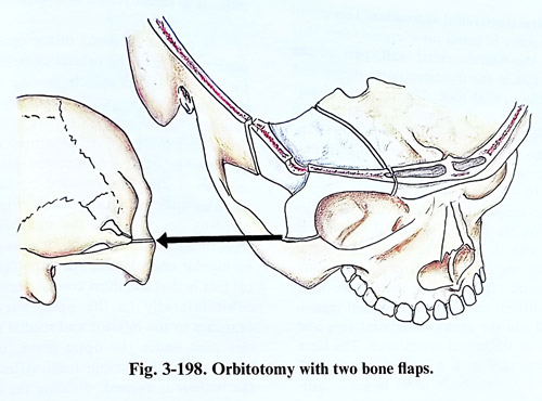
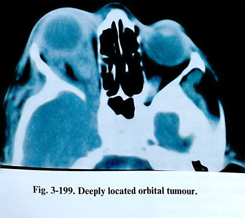
Lateral transcranial approaches. This group of approaches is based on a craniectomy that includes the lateral orbital wall, part of the cranial vault in the frontotemporal region and part of the orbital roof. The incision of soft tissues is done in the temporal region. It is a vertical and slightly curved incision, 2 or 3 cm behind the zygomatic process of the frontal bone and behind the hairline (Fig. 3-200). Its length is 10 - 12 cm and reaches the zygomatic arch. The temporal muscle is divided at its insertion on the superior temporal line and is detached with the periosteum from the lateral orbital wall, the frontobasal area and the anterior temporal region. After that, three burr holes are drilled: one in the frontobasal region, the second - in the anterior temporal area and the third - on the lateral orbital wall. The bone defect is enlarged with a nibbler merging all three burr holes. In order to get a sufficiently wide operative field, the lateral part of the orbital roof is removed also with opening of the superior orbital fissure (Fig. 3-201). This operative approach offers an adequate operative field upon broad areas of the lateral parts the orbital cavity. It is suitable for laterally located orbital tumours (Fig. 3-202). In the case of a cranioorbital tumour however, it is rather narrow for reaching its intracranial part. Besides this, a permanent cranial and orbital defect is produced.
Technique of orbital tumour removal. After opening the periorbit, the orbital tumour is located by palpation or intraoperative ultrasound. When the tumour's position is defined, dissection is carried out directly to it with the help of two or three fine self-retaining retractors.
The surgical technique of tumour removal is different for the neurosurgeon accustomed to work inside the cranial cavity. The dissection of intraorbital structures is often obstructed by the intraorbital fatty tissue coming out under pressure. Because of that the wide opening of the orbit, independently of the microsurgical technical advantages, is better and less traumatic to the intraorbital structures. After opening of the periorbit the first branch of the trigeminal nerve that lies on the levator and the superior rectus muscles is identified. The fourth nerve is more difficult to identify, as it is a very tiny structure.
In case of a glioma of the optic nerve, the penetration into the orbital content is between the medial rectus muscle on one side and the levator palpebrae and superior rectus muscle laterally on the other. The upper group of muscles must be well-separated as proximal as possible, and are retracted laterally. The annulus of Zinn is transected medially to them and the optic canal is opened (Fig. 3-203). If bleeding occurs from the ophthalmic artery small branches in this area, it is easily stopped by bipolar coagulation. An important anatomical fact is that the third cranial nerve enters the orbit laterally to the optic nerve and its branches to the inferior and medial rectus muscles pass under the optic nerve, or in other words under the tumour itself. After dissection, the tumour is excised, dividing the optic nerve anteriorly to the chiasm in unaffected parts of the nerve and behind the eye ball (Fig. 3-204; 3-205).
Optic nerve sheath meningiomas with certain visual acuity preserved are excised with extreme care, preventing damage of the nerve fibres. Lesser sphenoid wing meningiomas with cranioorbital extension are usually easier to remove. The orbital parts of the tumour are situated in the superior lateral quadrant of the orbit and are resected without any difficulties if the attachment area is small. The infiltration of the orbital walls and the greater wing of the sphenoid requires wide bone excision and meningiomas 'en plaque' cannot be removed completely.
Tumours of the lacrimal gland are common and they have a specific location. The lateral transcranial approach is the most suitable for their removal. The tumour is removed 'in one piece' with the gland, the periorbit, and the neighbouring tissue. If the clinical evolution of the tumour is less than 6 months, the tumour may be malignant. In such a case the prognosis is poor and after the biopsy result is confirmed, a more extensive excision is required. In some cases complete exenteration of the orbit with excision of the orbital roof and lateral wall are required.
Complications. During the postoperative period, besides the disturbances and complications usual for intracranial operations, oedema of the eyelids is almost always observed, associated with blood effusions on the skin. This oedema continues for few days, during which time the eyelid suture must not be removed.
Infections of the orbital contents and osteomyelitis are rare. Ptosis and limitation of eye movements are frequent and temporary. Decrease in the visual acuity is a serious complication, and can result from damage of the central retinal and/or posterior ciliary arteries. A permanent complication is slight enophthalmos that remains after removal of bigger intraorbital tumours.
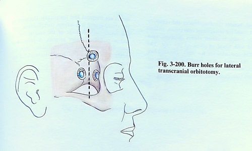
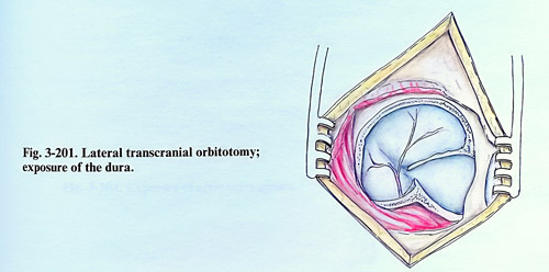
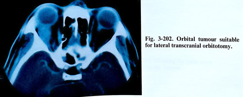
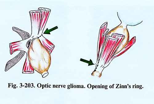
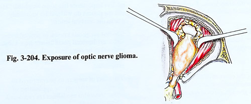
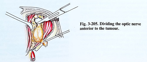
METASTASES
Only a small number of those patients with intracranial metastases come to the attention of the neurosurgeon. The most common sources of metastases of the brain are tumours of the lung, breast, kidney, and malignant melanomas. Other sources are relatively infrequent, such as tumours of the gastrointestinal tract, thyroid, uterus, ovary, pancreas, prostate, and sarcomas. About the half of the patients have neurological symptoms or signs as the first manifestation of malignant disease. The primary origin of the cancer is usually discovered later, but occasionally the primary tumour cannot be identified, even at autopsy. More than the half the patients in whom the primary tumour is unknown initially are subsequently found to have bronchial carcinoma. There is some predilection towards the finding a metastasis in the middle cerebral artery territory and the cerebellar hemisphere.
A metastasis is discovered either accidentally as being the cause of detected neurological symptoms and signs or as part of the total diagnostic work up of the patient with established malignancy. In the first case, there are some additional goals of surgery.
The differences are as follows:
Accidentally discovered -
Need of a biopsy to obtain histological proof of the lesion.
Symptoms/signs usually due to the brain lesion. Expected relief if removed.
Expected survival cannot be estimated before biopsy; removal preferred.
As the effect of other treatment modalities is unknown, preference is given to surgical removal.
In an established malignant disease -
Diagnosis established.
Morbidity due not only to brain lesion and its relative importance has to be weighted. "How will the patient do without his morbidity due to the cerebral lesion?
Survival prognosis known in general terms.
Expected morbidity and benefit from surgical removal to be compared to the natural course of the disease, and the benefits to be established.
The effect of surgery to be compared to those expected with optimal protocols, including the application of other treatment modalities (as radio-and chemotherapy).
The examination of the patient suspected of having brain metastasis should assess his neurological status and determine the degree of systemic cancer involvement. This cannot exceed the reasonable time for general assessment, especially when the patient is deteriorating neurologically. Neuroimaging studies are essential and must include high-resolution cerebral CT, supplemented by MRI to detect lesions that might go unnoticed by a CT and evaluation of the degree of brain oedema. Incidental, circumscribed nodular lesion with central necrosis can be supposed to be a metastasis. Contrast-enhanced MRI is the best study to detect small metastases and to confirm whether a metastatic lesion is truly solitary (Figs. 3-206; 3-207).
Selection of patients for surgery. The objectives in the treatment of metastases are achieved following the main rules for the management of an intracranial lesion. Modifications to these rules can be imposed as a result of the multiplicity of the lesion, its rapid and fatal evolution, and when at some stage the general condition of the patient is seriously affected.
There is consensus among neurosurgeons that surgical excision should be restricted to patients with a solitary intracerebral metastasis whose general medical status is satisfactory. The opinions are controversial concerning excision of an incapacitating metastasis even when it is known that the tumour is not solitary, or for the
metastases that are multiple, but surgically accessible. In those metastases located deep within the dominant hemisphere and the brain stem the removal can provoke very serious deficit and as a rule they are considered surgically intractable.
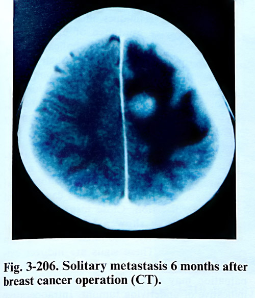
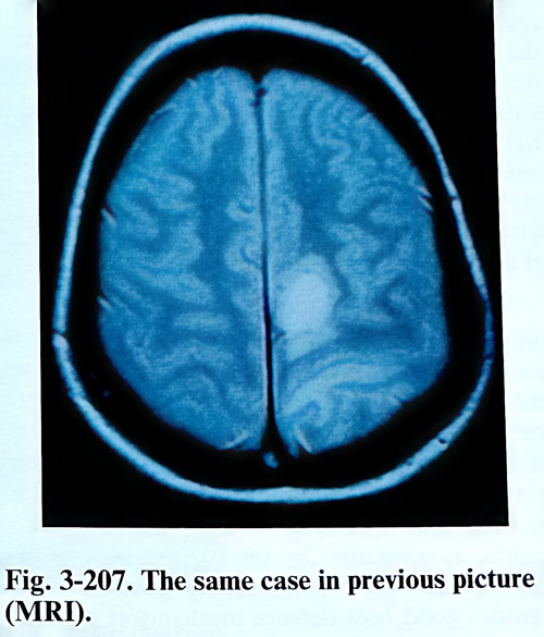
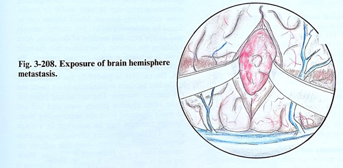
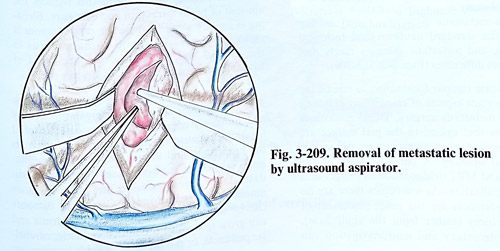
External stereotactic radiosurgery can be advised for patients with deeply located and multiple metastases, who are otherwise in good general condition and with good life expectancy according to the type of disease.
The radio- and chemosensitivity of the tumour may play an important role in the decision regarding surgery. Patients with a radioresistant tumour, such as renal cancer or melanomas are more likely than other patients to be offered surgical excision with or without postoperative radiotherapy. Surgery may also be favoured in patients with slow-growing tumours, as suggested by the natural history of the disease or by long latency because that signifies good host defence mechanisms against the primary tumour.
Surgical technique. The surgical technique depends on the aim of the treatment: removal of samples for biopsy or attempt to excise the metastatic tumour. Biopsy alone can establish a histological diagnosis without therapeutic benefit. Only if the therapy after histological diagnosis is radiation and chemotherapy, biopsy can be a satisfactory initial procedure.
Biopsy. Currently, in properly selected patients, image guided stereotactic biopsy is an accurate, highly diagnostic, and low morbidity method to obtain histological confirmation of the tumour type.
Craniotomy and excision. The surgical excision of a metastatic tumour is performed according the standard neurosurgical technical principles, and metastatic tumours rarely present serious difficulties (Figs. 3-208; 3-209).
Accurate tumour localisation is one of the most important aspects of craniotomy planning for brain metastasis surgery. Dural metastases of tumours that extend to the pial surface are uncommon and most metastases are not visible after dural opening. A detailed review of the axial CT and MRI (tridimentional) images aids tumour localisation. As sometimes there are no external references for the exact placement of the craniotomy (rather from the skull base), frameless stereotaxy and neuronavigation can be a useful technique for precise approach. Stereotactic techniques offer also an alternative for more precise guidance of craniotomy. Stereotactic guidance is best for small subcortical tumours that otherwise are occasionally impossible to find during craniotomy. Although stereotactic frame application and intraoperative CT scanning may slightly prolong the time of surgery, it can completely eliminate the problem of finding the tumour.
When localising techniques are not available, the neurosurgeon is confronted by a normal brain surface and a hidden subcortical tumour, and several practical tips can be used for tumour detection. Close inspection of the pial surface may show a localised area of increased vascularity, arterialized veins, or an expanded gyrus. These signs are indirect evidence of an underlying neoplasm. Gentle finger palpation may detect a subcortical firmness, indicative of underlying tumour. Intraoperative ultrasonography can be very useful to localise some subcortical lesions. Many lesions can be found by ultrasonography, but smaller tumours may evade even this method of detection. After localising the tumour, penetration into the brain tissue is through a small cortical incision. We prefer penetration through the depth of a sulcus. The excision is guided by the identification of the line between oedematous brain tissue and the tumour margin. The metastatic brain tumour often appears grossly separated and can be easily dissected from the surrounding brain.
Smaller tumours can be removed in a single piece, and that can be attempted for large tumours to reduce dissemination. Bleeding is usually minimal once the entire tumour is removed. Careful attention to haemostasis in the tumour bed will reduce the likelihood of haematoma formation in the operative site.
Operations for posterior fossa metastatic tumours have the tendency for greater morbidity and mortality compared to most supratentorial locations because of the tumour's proximity to the brain stem and the tendency to develop hydrocephalus. Tumours of the brain stem are rarely candidates for surgical excision and may be more amenable to radiosurgery. Cerebellar tumours can be located in the hemisphere or vermis and can grow to involve the fourth ventricle, cerebellar peduncle, or the brain stem. Such tumours are usually intraparenchymal and do not often grow primarily in the subarachnoid cisterns or extend through the tentorium (Fig. 3-210).
A generous, rather than a limited suboccipital craniectomy (with opening of the foramen magnum) aids in midline tumour exposure and in providing tonsilar decompression in the event of tumour recurrence or increased intracranial pressure. Because of the poor long-term survival and the probability of local recurrence, craniotomy (as opposed to craniectomy) has little place in metastatic tumour surgery in the posterior fossa (Fig. 3-211).
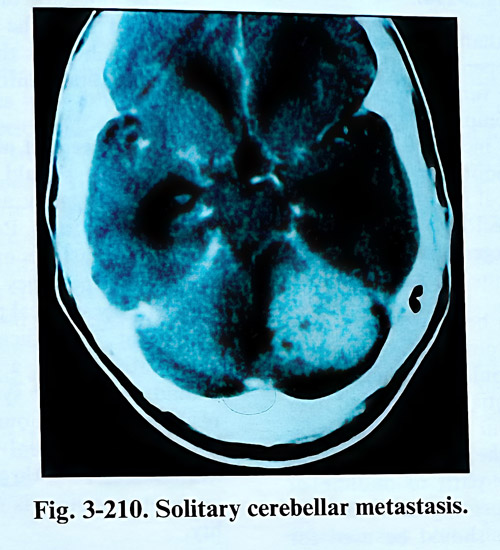
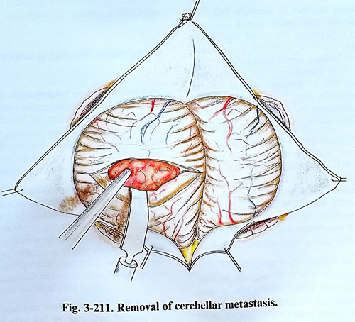
Lateral tumours can be approached via a lateral suboccipital or retromastoid exposure. A straight, direct route should be taken to the tumour to minimise potential neurological deficits associated with tumour excision. Coagulation and incision into the cerebellar vermis provide a low morbidity path to midline or paramedian lesions. A direct approach through the cerebellar hemisphere should be used for
more lateral tumours. The methods of resection further on are the routine ones.
The presence of hydrocephalus should be recognised from the preoperative imaging studies. In such cases the craniectomy is preceded by an occipital ventricular puncture through a burr hole. The placement of a shunt (usually ventriculoperitoneal should be indicated considering the life expectancy and the possible benefit of relief of symptoms. Patients who have advanced systemic metastases or multiple brain metastases and a life expectancy of 1 month or less do not warrant any surgical procedure, including shunt placement. The indication for a shunt operation in a patient with a metastatic brain tumour should be to treat symptomatic hydrocephalus resulting from tumour not amenable to surgical excision. Peritoneal seeding from malignant intracranial cells through the ventriculoperitoneal shunt system is only a theoretical possibility.