TUMOURS OF THIRD VENTRICLE
The most common tumours occupying the cavity of the third ventricle are those arising from its walls (astrocytomas of all grades, ependymomas), from the choroid plexus (papillomas, meningiomas, colloid cysts), and others with ectopic development or arising as secondary invasion from the skull base as germinomas, craniopharyngiomas, pituitary adenomas.
The third ventricle can be penetrated from three directions: through the foramen of Monro, enlarging the access through the velum interpositum, through the lamina terminalis especially when it is distended or invaded, and between the fornici at an extent of 20 millimetres. Which path to chose depends on the origin (attachment) and the type of the tumour.
For taking a biopsy or evacuation of cysts there are options of 'minimal invasive' techniques. The preference for stereotactic surgery in the past has been recently substituted by the alternative of neuroendoscopy.
Preoperative radiological investigations are directed towards the establishment of the exact size of the tumour and its relationships with surrounding structures. They must define as clearly as possible the following two characteristics: 1. Does the tumour have an origin and attachment inside or outside the third ventricle? 2. Is it a case of an intra- or extraaxial tumour?
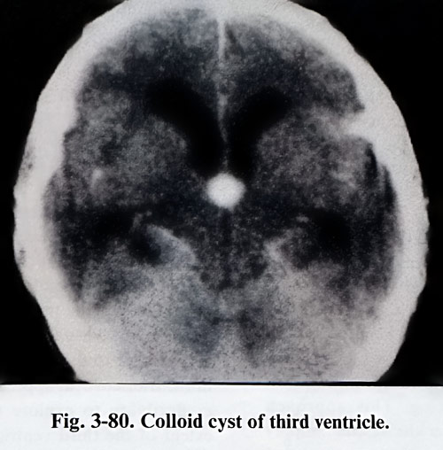
The solution can be found through the complete imaging of the structures by CT and MRI in axial, coronal and sagittal projections. The lesion must be described with respect to size, texture and which structures are bordering, with the added intention to detect its place of origin (Fig. 3-80). The presence of occlusions, disruptions of the walls, isolation of parts of the ventricle (with the dilatation that follows) as well as any perifocal brain oedema must also be detected. An angiographic study must demonstrate the vascularity and blood supply of the lesion, its relationship with the choroid plexus and the deep venous collectors, and for the specific requirements of the approach through the cortex or corpus callosum - the cortical venous drainage to the superior sagittal sinus.
Taking into consideration the characteristics of third ventricle space occupying lesions, with respect to the choice of a corridor for its removal, the lesions can be subdivided into three main categories. The first consists of extra-axial intraventricular lesions, the second – of intraaxial lesions with an intraventricular component, and the third - of basal lesions, with secondary penetration into the third ventricle.
The extraaxial intraventricular lesions are usually benign (colloids cysts, craniopharyngeomas, dermoids, teratomas, papillomas and others). They have well defined borders and are not so adherent to the wall of the ventricle's borders. On imaging studies they are surrounded by CSF, except for the area of attachment. Their margins are usually smooth. Intraaxial lesions with growth inside the third ventricle are most often gliomas, but metastatic tumours – medulloblastomas, germinomas - can have similar characteristics. The MRI and CT investigations detect a significant part with an intraaxial loca-tion. For the needs of the surgical strategy it is essential to discern which structures are affected. Basal tumours penetrate or only elevate the walls of the third ventricle. The majority of them are benign and are potentially excisable.
Those which disrupt the third ventricular wall penetrating from outside can be visualised through the foramen of Monro. The primarily basal origin of the lesion must be detected on the imaging studies. Very often bone changes are seen on the skull X-ray films and CT images. The integrity of the structures forming the inferior wall of the third ventricle must be reviewed thoroughly on the sagittal MRI images, as their affection can indicate penetration of the lesion inside the ventricular cavity.
The indications to the three most accepted paths for entry to the third ventricular cavity can be more precise after the lesion has been classified within one of the three categories mentioned above.
Extraaxial intraventricular tumours require adequate exposure of the ventricle before removing part of the lesion and their nature can very often permit total excision. The widest access to different areas of the ventricular cavity is provided by interforniceal or the transforaminal-transvelum interpositum approaches. Intraaxial lesions with secondary penetration into the ventricular cavity require access not only to the ventricle, but also to the primary site (origin) of the tumour. The involvement of the hypothalamus, thalamus, fornix or other structure will determine which path should be followed, but the surgeon should be able to take a biopsy or partially remove the lesion at least when reaching the ventricular cavity. Any one of the three paths can be chosen. Basal tumours always require a basal approach for the access to the ventricle. Any removal of the basal portion of the tumour preceding the approach to the ventricle provides histopathological proof and better access after debulking. This approach permits better control over the vascular supply of the tumour. If removal through the basal approach and lamina terminalis is not expected to be satisfactory, an additional option can be discussed for the same or another surgical session by one of the superior paths. The craniotomy is preplanned according to these needs.
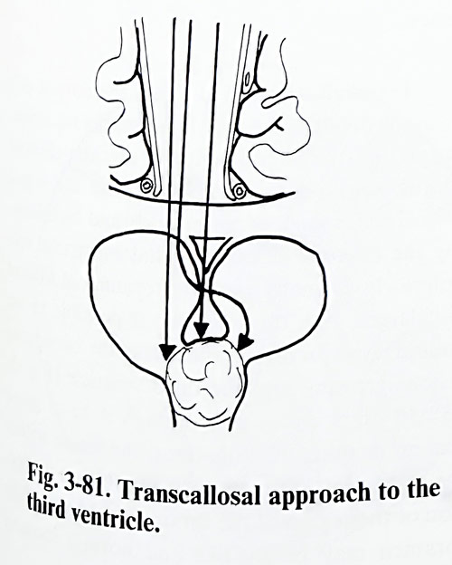
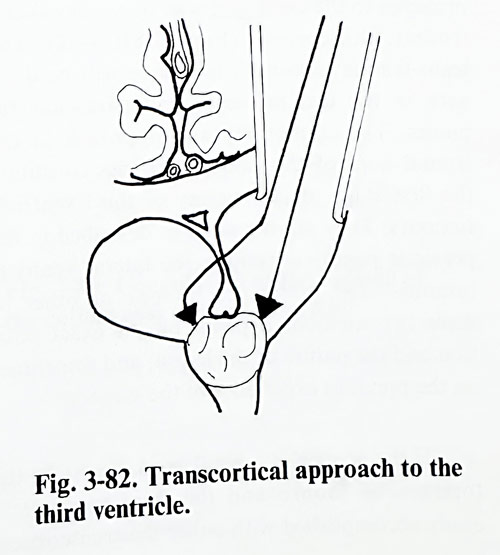
We will present two of the main approaches to the third ventricle: transcallosal and frontal transcortical (Figs. 3-81; 3-82). The trans-lamina terminalis technique will be dealt with in the text on sellar and parasellar tumours. The craniotomy and approach to the frontal horn of the lateral ventricles constitute the first stage of the surgery of third ventricle tumours. They are the same as described in the previous pages concerning the lateral ventricle tumours. The preference for one or other of these two methods depends on the exact position and the nature of the lesion, and sometimes on the previous experience of the surgeon.
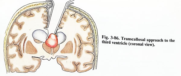
If the ventricles are enlarged, access to the foramen of Monro and the third ventricle is easily accomplished with either the transcortical or transcallosal approach. Access to both sides of the third ventricle is obtainable with either exposure, but because of the angle of vision the view of the ipsilateral portion of large lesions can be limited using the transcortical route. The line of vision to the depth of the anterior third ventricle is better with a transcallosal method
Indeed, if the ventricles are small, the transcallosal approach is certainly the better one. The use of the transcortical approach to the ventricle without hydrocephalus requires the disruption of a large amount of cortex and white matter, and the retraction can be difficult. The route also significantly limits mobility if departure from the initial plane of entry into the lateral ventricle is needed. The advantages of the transcallosal approach to the third ventricle are that the anatomy is constant, the distance to the third ventricle is shorter than in the transcortical approach, and there is greater flexibility to explore the anterior-posterior extent of the third ventricle with no disruption of hemisphere tissue, because no cortical incision is necessary. There is excellent unobstructed vision to the depth of the anterior third ventricle, and ventricular size is irrelevant. The advantages of the transcortical exposure are that there is less chance of compromising an essential draining veins going to the sagittal sinus or causing injury to the pericallosal arteries.
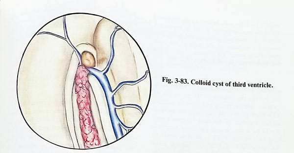
Transcallosal method. Once the corpus callosum is divided, the usual landmarks for orientation into the lateral ventricle are: the choroid plexus, the thalamostriate vein, and the septal vein. The foramen of Monro is found by following the choroid plexus and thalamostriate vein anteriorly. In many cases the foramen of Monro is enlarged and, through it, it is possible to see colloid cysts or other similar space occupying lesions growing into the third ventricle (Fig. 3-83). In other cases, even though the size on imaging investigations was apparently big enough, the lesion within the third ventricle may not be visible on initial inspection of the region of the foramen of Monto. The foramen may be slit-like and normal looking and the mass not seen. In such a case, the closed tip of the bipolar forceps should be introduced into the foramen and gently opened, acting as a temporary retractor on the walls of the foramen. That permits the surgeon to see further into the anterior part of the third ventricle.
When the colloid cyst or tumour is reasonably well seen, an effort should be made, working through the foramen of Monro, to free its anterior, inferior, lateral and posterior surfaces with blunt probes. At this stage the superior surface attached to the third ventricle's roof should be intact because the blood supply is from that surface, which is attached firmly to the ventricular roof. Unless the mass is small and has a tendency to deliver out itself, it is unwise to attempt removal of the lesion intact wothout some volume reduction because the pedicle may be disrupted and bleeding may occur from the roof of the third ventricle, unseen and unsuspected until the third ventricle is filled with blood. The mass is penetrated with a spinal needle and aspiration may then be made.
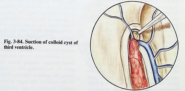
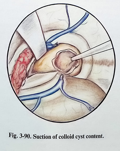
If a cyst content is being aspirated, the capsule shrinks simultaneously with the appearance of fluid in the syringe. If the material is too thick for aspiration through the needle, a larger needle or even a small suction tube may be used (Fig. 3-84). If the cyst content is semisolid, a crosswise incision into the wall allows the use of a small suction tip and small ring curettes to complete the evacuation.
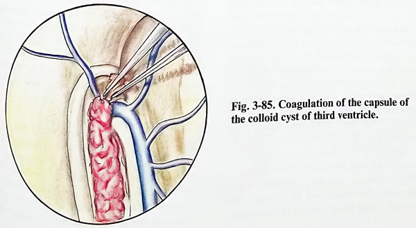
It should never be pushed on the cyst, beacuse of the risk of displacing it posteriorly into the third ventricle and losing sight of it. The complete evacuation of the cyst permits delivery of the capsule by pulling on it gently from various sites around the opening already made through the foramen of Monro. The pedicle will be the last part to be seen as it passes through the foramen of Monro which is then coagulated and divided (Fig. 3-85). If bleeding occurs within the foramen of Monro, coagulation should be used cautiously avoiding obstruction of the thalamostriate and internal cerebral veins. If bleeding continues, one may introduce a soft catheter into the foramen of Monro and then slide down a very small piece of oxidised cellulose between the catheter and the bleeding. point, the catheter serving as a support against the wounded vein. The catheter end is taken outside the wound and removed after 12 to 24 hours.
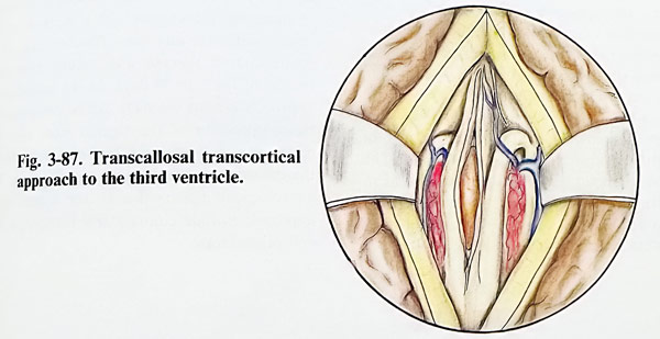
In many cases the pathological formation may be not visible through the foramen of Monro, or it will be difficult to manage the lesion through the foramen. In such a case the interforniccal approach may be indicated (Figs. 3-86; 3-87). After the clear identification of the anatomical structures in the exposed intraventricular cavity, bipolar coagulation forceps are used to fenestrate the septum pellucidum (septostomy), creating a single ventricular cavity and facilitate a possible shunting procedure in future.
The septum serves as a landmark for guidance to the midline union of the forniceal columns and the forniceal body. The raphe is identified at the site of the septum's attachment on the dorsal fornix. The incision starts at the level of the foramen of Monro with fine tipped bipolar forceps, and is carried posteriorly for 1,5 to 2 cm. The size and shape of the fornices in the midline are variable and are influenced by individual deformation by the pathological mass.
Good preoperative imaging studies help with proper orientation during the operative exposure. With completion of the interforniceal incision, the mass is identified. The normal structures in the diencephalic roof including the tela chorioidea, choroid plexus, internal cerebral veins, and posterior choroidal arteries are often thinner or displaced laterally by the presence of the mass. Retraction at the level of the fornix is often not necessary, or a very narrow retractor with minimum pressure may be used to maintain exposure either initially or after mass decompression is partially completed.
Once internal decompression has been achieved, lateral portions of the mass are separated gently from the third ventricle's wall. With lateral dissection, the internal cerebral veins may be identified and followed anteriorly to the foramen of Monro. The choroid plexus is often intimately adherent to the thalamostriate veins.
Finally, the posterior component of the mass is approached. For this, sometimes it is necessary to change the position of the microscope. With the mass excision extending from the third ventricle to the dorsum sellae, it is not unusual to visualise the clivus, basilar artery and branches, and prepontine cistern at the completion of lesion excision.
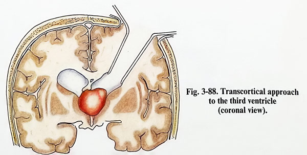
Frontal transcortical approach. The difference from the previous approach is that the foramen of Monro is approached at an angle to the sagittal plane (Fig. 3-88).
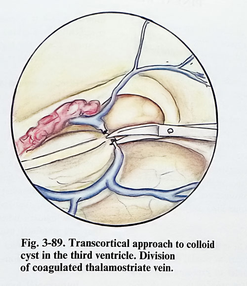
To enlarge the access to the third ventricle cavity, this approach can be expanded with trans-vellum interpositum separation. The choroid plexus near to the foramen of Monro can be elevated slightly and thalmostriate vein coagulated and divided from the internal cerebral vein (Fig. 3-89). The leptomeninges of the velum interpositum are detached from the foramen of Monro posteriorly about 1,5 to 2 cm. The medially inserted retractor is advanced so as to slightly dislocate the fornix with the choroid plexus and internal cerebral vein medially and the lateral retractor slightly depressing the thalamus. The tumour can then be approached between the internal cerebral vein and the thalamus using standard microsurgical instrumentation (Fig. 3-90).
Complications. Most of the complications seen with operation on a lesion expanding into the third ventricle have been related to the location and nature of the primary lesion rather than to the approach. Diabetes insipidus and akinetic mutism can be seen transiently. Aseptic meningitis has occurred in approximately 15% of patients who have undergone intraventricular operations. The signs of aseptic meningitis usually become apparent when corticosteroids are stopped. The CSF shows changes consisting of moderate pleocytosis with a decrease in the glucose content. Patients respond to corticosteroid therapy and require continued treatment for 5 to 30 days. In the transcallosal approach venous drainage can be compromised by compression of the sagittal sinus with the retractor or infarction of the frontal lobe due to the division of a significant draining vein. The neurological deficits due to the transcortical approach include contralateral hemiparesis and epileptic attacks.