TUMOURS OF CEREBELLUM AND FOURTH VENTRICLE
The tumours of the cerebellum and fourth ventricle have clear age related incidences. Infratentorial tumours in children are three times more frequent than those in the supratentorial space. In adult patients this relation is vice versa. The most common tumours in children are meduloblastomas, astrocytomas, and ependymomas. These types of tumours are rare in the adult patient and at the expense of predominant astrocytomas, metastases and haemangiomas.
There are several diagnostic features to be established before deciding the surgical technique. They are the location of the lesion regarding the midline (vermis) and the IVth ventricle, the type of displacement and involvement of the brain stem, the presence of hydrocephalus, the degree of vascularity, the presence of a cystic portion and finally the histopathological prediction for the lesion.
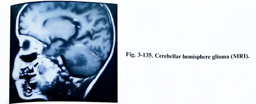
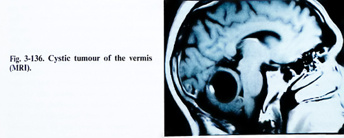
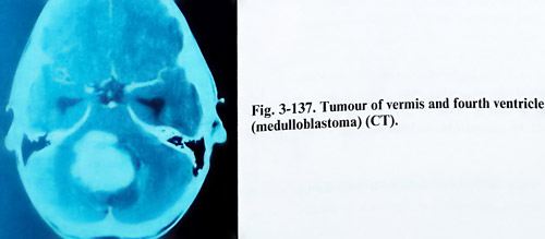
The non-enhanced and enhanced CT is usually the first investigation to do, and it confirms the existence of a posterior fossa mass lesion, and the displacement and obliteration of the fourth ventricle and cisterns. The density and enhancement of the lesion indicate its possible nature, especially the presence of cysts and a highly vascular nodule (astrocytomas and haemangioblastomas). MRI, however, is superior in its diagnostic value. It is able to demonstrate with better resolution the characteristics of the lesion, the patency of the CSF spaces and grey to white matter margins (Figs. 3-135; 3-136; 3-137). Angiography is reserved for the additional need to demonstrate a vascular pathology or highly vascular tumour.
Surgical planning entails considering the possibilities of current surgical techniques. Sub-cortical cerebellar lesions can be approached either by midline vertical incision of vermis or by a limited incision on the middle or lateral part of the hemispheric surface. Limited incisions are also possible on the lateral and (rarely) on the superior hemispheric surfaces. The lateral part of the hemisphere may be excised. Vermian incisions are possible after midline craniectomies or craniotomies and hemispheric ones - after lateral craniectomies or craniotomies. The choice of bone exposure area depends on the desired surface required for incision and manipulation of the tumour. Hydrocephalus, if clinically symptomatic, must be treated as a preliminary step for improvement of the general condition of the patient. Otherwise it can be controlled by an occipital horn puncture at or just before the time of tumour surgery, placing temporary external ventricular drainage. The drainage can be internalised at a later stage, as a permanent shunt should it be needed.
Another important goal of the approach is the decompression of neural structures, especially at the level of the foramen magnum. It is always done when displacement of tonsils and brain stem has taken place. This goal is achieved by removal of a sufficient part of the occipital squama, including the posterior rim of the foramen. In more advanced cases removal of lamina C1, and even C2 parts can be required.
The burr hole for ventricular drainage is placed 4 cm lateral and 6 cm above the inion on the right side. The lateral ventricle is canulated but rapid ventricular drainage should be avoided particularly if the intracranial pressure is significantly elevated, since it can cause the tearing of bridging cortical veins and create a subdural haematoma.
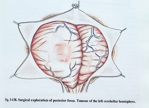
After dura is opened in a Y - shaped configuration, the cerebellar surface is inspected. When the tumour is located superficially and infiltrates the external cerebellar surface, it is seen immediately after opening of the dura. If it is located subcortically in the cerebellar tissue, exploration is made by first inspecting the cerebellar surface. Special attention is paid to the colour and the configuration of cerebellar sulci and the displacement of blood vessels. The cerebellar hemisphere, in which the tumour is located, seems enlarged in volume and causes a larger protrusion toward the craniotomy, than the opposite. Its sulci are broader and the cerebellar surface is smoother. The cerebellar tonsils are situated usually lower in the cisterna magna or are herniated down under the foramen magnum. The displacement of the vermis to one side also suggests the presence of a tumour in the hemisphere, and when it is bulging, a tumour along the cerebellar midline can be expected (Fig. 3-138). In few cases the cerebellar surface may be carefully palpated to get a tactile feeling about its consistency. The vicinity of the tumour has a consistency that may be more dense or softer than the normal and sometimes in cystic tumours can be felt fluctuating. After this, the cerebellar hemispheres are retracted and the cerebellopontine angles are explored. The exploration may be finalised with exposure of the fourth ventricle.
For this purpose the tonsils are separated from one another, until reaching the foramen of Magendie. A fine dissector can be inserted through the opening and a large part of the ventricular cavity is inspected.
Cerebellar astrocytomas may be solid, cystic, and their location in one of the cerebellar hemispheres makes total excision possible (Fig 3-139). The cystic tumour should be punctured first and the mass should be exposed through the cerebellar cortical incision. If the tumour is solid, dissection is carried out around the tumour or it can be debulked with the ultrasonic aspirator. In case of a cystic tumour with a mural nodule, a total excision of the nodule should be carried out, without removal of the wall.
When only one hemisphere is involved by the tumour, a unilateral approach is usually enough for manipulation. The approach was already described, and the size of craniotomy or craniectomy and extent to foramen magnum rim removal decided according to the space required for manipulation and the degree of downward shift of the hemisphere. The dura is usually opened in a stellate fashion, after assuring normalisation of the intracranial pressure.
Direct transdural puncture of a large hemispheric cyst is also acceptable for rapid reduction of the intracranial pressure. After inspection of the hemispheric surface, the craniectomy can be additionally extended after the location of the lesion.
Medulloblastomas are reddish-grey, friable masses that frequently distend the vermis and protrude from the foramen of Magendie. These tumours are easily aspirated, and their removal is carried out by penetrating between the cerebellar tonsils and after vertical incision of the vermis. The subarachnoid space at the foramen magnum is gently packed with cottonoids to reduce the possibility of seeding along the spinal subarachnoid space with tumour cells. Once the foramen of Magendie is identified, the vermis is split to reach the most superficial layer of the tumour. Tumour removal can be accomplished by gentle ordinary or ultrasonic suction and bipolar coagulation or laser evaporation.
Through all debulking, the surgeon should be aware of the position of the dorsal brain stem surface. Initially the obex is found following the dorsal columns, and later - the distorted inferior cerebellar peduncles and the IVth ventricular floor. Having these structures in view and then covered for protection, but not packed with pressure, will provide safe debulking and removal. Tumour removal is continued so that the lateral recesses of the fourth ventricle and aqueduct of Sylvius are visualised. It may not be possible to remove those portions of the tumour which are adherent to the floor of the ventricle. In many cases, the infiltrative growth of these tumours does not permit complete removal of some small portions.
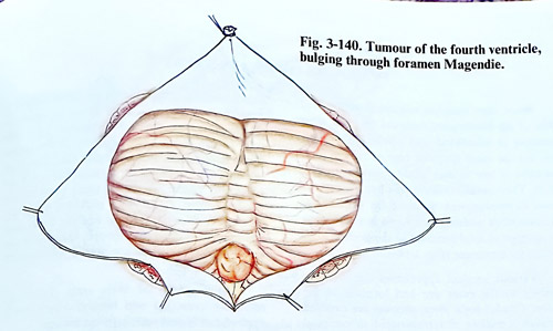
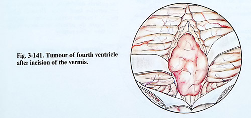
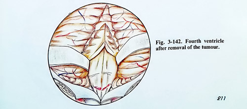
Ependymomas usually grow into the fourth ventricle and bulge out of the foramen Magendie, and extend along the medulla oblongata and upper cervical cord to at least the level of C1 (Fig. 3-140). After opening the cisterna magna, the tumour can be grasped and lifted up to the obex, exposing the floor of the fourth ventricle. At this point is possible to see whether the tumour is adherent to the floor of the fourth ventricle or can be freed from it (Figs. 3-141; 3-142). A cottonoid is slipped between the tumour and the floor of the fourth ventricle. The cerebellar vermis is then split, and with the aid of the ultrasonic aspirator, debulking of the tumour begins. In some cases fourth ventricle ependymomas can go out laterally through one foramen Luschka into the cerebellopontine angle and care is needed to dissect well the involved cranial nerves. The complete removal of these tumours is not usually possible because frequently they arise in the region of the hypoglossal and vagal triangle and are intimately adherent to the underlying tissue of the brain stem.
A variety of cystic masses may be encountered in the midline or near to the midline. They are symptomatic as a result of direct brain stem and cerebellar compression, and obstructive hydrocephalus. Such a cystic lesion should be fully exposed, and the cyst wall carefully examined.
The removal of choroid plexus papillomas from the fourth ventricle is relatively easy, as they are not invasive and can be excised with the bipolar coagulations and the ordinary suction or ultrasonic aspirator. The removal of the tumour attachment in proximity to the foramen of Magendie and from the fourth ventricular floor must be performed very carefully.
Cerebellar haemangiomas/ haemagioblastomas are vascular tumours often located in the cerebellar hemispheres. In 60% of the cases the mass exists as a cystic tumour with a mural nodule. Complete removal of the nodule is curative. The arterial feeders should be easily accessible and be interrupted before an attempt is made to remove tumour tissue. In a case of a very large, solid haemagioblastoma that cannot be removed as a single mass, the surgeon should attempt to interrupt the tumour's arterial supply before trying to remove the lesion.
Dermoid cysts located in the posterior fossa typically arise near the midline. These lesions are present from birth, although they may be not symptomatic until adult life, they grow slowly into a cyst as sebaceous material and desquamated epithelium accumulate inside.
Dermoids and epidermoids should be dissected free of surrounding cerebellum. Care must be taken not to spread the contents of the tumour liquid, because this may provoke irritation and give rise to a chemical meningitis and severe adhesive arachnoiditis.