TUMOURS INVOLVING THE CAVERNOUS SINUS
The tumours of the foramen magnum and craniovertebral junction occupy space in the posterior fossa and the spinal canal. Mostly these lesions are extracerebral tumours such as neurinomas, arising from the intracranial part of the cranial nerves and the upper cervical roots or meningiomas, attached to the mid- or lower clivus, foramen magnum or adjacent dura. In addition to these locations, technical difficulties of approach to these lesions are related to the jugular and hypoglossal foramina with the related to them neural and vascular strucrtures, and also the foramen magnum parts located anteriorly and/or laterally to the cord.
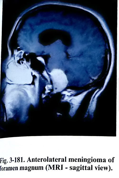
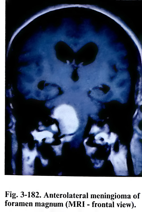
Most lesions in this area are investigated by imaging studies to demonstrate the relationship of the tumour to the rim of foramen magnum, the skull base foramina, and the structures passing through them, as well as the major vascular structures: vertebral and basilar arteries, their major branches, and the jugular bulb. Essential techniques of investigation are CT: enhanced, with images demonstrating bone and neural structures alternatively, in axial and coronal projections, and MRI. Bone destruction, erosion, sclerosis, foraminal enlargement and calcification are seen on the CT, but the more precise assessment of neural structures is left for the MRI imaging (Figs. 3-181; 3-182). Angiography should evaluate both carotid and vertebrobasilar circulations and should give the necessary data of collateral supply. It will also indicate the displacement or wall compromise of vascular structures, as well as the degree of vascularity of the lesion.
From the imaging studies the following must be decided upon: 1. If the vertebral or basilar arterial walls are affected by the lesion, immediate arterial reconstruction has to be possible through the surgical approach. 2. If the jugular bulb wall is affected, a venous sinus ligature must be prepared in case of rupture. 3. It should be decided which cranial nerves are encased and will pose a threat of iatrogenic damage or will be required to be sacrificed. The strategy decided on should implicate surgical planning in such a way that the surgical approach provides the necessary access to thenaffected structures and space for performing the manipulations.
When the tumour is located posterior to the brain stem and spinal cord, the posterior fossa is opened in a standard way and the laminae of the upper cervical vertebrae are resected. Meningiomas and neurinomas situated posteriorly are removed comparatively easily, applying the general principles for such removal. Laterally or mainly anteriorly situated tumours need the lower lateral craniocervical approach. For this approach the patient is placed in a semisitting or park-bench position.
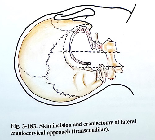
The skin incision is similar to that used for the retromastoid craniectomy but is extended caudally to the level of C2, exposing laminae of C1 and C2 unilaterally (Fig. 3-183). After transecting the muscles along the incision to the occipital squama, occipital muscles are detached from the occipital bone predominantly in a lateral direction. The incision of muscles can be performed along the midline with less bleeding, but retraction laterally can be difficult. The central and caudal part of the skin incision can be placed for these reasons also along the midline.
The separation is extended laterally to the tip of the mastoid and transverse processes of C1 - C2 are exposed. The deep muscle layers are detached with care to avoid injury to the vertebral artery. The C1 - C2 joint is identified, and the vertebral artery is exposed along the superior margin of the C1 lamina (the horizontal extracranial segment). If a broader space is required, the C2 spinal nerve can be transected close to its dural entry point. The suboccipital craniectomy is completed and extended as far laterally as the sigmoid sinus, including removal of the lateral rim of the foramen magnum and the lateral half of the Cl and C2 laminae with the posteromedial portion of C1 - C2 joint. If necessary the posterior half of occipital condyle can be removed and that has no effect on stability of the craniocervical junction. It is done with the high-speed diamond drill. It is important to expose the margin of the sigmoid sinus and to follow it caudally to the posterior border of the jugular foramen to the area of the tuberculum jugulare, That increases exposure and prevents unexpected injury. The vertebral artery entry area through the dura is dissected meanwhile, so the dura around it is safely accessible.
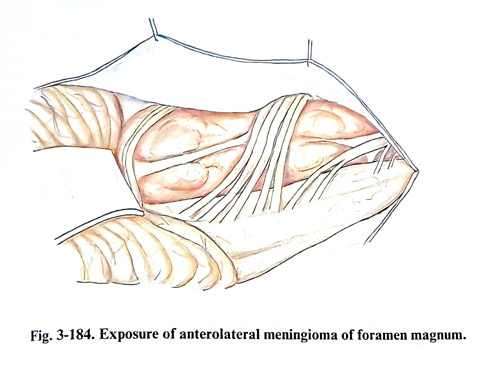
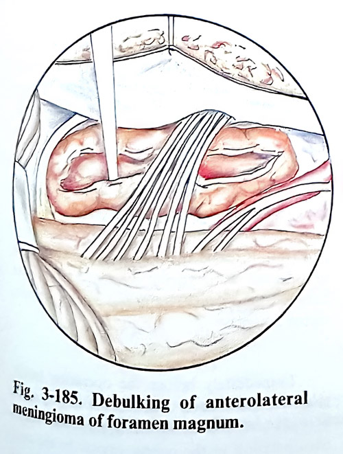
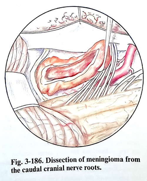
The dura is opened in a Y - shaped incision and the corresponding dural flaps are fixed with traction sutures (Fig, 3-184). Initially the tumor is enucleated by manipulation between the roots of the cranial nerves, cvhoosing the interspaces depending on the location of the tumour. The first denticulate ligament requires division. After the complete debulking follows the separation of the tumour capsule. In cases when the lower cranial nerves are encased by the tumour, neurophysiological monitoring and nerve stimulation can help with their identification and preservation. Dissection of the tumour capsule from the nerves is relatively easier than from the vertebral and basilar arteries and their branches. The preservation of the perforating vessels to the brain stem is extremely important for the outcome of these risky operations. Vascular compromise to neural tissue that occurs in this area can lead to catastrophic complications of lower brain stem dysfunction (Figs, 3-185; 3-186).
Particular difficulties arise in meningiomas attached to the dura at the intracranial entry of the vertebral artery, the jugular foramen and hypoglossal foramina. The vertebral artery can be mobilised opening its dural ring, the C2 transversal foramen and also be temporarily transposed until completing manipulation ventral to it. The parts of the artery immediately proximal and distal to the expected affected segment should be accessible for temporary trapping. The adventitial layer serves as a guide to the safe dissection of the wall. In case of true tumour infiltration of the wall of the vertebral artery, an attempt at total removal should be avoided (actually leaving only that portion, infiltrating the wall on a thin layer). The jugular bulb must be preserved. The hypoglossal nerve is sacrificed only if there is no accessible portion at the entry into the foramen.
The results of surgical therapy depend on the completeness of the resection, the involvement of various anatomical structures by the tumour, and the location and texture of the tumour. Functional recovery is related to disease progress and the promptness of the diagnosis. The earlier the diagnosis is established, the more complete resection is possible and the better results are obtained.
ORBITAL AND CRANIOORBITAL TUMOURS
The great variety of tumours and other mass lesions that occur into the orbit are of interest to several surgical specialities. Ophthalmologists deal with many of these problems by anterior direct approaches. The otolaryngologists manage many conditions arising within the sinuses and involving subsequently the superior, medial and inferior walls of the orbit. Neurosurgeons have access to those tumours, located deeply inside the orbital cavity and/or involving both the intracranial and intraorbital spaces.
When an orbital tumour is suspected clinically because of exophthalmos, visual impairment or limitation of eye movements, the patient needs a detailed examination. With good quality of the X rays of the skull, orbits, and optic canals, calcifications, hyperostosis, gross destructive lesions and sinus diseases can be seen.
An essential task of the imaging studies is to differentiate tumours from intraorbital inflammation. CT and MRI usually reveal the exact location and the size of the tumour, and in many cases the histology of the lesion can be predicted. CT should provide complete imaging of the orbital bony structures and contents in direct coronal and axial planes. Enhancement of the lesion by intravenous contrast is important for the differential diagnosis. MRI provides direct multiplane imaging, which is a very valuable contribution to the topography of the lesion. This modality also adds information about lesions close to the optic nerve and it is superior in visualising them inside the optic canal.
Many different surgical approaches to the orbital cavity have been introduced into practice by ophthalmologists, otolaryngologists, and neurosurgeons. There are controversial opinions on the indications for this large number of approaches. Several factors determine the choice of approach. These factors are related to the position of the tumour and its relationship to surrounding structures. We can specify them as follows:
1. Position of the tumour regarding the muscle cone - inside or outside.
2. Relation of the tumour to the optic nerve - superior, inferior, medial or lateral.
3. Presence of involvement of the optic nerve inside the canal.
4. Defining the lesion as primary of the orbit or secondary - penetrating from adjacent structures and cavities.
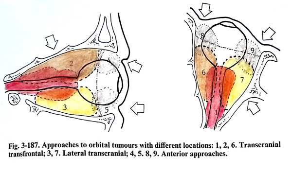
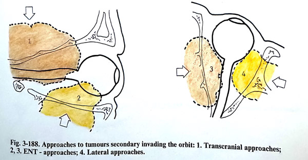
Personal preferences of surgeons have strongly influenced the selection and recommendations of approaches to the orbit. Irrespectively of that, we would like to suggest some guidelines. The lesions inside the muscle cone require a broader space for dissection, usually a transcranial approach, providing space for safe manipulation, compared to those mass lesions located outside the cone, which are usually quite accessible under the removed part of the orbital wall. Even more, if they are totally anterior to the equatorial plane of the bulb, they can be removed by an anterior orbitotomy. In those lesions, penetrating into the orbit from an adjacent structure or cavity, the approach should provide access simultaneously to that structure of origin to guarantee as much as possible radical removal (Figs. 3- 187; 3-188).
Tumours in close relation to the nerve have to be approached through broader transcranial orbitotomies and the position of the tumour regarding the optic nerve has a significant importance for the choice. The rationale of choice is that the nerve should not be on the way to the tumour when it is broadly splitting the extraocular muscles. When the tumour has mass effect and the configuration of muscles is grossly or moderately deformed, preferential ways of penetration inside the muscle cone are selected. Such small or intrinsic optic nerve tumours are removed between the medial rectus and levator/superior rectus muscles.
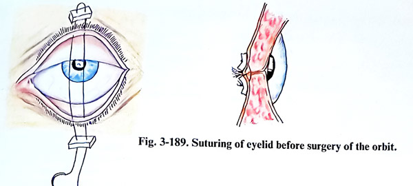
Immediately before the operation tarsorrhaphy is performed to ensure protection of the eyeball and especially of the cornea during manipulation in the orbital cavity. In the postoperative period it is left for a few days, until the oedema of the orbital tissue subsides (Fig. 3-189).
ANTERIOR SURGICAL APPROACHES
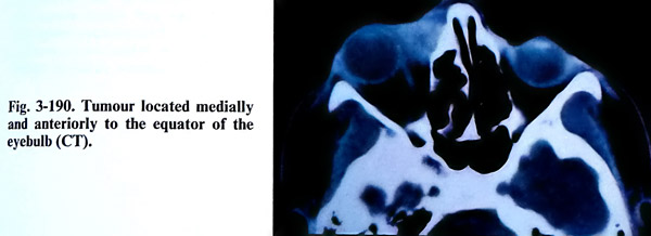
The anterior surgical approaches are suitable for tumours, located mainly anterior to the equator of the eye-bull. These tumours usually can be palpated through the orbital entrance (tig. 3-190). The anterior surgical approaches can be transcutaneous or transconjunctival. The transconjunctival approaches usually are performed by ophthalmologists.
ANTERIOR TRANSCUTANEOUS APPROACHES
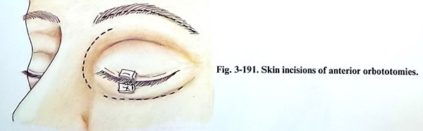
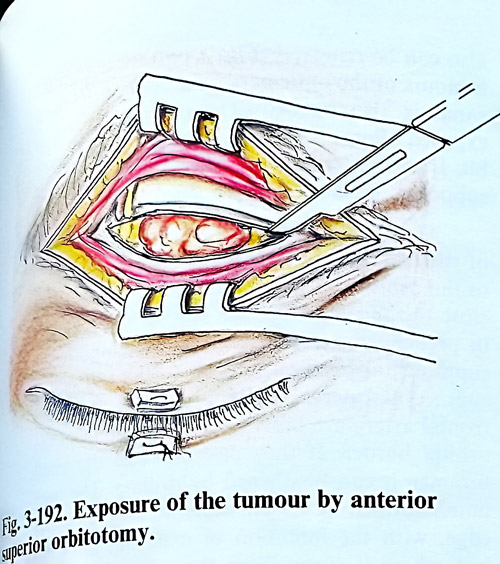
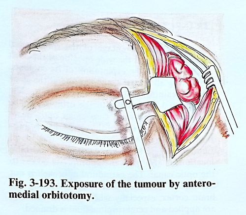
When the tumour is located superior to the eyeball and under the orbital roof, the skin incision is done through the eyebrow (Fig. 3-191). The orbicular muscle is split along its fibres, reaching the orbital edge of the frontal bone. The supraorbital incisura and foramen containing the frontal nerve and artery are preserved to avoid postoperative anaesthesia of the forehead. The fibrous septum of the upper eyelid is opened 2 - 3 mm away from its attachment to the bone. Its transection is cautious and should avoid injury to the levator palpebrae superioris muscle (Fig. 3-192). This approach is suitable for the removal of haemangiomas, dermoids, and mucoceles. After the bone has been exposed, the periorbit is separated from the bone and the tumour surface is reached. When the tumour is situated medially and superiorly to the eye-ball, the skin incision can be extended medially between medial cantus and the midline at the base of the nose. The deep penetration into the orbital cavity should avoid the damage of trochlea and the lacrimal sac (Fig. 3-193).
The closure of the operative wound is in three layers, suturing the septum of the eye-lid, the orbicular muscle and the skin. In the majority of cases placement of drainage is not necessary.
If the tumour is located inferior to the eyeball, the incision is along the inferior border of the orbital entrance. It should reach the periosteum close to the edge of the orbital entrance and its separation from the edge allows penetration into the orbital cavity. The periorbit is opened according to the pathology expected and found. The inferior rectus muscle and the lacrimal sac are the anatomical structures that need protection.
There are no differences in the closure of the operative wound compared to the previous method.
LATERAL EXTRACRANIAL ORBITOTOMY WITH BONE FLAP
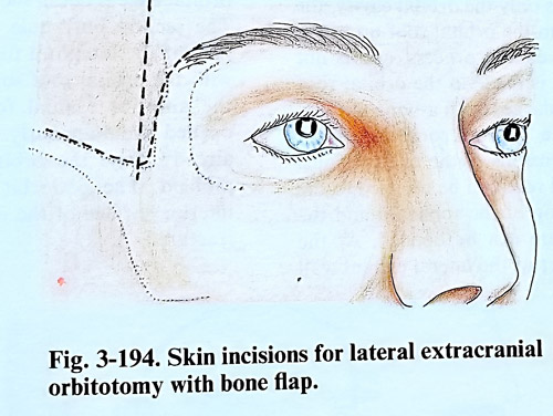
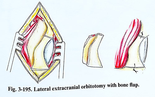
This approach is suitable for removing laterally and anteriorly located tumours into the orbital cavity. The skin incision is horizontal and along the superior border of the zygomatic arch. It begins 2 cm from the lateral end of the eye-lid and reaches near to the tragus. There is a second option for the skin incision as is shown in fig. 3-194. The lateral ligament of the eyelids is separated from the bone. The orbicular muscle is detached from the lateral orbital edge and the neighbouring area. The periosteum is cut near to the border of the orbital entrance and after that the periorbit is separated from the bone of the lateral orbital wall and adjacent surfaces of the orbital roof and floor. The lateral rim and the wall of the orbit are cut in two lines: the first a little superior to the zygomatico-frontal suture, and the other at the level of the superior border of the zygomatic arch. The flap is fractured across its base and the bone piece is preserved in saline. The opening of the orbit can be enlarged, nibbling additionally the greater wing of the sphenoid. The periorbit is opened in a way suitable for manipulation inside the orbital cavity. At the end of surgery the bone flap is fixed in its place by wiring or small metallic osteosynthetic plates (Fig. 3-195).
Complications: Extensive orbital oedema can complicate the postoperative period. In such cases corticosteroids are applied. They are continued according to the response to the treatment. Postoperative infection is rare. Visual impairment or loss is also rare and is due to optic nerve ischaemia associated with occlusion of posterior ciliary arteries or the central retinal artery. Ocular movements are affected consequent to the retraction of the intraorbital (extraocular) muscles and almost always this deficit is temporary. Postoperative enophthalmos due to atrophy of orbital fat is common. but usually is not a cosmetic defect of significance.
TRANSCRANIAL APPROACHES
The main advantage of the transcranial approaches is that the wide opening of the orbit makes possible the exploration of its contents with preservation of all orbital structures and at the same time obtaining a good cosmetic effect. The transcranial approaches to the orbit are to be preferred, when it is a case of an optic nerve tumour, meningiomas of the orbit, neurofibromas, osteomas or encephalocele, and in all tumours, where there are data of cranioorbital location. It also gives an appropriate access to the optic canal. The extradural approach is not associated with a significant risk of injuring the brain cortex, especially when osmotic diuretics are applied and CSF is drained.
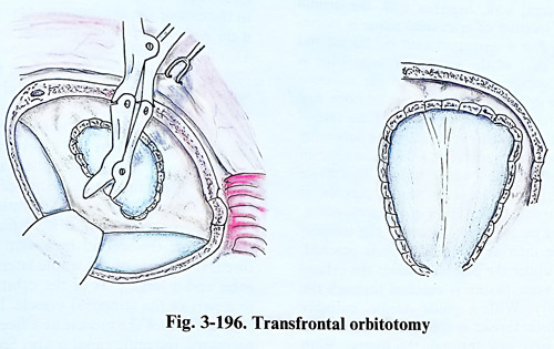
Transfrontal approach. The craniotomy is similar to the described in the approaches to the pituitary region. When it is a case of a tumour that occupies only the orbital cavity, the dura is detached from the orbital roof up to the base of the anterior clinoid process, and is not opened. A burr hole is made in the orbital roof and it is enlarged with a bone nibbler. The excision of the orbital roof anteriorly depends on the size of the frontal sinus. Backward the orbital roof can be resected to the edge of the lesser wing of the sphenoid and the superior orbital fissure can be opened if needed. At the same time a large part of the lateral orbital wall also can be removed (Fig. 3-196). In cases with tumours of the optic nerve, the roof of the optic canal is carefully drilled out. The orbital content is exposed after a Y-shaped incision of the periorbit. If an intracranial extension of the tumour is supposed, the dura is also opened.
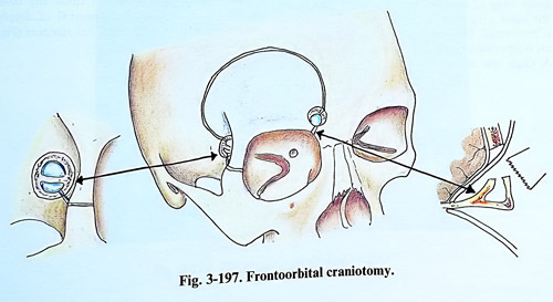
The frontal flap can be enlarged with part of the orbital roof, gaining additional exposure to the orbit. Two burr holes drilled close to the skull base are placed very precisely in predefined positions. The medial one has a supraorbital placement, medial to the exit of the supraorbital nerve. The supraorbital nerve and frontal artery are previously separated from the orbital edge. If they pass through a small foramen, it can be opened by drilling. This burr hole is drilled at about 1,5 cm superior to the orbital edge, with the intention of exposing the dura just where it turns from the convexity to the base over the orbital roof. The exposed dura is retracted and the orbital roof - perforated. The orbital edge is divided at that location (Fig. 3-197). The second burr hole is in the most anterior part of the temporal fossa and should coincide with the orbital roof in a way that after being drilled, the anterior cranial fossa and the orbit are opened simultaneously. Through this burr hole the orbital roof is additionally nibbled. The periorbit is separated from the inferior surface of the orbital roof and the flap fractured along a line on the roof, connecting the burr holes.
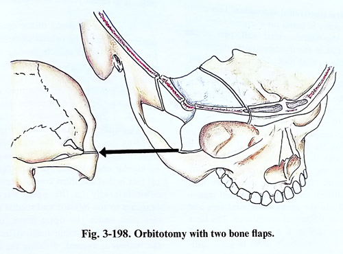
Orbitotomy with two bone flaps. After a craniotomy in the frontotemporal region, a second bone flap from the orbital roof and its lateral wall is lifted, together with the orbital edge. The incision of epicranial tissue is bitemporal. The flap is separated until the lateral and the major part of the superior orbital edge is exposed together with a part of the superior edge of the zygomatic arch. The second bone flap consists of the orbital roof and the lateral wall of the orbit. This is done in the following way: the anterior part of the superior edge of the zygomatic arch - where it forms an angle with the zygomatic process of the maxilla, is dissected through an incision of the periosteum. Next sliding with a fine periosteal elevator on the bone surface of the temporal fossa is reached the inferior orbital fissure. Separating under the periorbit inside the orbital cavity, the inferior orbital fissure is reached through the orbital cavity. With a curved guide needle or hook a thick thread is inserted for passing a Gigly saw through the fissure. With the saw the lateral orbital wall is cut as low as possible, usually at the level of the superior margin of the zygomatic arch. The same bone division can be done easily with a vibrating saw (Fig. 3-198).
Next the frontobasal dura is separated from the orbital roof and is retracted until the lesser wing of the sphenoid is exposed. Behind the lesser wing edge the superior orbital fissure is penetrated. Under the orbital roof, the periorbit is separated, beginning from the orbital edge. A Gigli saw is introduced under the superior orbital edge until it comes out from the superior orbital fissure. The orbital roof is transected together with the orbital edge in the sagittal plane. This incision is best done just lateral to the exit of the supraorbital nerve or medially if the nerve is separated from the bone foramen. This bone incision can be made easily and safer now by the vibrating saw. These two incisions separate a large part of the orbital roof and the whole lateral orbital wall, and they remain fixed to the cranial base only by the greater wing of the sphenoid between the superior and inferior orbital fissures. This part of the bone is nibbled partially beginning at the edge of the craniotomy and reaching in a basal direction as close as possible to the inferior orbital fissure. With a slight effort the second bone flap base is fractured. It consists of the whole lateral wall of the orbit and a large part of the orbital roof, fixed to a part of the temporal muscle. It can be also separated from the muscle as a free bone flap. If necessary, the optic canal is also opened, removing its superior wall. When the work inside the cranial and orbital cavities is completed, both flaps are repositioned and fixed by wiring or metallic osteosynthetic plates.
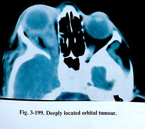
Comparing it with other methods, this operative approach provides probably the widest operative field, both for the cranial and orbital cavities and does not cause any bone defect. It is suitable for removal of deeply located complex orbital and cranioorbital tumours (Fig.3-199).
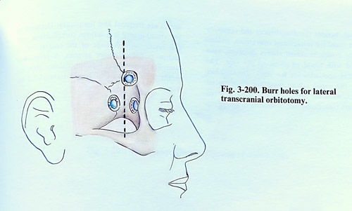
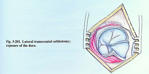
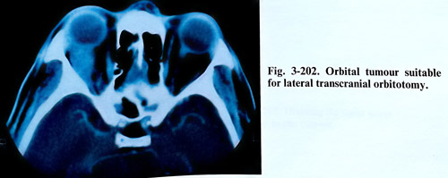
Lateral transcranial approaches. This group of approaches is based on a craniectomy that includes the lateral orbital wall, part of the cranial vault in the frontotemporal region and part of the orbital roof. The incision of soft tissues is done in the temporal region. It is a vertical and slightly curved incision, 2 or 3 cm behind the zygomatic process of the frontal bone and behind the hairline (Fig. 3-200). Its length is 10 - 12 cm and reaches the zygomatic arch. The temporal muscle is divided at its insertion on the superior temporal line and is detached with the periosteum from the lateral orbital wall, the frontobasal area and the anterior temporal region. After that, three burr holes are drilled: one in the frontobasal region, the second - in the anterior temporal area and the third - on the lateral orbital wall. The bone defect is enlarged with a nibbler merging all three burr holes. In order to get a sufficiently wide operative field, the lateral part of the orbital roof is removed also with opening of the superior orbital fissure (Fig. 3-201). This operative approach offers an adequate operative field upon broad areas of the lateral parts the orbital cavity. It is suitable for laterally located orbital tumours (Fig. 3-202). In the case of a cranioorbital tumour however, it is rather narrow for reaching its intracranial part. Besides this, a permanent cranial and orbital defect is produced.
Technique of orbital tumour removal. After opening the periorbit, the orbital tumour is located by palpation or intraoperative ultrasound. When the tumour's position is defined, dissection is carried out directly to it with the help of two or three fine self-retaining retractors.
The surgical technique of tumour removal is different for the neurosurgeon accustomed to work inside the cranial cavity. The dissection of intraorbital structures is often obstructed by the intraorbital fatty tissue coming out under pressure. Because of that the wide opening of the orbit, independently of the microsurgical technical advantages, is better and less traumatic to the intraorbital structures. After opening of the periorbit the first branch of the trigeminal nerve that lies on the levator and the superior rectus muscles is identified. The fourth nerve is more difficult to identify, as it is a very tiny structure.
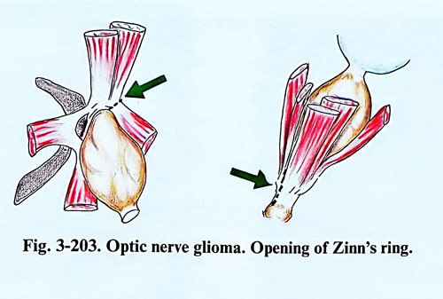
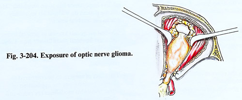
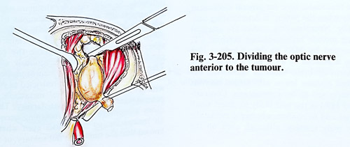
In case of a glioma of the optic nerve, the penetration into the orbital content is between the medial rectus muscle on one side and the levator palpebrae and superior rectus muscle laterally on the other. The upper group of muscles must be well-separated as proximal as possible, and are retracted laterally. The annulus of Zinn is transected medially to them and the optic canal is opened (Fig. 3-203). If bleeding occurs from the ophthalmic artery small branches in this area, it is easily stopped by bipolar coagulation. An important anatomical fact is that the third cranial nerve enters the orbit laterally to the optic nerve and its branches to the inferior and medial rectus muscles pass under the optic nerve, or in other words under the tumour itself. After dissection, the tumour is excised, dividing the optic nerve anteriorly to the chiasm in unaffected parts of the nerve and behind the eye ball (Fig. 3-204; 3-205).
Optic nerve sheath meningiomas with certain visual acuity preserved are excised with extreme care, preventing damage of the nerve fibres. Lesser sphenoid wing meningiomas with cranioorbital extension are usually easier to remove. The orbital parts of the tumour are situated in the superior lateral quadrant of the orbit and are resected without any difficulties if the attachment area is small. The infiltration of the orbital walls and the greater wing of the sphenoid requires wide bone excision and meningiomas 'en plaque' cannot be removed completely.
Tumours of the lacrimal gland are common and they have a specific location. The lateral transcranial approach is the most suitable for their removal. The tumour is removed 'in one piece' with the gland, the periorbit, and the neighbouring tissue. If the clinical evolution of the tumour is less than 6 months, the tumour may be malignant. In such a case the prognosis is poor and after the biopsy result is confirmed, a more extensive excision is required. In some cases complete exenteration of the orbit with excision of the orbital roof and lateral wall are required.
Complications. During the postoperative period, besides the disturbances and complications usual for intracranial operations, oedema of the eyelids is almost always observed, associated with blood effusions on the skin. This oedema continues for few days, during which time the eyelid suture must not be removed.
Infections of the orbital contents and osteomyelitis are rare. Ptosis and limitation of eye movements are frequent and temporary. Decrease in the visual acuity is a serious complication, and can result from damage of the central retinal and/or posterior ciliary arteries. A permanent complication is slight enophthalmos that remains after removal of bigger intraorbital tumours.