INTRACRANIAL ANEURYSMS: GENERAL PRINCIPLES OF MANAGEMENT
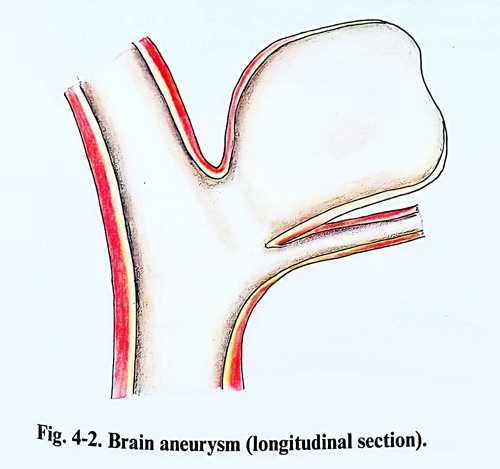
The aim of the surgical treatment of intracranial aneurysms is prophylactic: to prevent new rupture of the aneurysmal sac, and only in some cases to evacuate an intracerebral haematoma, which endangers the patient's life (Figs.4-1; 4-2).
GENERAL PRINCIPLES OF ANEURYSM SURGERY
Complete angiographic study is required of the four main vessels with multiple views. Collateral circulation is evaluated on static images or by dynamic tests as temporary occlusion with balloon. These last tests are compulsory when temporary occlusion of major arteries or 'trapping' can be expected.
The direct surgical procedure with exclusion of the aneurysmal sac from the brain circulation is universally aimed. Alternative methods are trapping of the aneurysm, by proximal and distal occlusion of the parent artery, wrapping of the aneurysmal sac, proximal endovascular occlusion, or intra aneurysmal endovascular occlusion, with induction of thrombosis inside the sac.
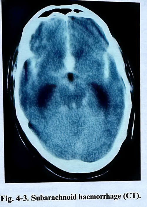
When multiple aneurysms are discovered, the ruptured one can be identified by localisation of the haemorrhage on CT, assessment of haemosiderin deposition on MRI, aneurysm dome configuration on the angiography, or clinical neurological findings (for example sudden IlIth nerve palsy) (Fig. 4-3). If more then one aneurysm should be treated in one surgical session, the first should be the ruptured one.
During the surgical approach, brain relaxation is achieved by appropriate use of hyperventilation, osmotic agents, diuretics, and evacuation of CSF (preoperative placed lumbar drainage or aspiration after the craniotomy). The surgeon should be prepared to delay the operation if relaxation is not sufficient.
Proximal vascular control must be established on the parent artery if possible, if not proximal vascular supply on the neck has to be isolated and occasionally occluded or a balloon is inserted in the internal carotid artery. Giant aneurysms of the basilar artery during the surgery are best controlled by temporary intra-vascular occlusion with balloon catheter.
The sequence of vascular exposure is the proximal parent vessel, then the distal parent vessel, and finally the aneurysmal neck. Sharp dissection is preferable to blunt one, intending to reduce traction forces applied to the aneurysm. Once the aneurysmal neck is isolated, critical vessels adjacent to the aneurysm are defined and dissected away when possible. This dissection can be often postponed until the aneurysm is collapsed after clip application.
Local hypotension by trapping with low closing force temporary clips is preferable to systemic arterial hypotension. Temporary trapping should by used to avoid aneurysmal rupture anticipating the complication when the anatomical conditions create difficulties. If vascular control cannot be achieved within the operative exposure, a temporary balloon can be navigated into the appropriate location.
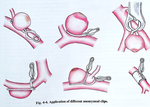
The clipping axis is preferable to be along the axis of the parent artery. Clipping across the axis of the parent artery increases the risk of stenosis and bending of the artery, or tearing close to the aneurysmal neck. Application of multiple clips is necessary either to enforce the single already applied clip or in case of a complex aneurysm (Fig. 4-4). Once the permanent clip is placed and temporary clips are removed, the aneurysm is punctured with a fine needle and aspirated to check for residual filling. In case of large or complex aneurysms may be required temporary trapping, aspiration of the sac before placing the final clip, and only than the dissection is completed. Residual filling of the aneurysm requires repositioning of the clip, multiple clips, or a reinforcing clip to increase the closing pressure. Patency of the parent artery is evaluated by assessing the vessel diameter, colour and pulsation, intraoperative doppler study or intraopreative angiography.
SELECTION OF PATIENTS FOR OPERATION
The proper selection of patients with intracranial aneurysms for surgical treatment is essential for the safe course of the operation and the operative results. In considering the indications for surgical treatment it is necessary to assess the patient's general and neurological condition, localisation of the aneurysm and its anatomical peculiarities, conditions of the cerebral arteries, patient's age and the presence of intracranial haematoma.
The patient's neurological and general condition with intracranial aneurysms plays a decisive part in determining the indications for operative treatment. Different scales for grading the patient's condition after subarachnoid haemorrhage have been introduced. All of them intend to select those patients, whose condition is not to much affected to tolerate without additional deterioration and complications the preventive surgery proposed. Such is the scale of Hunt and Hess subsequently modified on different occasions:
Grade I (minimal bleeding). The patient is alert without neurological deficit. Grade II (mild bleeding). The patient is awake but with headache, neck rigidity or minimal neurological deficit, like paresis of the third cranial nerve. Grade III (moderate bleeding). The patient is drowsy or confused with or without focal signs. Grade IV (moderate or severe bleeding). The patient is semicomatous with or without neurological focal signs. Grade V (severe bleeding). The patient is in coma and decerebrate, failing vital signs. At any moment every patient with a SAH can be evaluated and scored according to one of the existing scales. When the patient is over 50 years of age, the gravity of the condition is assessed with one grade higher than the observed signs would indicate. Usually over the age of 65 operative treatment is not recommended, but in many countries with achievements in population longevity these limits have been moved forward. If the patient has serious heart, lung, kidney or liver diseases, one more grade is added too, as these conditions represent an additional risk for operative treatment. The best surgical results are achieved with patients in grade I and II.
The condition of the cerebral arteries is of great importance in the assessment of the indications for operative treatment. Arterial atherosclerotic plaques may discredit the technical performance of the operation. They oblige the surgeon to be more careful when working on the vessels and especially when he has to clip or ligate, as at this time the delicate and sometimes rigid arterial wall may be torn.
The collateral circulation has to be taken into consideration, when the indications for operative treatment of an aneurysm are evaluated. All segments of the circle of Willis have to be studied as size and direction of flow. The pre-operative planning should assess the option of collateral supply if the parent vessel is occluded. For instance, when aneurysms are in the region of the anterior communicating artery, the surgeon must carefully establish the patency of the anterior cerebral arteries and what is the direction of flow.
Arterial spasm, following subarachnoid haemorrhage, is one of the major complications that has to be carefully evaluated in considering the indications for operative treatment. Practice shows, that when an operation is performed in patients with severe arterial spasm, the results are poor and the mortality is increased. In such cases it is advisable to postpone the operation until the spasm resolves. In the majority of patients with clinically and angiographically significant diffuse spasm, their condition improves after a period of three weeks.
The localisation of the aneurysm also influences the selection of patients for operation.
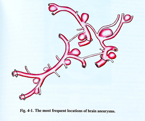
From surgical point of view the aneurysms of the middle cerebral artery are the most accessible, because they are superficially situated in the Sylvian fissure. Aneurysms of the internal carotid artery require greater retraction of the frontal and temporal lobes, and the dissection of arteries and the aneurysmal sac need more time and attention. When it is a case of an anterior communicating artery aneurysm, the operative approach is even more difficult, due to the close relation of the aneurysm to important vessels and the greater depth of its localisation. The favourable localisation of the aneurysm on middle cerebral artery makes surgery to be done earlier than in the cases of internal carotid artery aneurysms, while in aneurysms of the anterior communicating artery and the vertebrobasilar circulation the delay is usually the longest.
The anatomic characteristics of the aneurysm are also important for patients’ selection for surgery. The size of the aneurysmal neck and its anatomical features influence the selection of the technical approach for isolation of the aneurysm from the circulation. In the event of a narrow, well formed aneurysmal neck, without arterial branches in its vicinity, the dissection and clipping are performed easily, without additional trauma on the brain. When there are large blood vessels around the aneurysmal sac and its neck, then the dissection is much more difficult and requires greater retraction of brain surface. If the neck of the aneurysm is wide, the clipping becomes more difficult, even impossible without narrowing the parent artery. With large aneurysms (giant aneurysms) it is very often necessary to interrupt temporary the circulation in the parent artery, and at the end it may be necessary to make microsutures of the walls.
The more complex the anatomical topography around the aneurysmal neck, the more indications for operation have to be carefully considered. The finding of subdural, intracerebral or intraventricular hematoma in cerebral aneurysms rupture has to be taken thoroughly into consideration in the indications for operation. When the patient's condition obviously is deteriorated and that is due to the intracranial haematoma as a mass lesion effect, there are indications of urgent surgery and the operation has the task not only to exclude the aneurysm from the circulation, but to evacuate the haematoma and save the life of the patient.
The rupture of an aneurysm during pregnancy sets special requirements in deciding in favour of operative management. There are no contraindications on the part of pregnancy for angiographic investigation of the brain. Preventive measures must be taken against irradiation of the foetus, especially during the first three months of pregnancy. When an aneurysm is established in a pregnant patient, many authors consider that the surgical decision has to be the same as in a non pregnant, i.e. have operative treatment performed irrespective of the pregnancy. During the last weeks of pregnancy, the surgery can be anticipated by a Caesarean section and the operative treatment performed immediately afterwards.