ARTERIOVENOUS MALFORMATIONS OF THE BRAIN
Arteriovenous malformations (AVM) require surgical treatment because of their three major clinical manifestations: haemorrhage (subarachnoid and intracerebral), epileptic seizures and progressive neurological deficit.
About 50% of the clinical cases present with haemorrhage, another 25% - with epilepsy, and the rest (25%) with neurological deficit, other minor complains or are accidentally discovered on MRI.
Haemorrhage due to AVM has much lower incidence of early rebleeding than the aneurysmal, thus permitting full investigation and surgery in an already improved condition of the patient. Related to the presence of AVM arterial aneurysms have also to be clipped. The resulting from AVM bleeding vascular spasm is much less severe. The rebleeding rate for different types of AVM is estimated as an average of 4% per year and is higher for AVMs which already bled, especially through the first year after bleeding, and increases with age. Epilepsy is controlled by surgery with the techniques of functional neurosurgery, with the same indications as in epilepsy of other aetiology. The progressive neurological deficit, considered to be provoked by "steal" effect of the AVM circulation on the surrounding brain tissue is expected to improve after surgery.
SELECTION OF PATIENTS FOR OPERATION
Current indications are the result from the analysis of the natural outcome of the disease considering rebleeding rate with its associated morbidity and mortality. All patients whose general condition permits (and provided their life expectancy is long enough to justify it) have to be evaluated for surgery. The same refers to all patients with epilepsy or other complains, taking into account their natural prognosis. The risk of treating them conservatively has to be compared to the surgical risk of total excision of the malformation by craniotomy.
At present from existing three methods of surgery: "open" surgical excision through cra-niotomy, endovascular obliteration and radio-surgery (stereotactic radiosurgery), only the first can provide high degree of certainty for radicalism. Therefore, we consider the other two as a second line or adjuvant to the surgical excision.
For the evaluation of surgical risk for open excision of an AVM, recently have been identified three more important factors. They are: 1. The size of malformation nidus. 2. The part of the venous system (deep or superficial) participating in the drainage of the AVM, and 3. The functional importance of the brain areas occupied by the nidus, particularly its functional importance or "eloquence". With the contribution of leading neurosurgeons, AVM surgical cases, evaluated retro- and prospectively, have shown that the affection of the deep venous system by the high pressure drainage, by big AVMs in "eloquent" areas of cortex or brainstem, is associated with clearly significant increase of mortality and morbidity.
Only by balancing the natural risk with the surgical risk according to these factors can be achieved the proper decision for surgery. For these purposes have been developed several grading systems, from which that of Spetzler and Martin appears at present to be the most universally applicable. As an addition to the evaluation, factors as coexisting aneurysms or "low flow" type of the AVM can influence the decision more in favour of surgery.
EXCISION OF THE ARTERIOVENOUS MALFORMATIONS
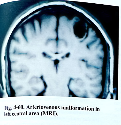
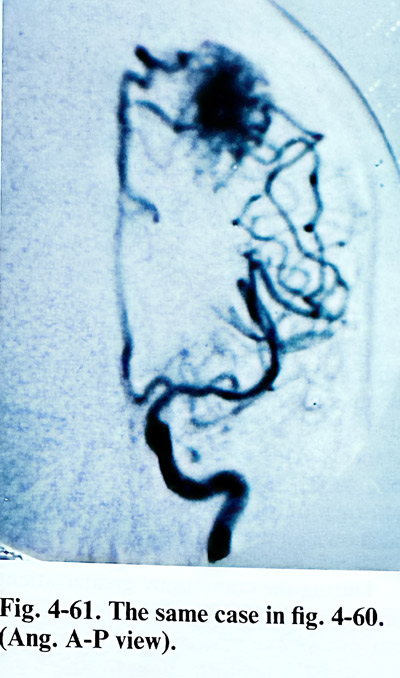
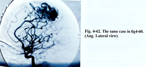
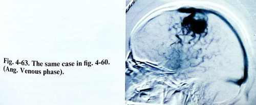
When planning the AVM surgery it has to be decided if the feeding can be reduced by endo-vascular procedures in advance. That procedures have to precede shortly the day of surgery, usually at the time of superselective angiographic study. On this last study the position of all major feeders and drainers has to be identified in relation to bony, dural and cortical reference points. Although the angiography can be very precise, additional space for hidden feeding vessels and free manipulation upon them has to be provided, so larger craniotomies with generous distances to the nidus are recommended (Figs.4-60 - 4-63). Very often during the operation the malformation proves to be of larger size than that shown on the angiography.
During the craniotomy greater attention is paid to haemostasis as in many cases vessels from of the external circulation communicate freely with the malformation. In such a case, the opening of the dura is made with an incision that gets around such communications. Frequently after opening of the dura only enlarged arterialized veins are seen in the cortical surface.
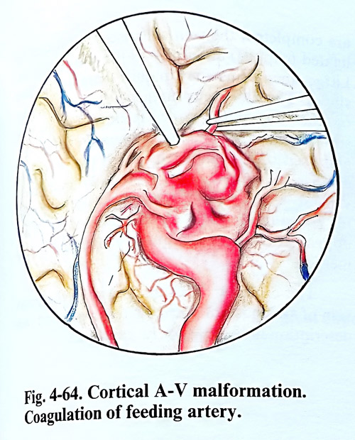
The arachnoid around these veins is opened. The dissection of the malformation begins sub-pial and if the feeding arteries are not seen on the surface, should be searched carefully for them considering the angiographic data (Fig. 4-64).
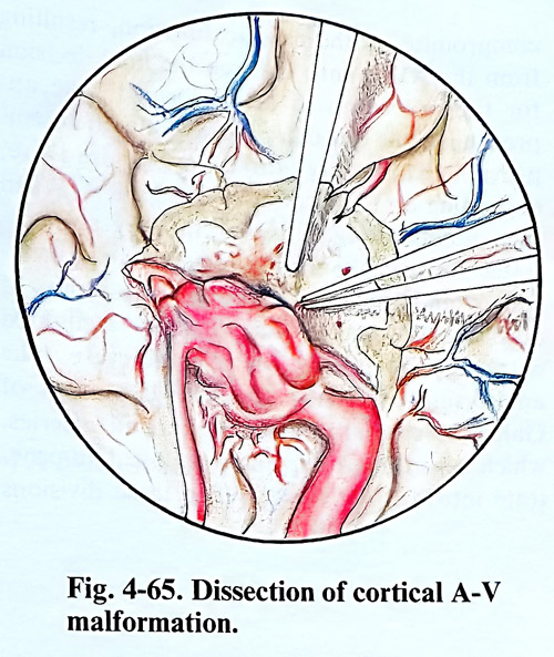
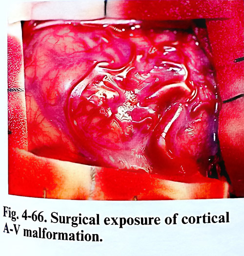
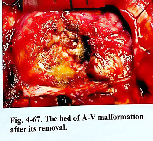
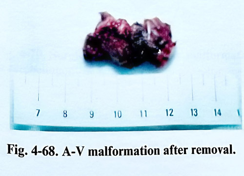
After clipping and coagulation of the feeding arteries, they are divided near to the nidus of the malformation. The interruption of the supplying arteries causes partial collapse of the malformation and this facilitates the dissection to follow (Fig. 4-65). In case of perinidal haematoma, the residual cavity around the malformation makes the dissection easier. If there are difficulties to follow the plane of cleavage around the malformation, the thin layer of gliosis around the nidus is a good indicator. When it is impossible to find a feeding artery near the malformation and there is a risk of injury of apparently normal brain tissue, temporary clipping of some branches of the circle of Willis more proximally is an option. After interruption of the feeding arteries and dissection of the nidus, the draining veins are clipped and divided last. The malformation bed is inspected carefully for residual pathological vessels and final haemostasis is achieved. If arterial hypotension is applied, the blood pressure should be raised to normal before deciding on safety of haemostasis (Figs. 4-66; 4-68).
Rupture during the dissection causes at times dramatic bleeding and in these circumstances manipulation may cause destruction of brain tissue or considerable loss of blood with abrupt decrease of the systolic blood pressure. In such cases one of the malformation walls must be dissected very fast and a broad clip be applied on the bleeding area until bleeding is controlled and dissection continues in the usual meticulous way.
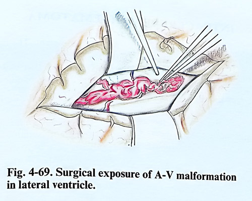
The dissection of a deep arteriovenous malformation has some particular points for consideration. The removal of a para- and intraventricular malformation carries risk of producing severe intraventricular haemorrhage. The feeders in completely intraventricular AVMs originate from the anterior or posterior choroidal arteries, and can be accessible in the ventricular cavity (Fig. 4-69).
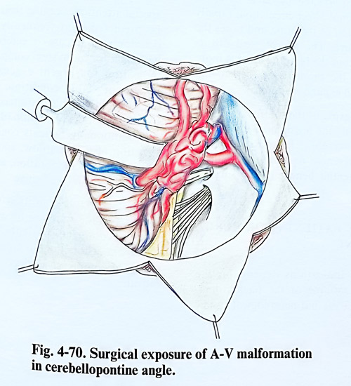
The excision of an arteriovenous malformation from the cerebellum is requiring the clipping of some cerebellar arteries and when it is situated in the cerebellar hemisphere, partial lobectomy can safely be performed, without causing any cerebellar symptoms (Fig. 4-70). The removal of an arteriovenous malformation from the vermis usually does not present any special difficulties.
The malformations of the great vein of Galen are particular type of AVM. In spite of the fact that they are rare, the decision for surgery has been always a major point of controversy. In this type of malformations due to high intravenous pressure, the great vein is dilated to the shape of a sphere or ellipsoid with diameter 3 or more centimetres. The feeding vessels are branches of the anterior and middle cerebral arteries and enter directly into the vein uni- or bilaterally.
This type of arteriovenous malformations can be treated in very rare occasions by direct attack despite the existing controversies. That is not the first choice at present, and endovascular procedures have to be attempted after complete selective angiographic studies. On the basis of these last investigations and the assessment of the general condition (often impaired due to compromise of the cardiac function, resulting from the AV shunt), is taken the final decision for the procedure to be performed. The approach is done through parietal and parietotemporal craniotomy close to the midline, depending on the location of the feeders. When the parietal approach is used, the surgeon penetrates between the medial surface of the hemisphere and the falx, followed by division of corpus callosum. The approach is facilitated a lot by lateral ventricle puncture. The aneurysmal wall (the widened great vein of Galen) is carefully dissected and all arteries, which according to the angiographic data penetrate into it are divided. When these divisions are completed, the vein collapses, and it can be ligated (clipped) anteriorly to the straight sinus.
Large aneurysms may require surgery in two stages. The first stage undertakes a craniotomy on the side of the wider feeders. During the second stage the same is made on the other side and excision is completed. When the supplying arteries are located more posteriorly, more convenient is the parietooccipital craniotomy with or without incision of the tentorium. This approach does not require corpus callosum incision.
The excision of an aneurysm of the great vein of Galen is not a routine technique and this description indicates only its guidelines.