ANEURYSMS OF ANTERIOR CIRCULATION
ANEURYSMS OF INTERNAL CAROTID ARTERY
Internal carotid artery aneurysms present different technical problems according to the segment of the artery they involve. The following segments are currently considered: intracavernous, ophthalmic, posterior communicating and anterior choriodal, and internal carotid bifurcation.
The relation of the aneurysmal neck to the optic nerve, anterior clinoid process and the distal dural ring determine the principles of the technique to be applied also and the expected complications. Proximal arterial control can be dificult in aneurysms of the cavernous and ophthalmic segments and preoperative precise location of the aneurysmal neck, can allow the surgeon to choose the safest technique (at the neck, in the Glasscock triangle or with endovascular balloon). To assess the consequences of elective or emergency trapping of a part of the internal carotid has to be investigated the functional effect of a temporary balloon occlusion.
ANEURYSMS OF THE INTRA-CAVERNOUS CAROTID ARTERY
The options for the treatment of intra-cavernous aneurysms include endovascular intra-aneurysmal placement of a balloon or thrombogenic substances, trapping procedures by direct or endovascular technique, and direct intracavernous aneurysmal clipping. The direct clipping of cavernous internal carotid artery aneurysm is a suitable when the aneurysm is paraclinoid or intracavernous, expands the distal dural ring and enters the intradural space.
The intracavernous aneurysms not amenable to indirect technique can be also managed directly.
Fortunately, most intracavernous carotid aneurysms arise from the C3 - C4 segment (proximal anterior loop) and can be clipped directly after opening the distal dural ring, opening the roof of the cavernous sinus, and partially mobilising the lateral wall of the cavernous sinus.
DIRECT CLIPPING OF INTRA-CAVERNOUS ANEURYSMS
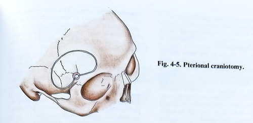
The patient is placed in a supine position on the operating table with the head rotated 30 degrees to the opposite side. The pterional craniotomy is the most suitable, but has to be extended anteriorly to the midpoint of the orbital entrance at the level of orbital roof. Laterally temporal fossa is opened to its floor (Fig. 4-5).
Carotid bifurcation is exposed in the neck after skin incision along the skin creases. Internal and external carotid arteries are dissected and isolated with vessel loops to establish proximal vascular control. The internal carotid artery is also available for catheterization, balloon occlusion and angiography. An alternative option exists either by using endovascular technique and balloon occlusion, or by exposure of the artery in the Glasscock's triangle. CSF is aspirated through the previously placed lumbar catheter or the exposed cisterns. The dura is retracted to expose the orbital roof, which is entered by high speed drill. The posterior half of the orbital roof is removed by bone nibbler. The superior orbital fissure is exposed removing additional bone from the lesser wing of sphenoid. A high speed diamond drill is used to remove the anterior clinoid process and the roof of the optic canal. Care is needed to avoid entering the ethmoidal sinus, which is medial to the optic canal and can produce postoperative CS fistula and infection. The bone lateral to the superior orbital fissure and medial to the foramen rotundum, is thinned with the drill and fractured laterally to expose fully the superior orbital fissure. An incision of the dura is made medial by and parallel to the superior orbital fissure. The dural incision is extended medially and laterally to provide access to the floor of both anterior and middle cranial fossae (fig. 4-6)
The distal dural ring that encircles the internal carotid is incised sharply, using an arachnoid knife in direction away of the arterial wall. Beginning at the oculomotor foramen, the lateral wall of the cavernous sinus is incised along the axis of the optic nerve and the mid-portion of the oculomotor triangle. The next dissection reflects the dura propia of the lateral wall to expose cranial nerves III and IV and the first division of V. The inner membranous layer formed by the perineural sheaths (of cranial nerves) when preserved allows mobilisation of dura propia without disruption of the venous channels of the cavernous sinus. Beginning at the falciform ligament, the optic nerve sheath is incised along the course of the nerve and the dura on the medial side is exposed.
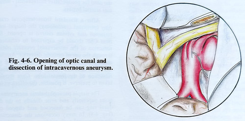
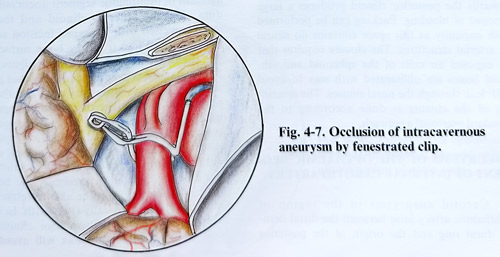
The optic nerve is retracted medially to expose the proximal dural ring and it is opened along the medial edge of the oculomotor nerve, retracting the nerve laterally to expose the fundus and the aneurysmal neck. Using the dissector, the relationship of the aneurysmal neck to the carotid artery and the distal dural ring can by established. A right-angle fenestrated clip is applied along the axis of the carotid artery preserving its patency (Fig. 4-7). In some circumstances aneurysms of the carotid cavernous region may require collapse of the aneurysmal sac for effective dissection and clipping. The collapse of the aneurysm can be obtained by temporary occlusion of the internal carotid artery in the neck and distally to the intracranial dissection site. Aspiration of the sac will permit dissection and precise placement of the clip. Intraoperative angiography is desirable in aneurysms in this area because direct inspection for completeness of clipping is often difficult.
At the completion of the intradural part of the procedure, haemostasis should be complete. Small pieces oxidised cellulose may be inserted in the anterior triangle between the internal carotid and the optic nerve, around the carotid siphon tip, and pressing against the medial wall of the superior orbital fissure dura. Oxidised cellulose must be modestly used to avoid excessive pressure on the oculomotor nerve that is running closely. Opening of the medial triangle towards the posterior clinoid produces a large amount of bleeding. Packing can be performed with impunity as this space contains no neural or arterial structures. The closure requires that all exposed air cells of the sphenoid and ethmoid bones are obliterated with wax to avoid CSF leak through the nasal sinuses. The remaining of the closure is done according to the standard pterional approach.
ANEURYSMS OF THE OPTHALMIC SEGMENT OF INTERNAL CAROTID ARTERY
Carotid aneurysms in the region of ophthalmic artery arise between the distal carotid dural ring and the origin of the posterior communicating artery. The aneurysmal neck can be located: next to the ophthalmic artery origin, lateral to the ophthalmic artery or juxta-ophthalmic near the origin of the superior hypophyseal artery. Superiorly and medially projecting aneurysms are under the optic nerve and anterior clinoid process, leading early to visual field defect. Aneurysms at the origin of the superior hypophyseal artery usually project posteriorly or laterally. Endovascular intra-aneurysmal occlusion must be considered as alternative treatment to direct open surgery. If occlusion of the carotid artery is anticipated, the patient must pass a preoperative test by tolerating internal carotid occlusion for 10 to 30 minutes with a quantitative study of the cerebral blood flow. Failure to tolerate occlusion shows the need for preoperative extracranial - intracranial arterial bypass.
After a standard frontotemporal craniotomy (pterional craniotomy) the frontal lobe is retracted and sylvian cistern is opened. Sufficient CSF should be aspirated to enhance the reduction of the intracranial pressure obtained through the use of osmotic diuretics. Arachnoid around the internal carotid artery and the optic nerve is opened widely to enhance further retraction of the frontal lobe. Care should be taken to preserve, if possible, the olfactory tract.
The simple paraophthalmic aneurysms that point superiorly and arise distally to the distal dural ring can be dissected and clipped easily (Fig. 4-8). In many occasions the dissection of the aneurysmal neck needs removal of the anterior clinoid process. This removal is one of the most critical procedures to the successful management of ophthalmic segment aneurysms. The dura over the anterior clinoid and the optic canal is incised with T-shaped incision and two flaps are separated from the bone surface (Fig. 4-9). Before that careful dissector palpation over the optic canal is needed to identify that portion that may be covered only by dura and therefore may allow inadvertent thermal or compression injury to the optic nerve. It is advantageous to remove the entire clinoid process to expose the proximal cavernous sinus and the distal carotid dural ring. It is also important to unroof the optic nerve completely, especially the bone on the medial border, so to be able to retract optic nerve medially or laterally (Fig. 4-10). In case of excessively pneumatised anterior clinoid pro-cess, packing with bone wax will avoid post-operative CSF rhinorrhoea. If sphenoid sinus is opened and, especially when decompressing the medial aspect of the optic canal and mucosa is opened, exenteration of all mucosa, packing with piece of muscle and sealing with glue are mandatory.
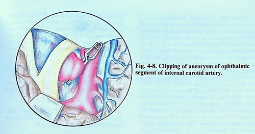
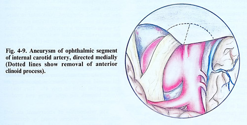
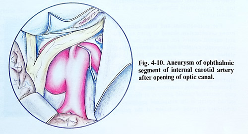
Opening of the inner layer of dura (dura propia) along the lateral aspect if the optic nerve is necessary. This exposes the carotid artery more proximaly and allows better exposure of the origin of the ophthalmic artery. If the neck of the aneurysm extends further proximal then the opening of the distal carotid ring, dissection of superior part of cavernous sinus will be necessary. Once the appropriate segment of carotid artery has been exposed, the extent of the aneurysm neck should be identified and dissection begins. The ophthalmic artery should be dissected free of the aneurysmal neck.
Superior hypophyseal branches as well as the branches to the optic chasm should be identified and preserved at the time of clipping. Intermittent retraction of the optic nerve may be necessary. Depending on the size of the aneurysm, the origin of the posterior communicating artery and anterior choroidal artery may need to be dissected. Prior to clip application, a fine dissector should be passed completely behind the neck of the aneurysm to ensure an easy passing of both aneurysm clip blades. Any apparent resistance should be immediately followed by further dissection technique. Following complete mobi-lisation and dissection of the aneurysm, the decision is made regarding clip selection. Except in aneurysms with very narrow necks, the clip should be placed with the blades along the axis of the parent vessel to avoid kink or tearing of the neck. Sometimes, several sizes or configurations of clips may need to be tried in an effort to obtain complete aneurysm obliteration while maintaining the appropriate lumen dimension of the carotid artery.
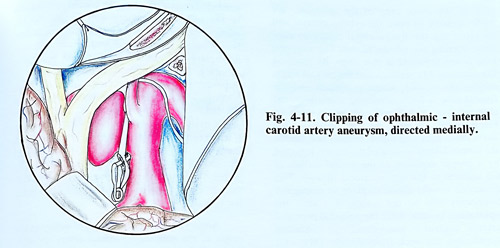
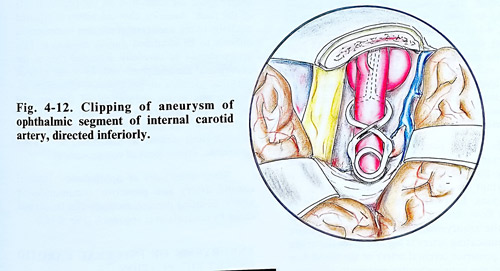
For polilobulated and large aneurysms, strong consideration should be given to proximal carotid control for managing inadvertent intraoperative rupture and as a definitive manipulation to evacuate the aneurysm prior to clip placement (Figs. 4-11; 4-12).
Proximal control however, can be accomplished at three sites as was described in "Tumours invading cavernous sinus". The easiest control of the carotid artery is at the neck. A second option is the extradural exposure and temporary clipping occlusion of the petrous portion of the carotid artery (Glasscock triangle). A third and more distal site for carotid control is the short segment of carotid between the two carotid rings. It is important to leave a small amount of aneurysmal neck between the clip blade and the parent vessel to avoid constriction of the carotid artery. In cases of bilateral carotid ophthalmic aneurysms the contralateral aneurysm occasionally can be clipped through the same craniotomy. This is easyly accomplished, when the contralateral aneurysm is reasonably small and projects medially or posteriorly from the carotid artery.
ANEURYSMS OF THE POSTERIOR COMMUNUCATING ARTERY AND ANTERIOR
CHOROIDAL ARTERY REGION
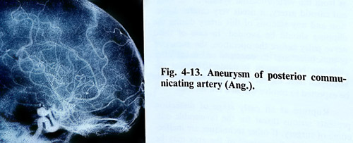
Aneurysms that arise at the junction of the carotid and posterior communicating arteries can be round, elongated, polilobular in shape, varying in size, although aneurysms grater than 2,5 cm in diameter are rarely seen. The most of the aneurysms with this location (86%) project posterolaterally and involve the III cranial nerve in about one third of the cases (Fig. 4-13). In a small group of these patients the aneurysm extends above the tentorial edge and the aneurysmal sac may adhere to the temporal lobe.
Medially directed aneurysms arising at the internal carotid artery-posterior communicating artery junction occur in only 4%. They tend to extend beneath the optic nerve and produce visual symptoms.
The relationship of aneurysmal sac with anterior clinoid process should be determined on the angiography to decide the need of drilling this process.
The anterior choroidal artery aneurysms are rare and arise from the inferior aspect of the internal carotid artery 3 - 6 mm proximally to the carotid bifurcation. They project laterally, while the anterior choroidal artery runs medially.
Surgical technique. With the patient in supine position and the head rotated at 45 degrees to the opposite side, the standard pterional craniotomy with a half-and-half exposure of the frontal and temporal lobes is performed. After opening the dura, a self-retaining retractor is placed under the orbital surface of the frontal lobe and the surgeon follows the edge of the lesser wing of the sphenoid. We prefer initially to open the sylvian cistern, and by retracting the frontoorbital cortex to continue opening the sylvian fissure. The temporal lobe is not retracted at this stage. By reaching the proximal Ml segment and the carotid bifurcation, the carotid cistern is opened and the frontal lobe is freely separated medially to the chiasmatic cistern, allowing additional space for manipulation.
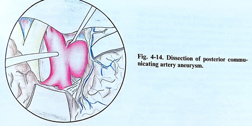
Considering the position of the aneurysmal neck and dome, initially is dissected the antero-lateral surface of the parent artery, followed by a proper exposure of the medial surface of C2 segment in the optico-carotid triangle. This dissection usually allows to place an emergency temporary clip on the carotid just after the ophthalmic branch. After the potential proximal arterial control is guaranteed, the dissection approaches the neck from distally (M1 - C1 segments) and if the aneurysm is not in contact with the medial temporal lobe, uncus can be retracted. The dissection of the neck is completed to allow free passage of the clip blades (Fig. 4-14).
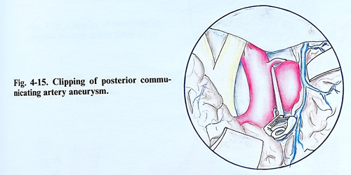
At times a shorter C2 segment or a bigger dural fold laterally to the clinoid may require incision or coagulation of dura to produce safe space for dissection and the proximal clip blade. Clip is positioned preferably along the artery, not stretching the walls of the carotid and not obliterating the posterior communicating artery (Fig. 4-15).
In cases of posterior communicating artery aneurysms, the establishment of the size of this artery and the direction of the blood flow is important for the dissection and clipping of the aneurysm. In cases of a small artery with insignificant blood flow, it can be clipped together with the aneurysmal neck. When the posterior communicating artery is big and gives the origin of the posterior cerebral artery (“fetal” type) or the blood flow is from the vertebrobasilar system to the internal carotid artery (on the Alcock test), it needs very careful dissection and any stenosis of this artery during the clipping should be avoided. In case of third nerve palsy before the operation, the aneurysm is punctured and its contents evacuated after clipping. No attempt is made to dissect the aneurysm from the nerve. Third nerve palsy can be expected to resolve within a few months.
Rupture at an early stage of dissection presents serious threat for the favourable outcome of surgery. If other techniques are ineffective, temporary "trapping" of the area may be needed. Temporary clips are placed just distally to the ophthalmic artery and distally to the aneurysmal neck area, as often retrograde flow is significant. Even then posterior communicating artery can sometimes produce persisting bleeding, but permanent clipping should be done better under these circumstances. Then the temporary ICA clips are removed, starting from the distal one. Ruptures close or at the aneurysmal neck require always temporary trapping.
If a clip cannot be placed effectively on the neck, the artery is clipped by a specially designed fenestrated clip or is wrapped preserving its patency as suture can be time consuming. Permanent carotid occlusion at the neck is the last remedy.
Aneurysms of internal carotid artery at the origin of the anterior choroidal artery are situated a little higher and at 3 - 6 mm from the internal carotid artery bifurcation. That allows bigger space for dissection on the proximal side of the neck. Anterior choroid artery is situated medially to the aneurysmal sac and sometimes is difficult to be identified during dissection. This artery should be carefully dissected free of the aneurysmal sac and preserved. In about 15% of patients, sacrifice of the anterior choroidal artery results in the infarction of the internal capsule. This artery can arise from the aneurysmal sac and its clipping may provoke acute neurological deficit as hemiplegia, hemianaesthesia, even impairment of consciousness.
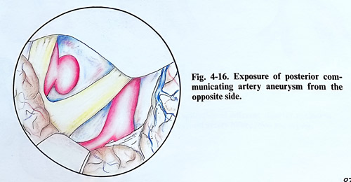
Some aneurysms of ophthalmic - internal carotid artery and posterior communicating artery, pointing postero-medially can be clipped easily by contralateral approach (Fig. 4-16).
ANEURYSMS OF INTERNAL CAROTID ARTERY BIFURCATION
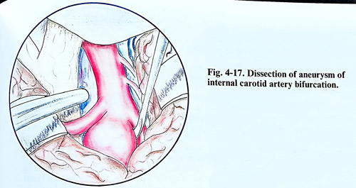
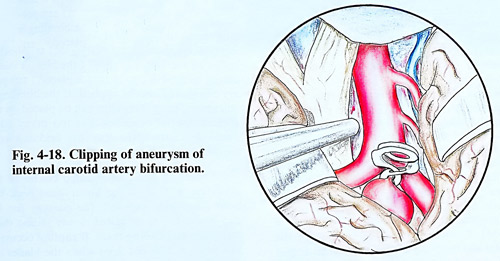
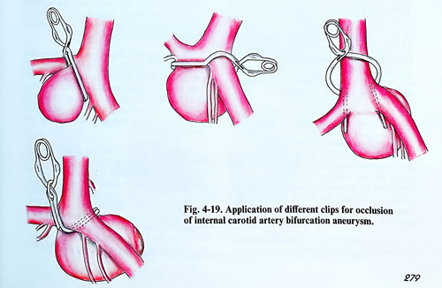
They represent 5% of all intracranial aneurysms and are located between the anterior and middle cerebral arteries. Most often project superiorly into the anterior perforate substance or posteriorly, distorting the optic tract. Frequently these aneurysms excavate bed in the anterior perforate substance. Sometimes they are direct continuation of internal carotid trunk or have a broad-based neck that incorporates the junction of the internal carotid artery together with part of the anterior cerebral or middle cerebral arteries trunks. These anatomical peculiarities demand fine and cautious dissection of the aneurysmal neck to avoid any damage of the central arteries, arising from the proximal segments (Al and M1) of anterior and middle cerebral arteries. The dissection should be sufficient to insert and then inspect the clip for inclusion of any perforators prior to its final placement (Figs.4-17; 4-18; 4-19).
Further inspection must confirm that the origin of anterior and middle cerebral arteries is not compromised by clip placement and that the aneurysmal neck is completely obliterated. The majority of surgical complications are attributed to the occlusion of perforating arteries that arise from the proximal segment of the anterior and middle cerebral arteries or adherence of anterior choriodal or the recurrent artery of Heubner to the aneurysm.
Tearing or rupturing of the aneurysmal neck should be avoided by all means because there are no effective options for management of this complication. An encircling clip will occlude the perforators. The only option might be trapping the aneurysm, which deprives circulation to the hemisphere and requires an emergency bypass. Intraoperative angiography is advisable for complex aneurysms of the region because of the difficulty in accurately visualising the anatomical details.
ANEURYSMS OF THE MIDDLE CEREBRAL ARTERY
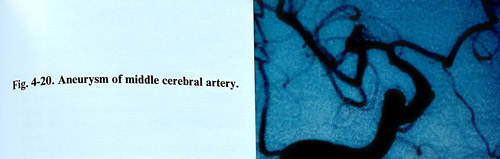
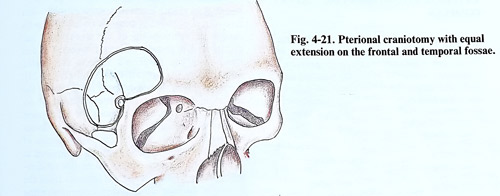
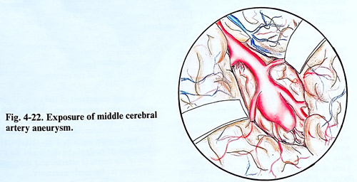
Commonly middle cerebral artery aneurysms arise at the bifurcation or trifurcation of this artery and are often partially embraced by the distal insular branches (Fig. 4-20). In a few cases the aneurysms arise from the more proximal part of the middle cerebral artery, often at the origin of the anterior temporal branch. The frontotemporal or "pterional" craniotomy with equal extension on the frontal and temporal fossae is the most suitable to reach middle cerebral artery aneurysms (Fig. 4-21). The dura is opened with a flat half-circle shaped line based anteriorly. The next step is to elevate the frontal lobe and identify the ipsilateral optic nerve, using gentle retraction. The chiasmatic and carotid arachnoid cisterns are opened, and CSF is removed from the basal cisterns, as this provides yet more brain relaxation prior to sylvian dissection. Beginning from the level of internal carotid artery, using a sharp arachnoid knife, the arachnoid bands connecting both lobes are divided. Continued opposing traction of the frontal and temporal lobes facilitates splitting of the Sylvian fissure. The small veins bridging the fissure can be coagulated and divided, and the main venous trunk (Sylvian vein) is usually retracted together with the temporal lobe (Fig. 4-22).
The more common distal bifurcation aneurysms can be approached after directly exposing the Ml segment if it is long enough or following carotid bifurcation if it is short. During the dissection blood clots should be washed out from within Sylvian fissure in an effort to minimise postoperative vasospasm.
When a large temporal haematoma is present, a cortical incision is made where the clot is close to the surface, usually through the superior temporal gyrus, until the haematoma is entered. The haematoma can be removed completely until the aneurysm is reached. This manipulation should be very cautious to prevent aneurysm rupture, or the haematoma is removed partially enough to slacken the brain and then the surgeon turns to a transsylvian exposure of the aneurysm.
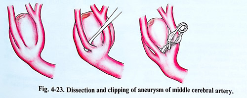
Once the aneurysm is reached the anatomy of the area should be exposed in a systematic way. Since the Ml segment at the level of the aneurysm bifurcates or trifurcates, all branches need to be identified. It is not possible absolutely to predict from the angiograms how many branches arise from the main divisions of the middle cerebral artery (Fig. 4-23). When these vessels are adherent and complete dissection of the aneurysm needed, or when the aneurysm dome is very thin walled, Ml temporary occlusion may be carried out for repeated short intervals as required to facilitate dissection by softening the aneurysm. If the aneurysm bleeds during its final dissection, proximal Ml temporary clip should also be applied. If the bleeding is still severe, temporary clips can be placed on the efferent arterial branches, but too many additional clips narrow the operative field and disturb further dissection. Otherwise in uncomplicated course of the operation and when the aneurysmal neck has been adequately exposed, the passage for the clip blades has to be tested slowly sliding them into place and closing them. If rupture occurs at this point and it stops when the blades are filly closed, it means that the hole was distal to the clip and the blades completely occluded the neck of the aneurysm. If bleeding originating proximal to the clip continues, the surgeon resorts to temporary clipping. The place of rupture is found and the clip positioned to isolate the aneurysm and rupture site.
Some large MCA aneurysms which project anteriorly from the bifurcation can almost reach the surface of the Sylvian fissure. Such aneurysms should be suspected from the angiograms, and because of the blood in the subarachnoid space, the red aneurysmal dome can be difficult to identify during dissection and can be damaged inadvertently. Large aneurysms of this type can block proximal Ml exposure in some cases and this relation may need the aneurysm to be approached from the distal part of the Sylvian fissure. If during the dissection of the Sylvian fissure the aneurysm ruptures prior to its identification, a temporary clip should be placed upon Ml and arterial blood pressure maintained in normal or slightly hypertensive values. Dissection must be quick, but safe in exposing M1, and an area for tentative clipping at a more distally should be considered as early as possible through the dissection. After clipping, the entire aneurysm and clip should be visualised from all sides, ensuring complete obliteration and absence of any branches included within the clip blades, with clip repositioning (definitive clipping) if necessary. On certain very rare occasions a small residual part of the aneurysm can be left unobliterated in order to preserve some major vessel origin patent. In such a case the residual part can be only wrapped.
ANEURYSMS OF ANTERIOR CEREBRAL - ANTERIOR COMMUNICATING ARTERY
COMPLEX
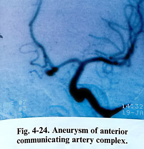
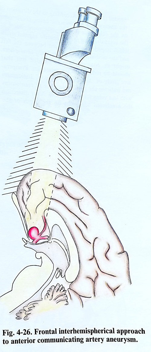
The aneurysms of the complex of the anterior cerebral and anterior communicating arteries are reached through a pterional craniotomy. For some aneurysms pointing anteriorly and adherent to the chiasm and tuberculum sellae dura can be used an interhemispherial approach or frontoorbital craniotomy (Figs. 4-24; 4-26).
The dissection in a pterional approach begins from the internal carotid artery, continues with its bifurcation, and anterior cerebral artery without uncovering completely or interrupting any branches. At opening the internal carotid cistern the arachnoid is additionally divided distally to the sylvian fissure, providing larger field and access to the anterior communicating artery area with less retraction pressure applied to the cortex. Frontal lobe retraction in anteriorly directed aneurysms should be cau-tious, as adherence of the dome to the basal dura can provoke premature rupture. Aneurysms with more complex anatomy requiring broader dissection area may need an additional orbitozigomatic osteotomy to reduce retraction.
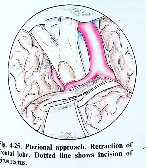
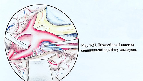
Once carotid is fully exposed, the surgeon follows Al segment and carefully separates gyrus rectus and olfactory tracts from the chiasm to gain approach to the opposite Al, and by that complete proximal vascular control. For broader exposure of the anterior communicating artery complex aneurysms the ipsilateral gyrus rectus can be partially removed under the pia outside the interhemispherical fissure, usually to avoid premature rupture (Fig.4-25). In that way, the interhemispherical fissure, usually very tight after SAH, becomes easier to dissect. The interhemispherial space, however should be opened following the Al and anterior communicating artery, and than the initial part of A2. This sequence of structures to expose should give access to the neck of the aneurysm before the fundus, with secured proximal control (Fig. 4-27).
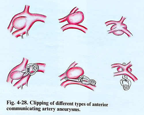
As in other locations of cerebral aneurysms, a great variety is observed in respect to the size, shape and interrelation to large arteries. Most often segments A2 arise from only one Al segment. The aneurysm is most frequently found at the bifurcation of Al and is usually directed upward. During the dissection of the aneurysm the surgeon must be guided by the angiography. From the angiographic data however, is not always possible to establish with details the relationship of the aneurysmal sac and its neck in respect to the main vessels and in these cases the surgeon's actions are guided primarily by the operative findings (Fig. 4-28).
Great attention should be paid in dissecting the proximal portions of the anterior cerebral arteries not to approach too near the bifurcation of the internal carotid artery, as some perforating arteries may be injured. The appearance of a recurrent (Heubner) artery should be noticed in advance. At clipping care is taken not to compromise the hypothalamic perforators from the posterior surface of the anterior communicating artery complex.
Large aneurysms of the anterior communicating artery aneurysm and those with intraoperative rupture on the neck, may require special technique of occlusion: special clips, trapping, even suture. For this purpose the dissection of both distal and proximal parts of the cerebral arteries and their temporary occlusion is absolutely necessary.
ANEURYSMS OF THE PERIPHERAL PART OF THE ANTERIOR CEREBRAL ARTERY
The aneurysms of the peripheral part of the anterior cerebral artery are most frequently found at the origin of the callosomarginal artery. There are also aneurysms at the origin of the frontopolar artery, in the region or branching of the callosomarginal artery and in the distal part of the pericallosal artery.
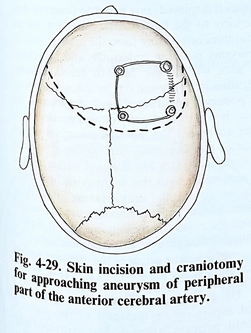
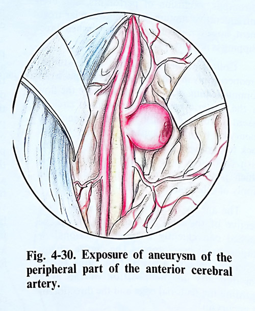
For surgical approach of these aneurysms, craniotomy close to the midsagittal line is needed (Fig. 4-29). According to the direction of the aneurysmal sac, in some cases, craniotomy on the other side is more convenient. Some of the veins draining in the superior sagittal sinus, which impede the penetration between the falx and the medial surface of the brain hemisphere, might be coagulated and divided but that should be reduced to the minimum. Preoperative study of the venous phases of angiograms can indicate the preferable place of craniotomy with less veins for interruption. After dissection the arachnoid adhesions on the depth of the interhemispherial fissure, one or both pericallosal arteries and the aneurysm are reached (Fig.4-30).
Very often these aneurysms do not lie freely in the subarachnoid space, but are buried into the medial surface of the frontal lobe or in some cases into the opposite hemisphere. There can be an anastomosis at the level of the aneurysm between the pericallosal arteries similar to the anterior communicating artery. In such cases the surgeon should protect it.