EXTRA-INTRACRANIAL ARTERIAL ANASTOMOSIS
The results of extra-intracranial bypass study published in 1985 indicated that this technique was not effective in reducing stroke incidence or stroke related death. Currently the operation is applied rarely in selected patients who have a vascular lesion directly related their symptoms and who have decreased blood flow in the corresponding hemisphere on cerebral blood flow study. The same patients should not have effective collateral circulation on evaluation of their angiograms and functional tests.
Extra-intracranial anastomosis is applied also with prophylactic aim when an ischaemic lesion is expected in the territory of the internal carotid or the middle cerebral arteries before the removal of big basal tumours, which envelop the internal carotid artery. In view of all this, cerebral revascularization with different EC -IC techniques is not any more an accepted routine in cerebral ischemia.
Once the patient has been decided to be in need of cerebral revascularization, his angiographies are evaluated for donor and recipient arteries and the possible site of anastomosis. If donors are deficient in quality, vein grafting is considered. The general condition of the patient is evaluated regarding intracranial surgery under general anaesthesia with several hours duration and coagulation profile is adjusted to the required, as surgery is performed either with normalised or heparinized patient. That will also demand careful monitoring of coagulation after surgery.
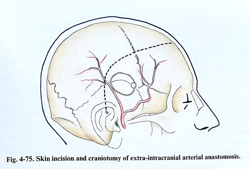
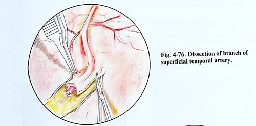
The skin incision is linear, along the course of the superficial temporal artery (Fig. 4-75). If the position of the artery cannot be palpated, the portable Doppler device can by very useful. The dissection should be done in a way not to damage the small branches or the superficial temporal artery itself. Its anatomical location is between the skin and galea, so once it is exposed distally, the dissection proceeds following it at the above mentioned anatomical plane. Either the parietal or the frontal branch of the artery is selected for anastomosis (Fig. 4-76). Together with the arterial branches are also running veins, which are recognised by the lack of pulsations. The temporal muscle is incised in a linear or " T " shape and the bone surface is exposed. A burr hole is placed and is enlarged with a craniotomy as a small free flap.
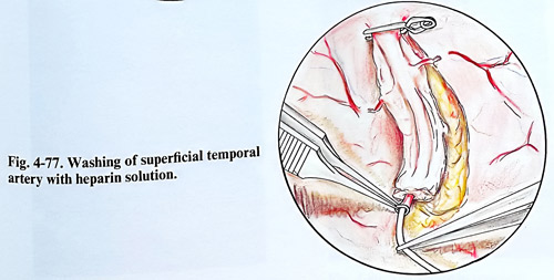
The dura is opened with a stellate incision and the tips of the flaps are lifted with traction sutures. A suitable recipient artery (about 1 mm in diameter) is identified on the cortex. The artery may be any branch of the middle cerebral artery, but a temporal one is preferable. If a suitable branch cannot be found or it appears very close to the edge of the craniotomy, small additional nibbling is required. If the cortical surface is tending to protrude, the arachnoid is opened. Seldom has to be resorted to a lumbar puncture. More often at completion of the anastomosis, the cortex is separated from dura and care is taken to avoid subdural collections postoperatively. When dissecting the donor, the periarterial tissue is left around it, coagulating and cutting all branches. The length of the dissected artery must be about 5 cm. The adventitia is removed only from the isolated end of the superficial temporal artery and its proximal part is clipped. A thin catether is inserted into the arterial cavity for washing with heparin solution (Fig. 4-77).
Next is isolated the cortical artery and the arachnoid around it is opened with microscisors. Small branches are coagulated and divided. A segment from the artery up to 1 cm long must be isolated. Its diameter should not be less than 0.8 mm. A fine dissector or hook is passed under the artery, to verify whether it is free and after this a rubber plate (with a thickness of a surgical glove material) is inserted under it. The artery is lifted on the plate to protect the underlying brain cortex and permit easier manipulation with the microinstruments on the artery.
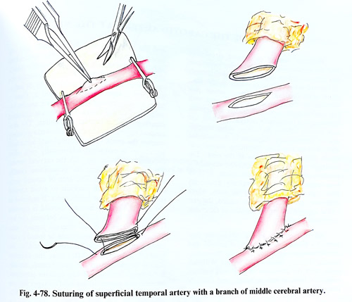
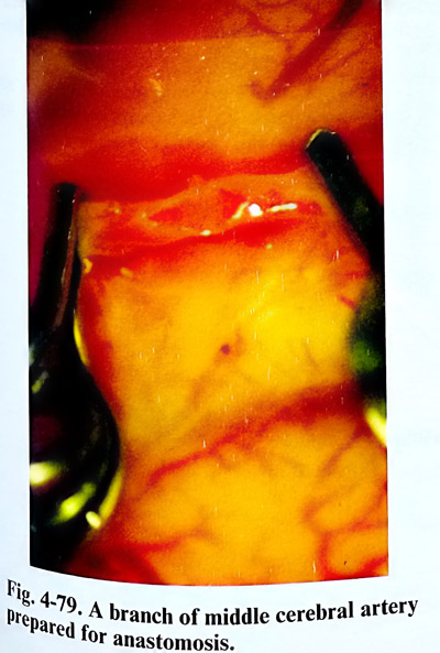
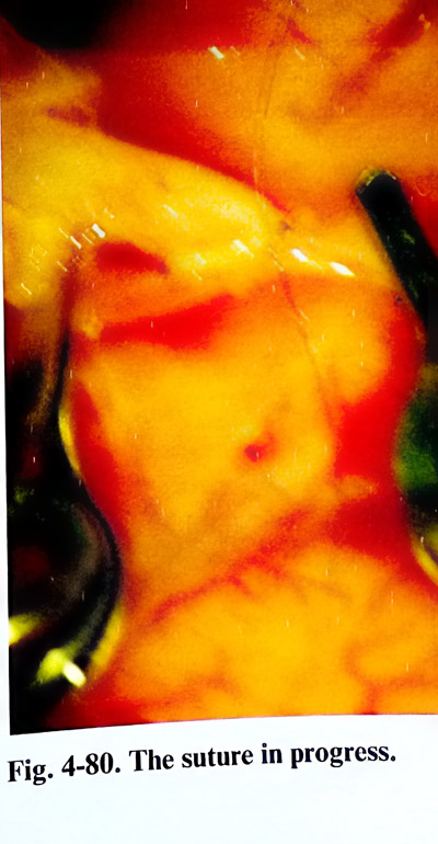
Two clips are placed at both ends of the isolated part, clipping the cortical artery together with the rubber plate. The arterial cavity is opened with a small blade or curved microscissors. An elliptic opening is completed with the curved microscissors. The cavity is washed with heparin solution. The peripheral end of the superficial temporal artery is cut obliquely, to get wider aperture to the anastomosis. The later is performed with about 10 to 12 single sutures with a needle and material 10 - 0 (Fig. 4-78). Before the last suture, the clip on the proximal part of the superficial temporal artery is slightly released so that air bubbles are pushed out. All clips are removed, starting with those on the cortical artery and after that on the superficial temporal artery. The place of the anastomosis is slightly pressed with cottonoid and oxidise cellulose, as very often minor bleeding occurs at the place of the sutures, which stops in a few minutes only with this technique. If the bleeding continues, clips are applied anew and an additional suture is placed (Figs. 4-79; 4-80).
The dural flaps are turned back on the cortical surface and closed with single sutures.
The temporal muscle and its fascia are closed separately, taking care not to strangulate the donor artery. The skin is sutured, applying fine drainage in the epidural space.
Operative variants. A double anastomosis can be performed, using both branches of the superficial temporal artery and each of them is connected with a separate cortical branch. No obvious benefit has been shown yet from this technique.
The occipital artery can be used for anastomosis with a branch of the middle cerebral artery, when the superficial temporal artery and its branches are inadequate.
Another option for the high-flow requirements compensation of the middle cerebral artery circulation after carotid occlusion are the vein grafts. Two types have been used: "short" - between the proximal superficial temporal artery and a cortical branch, and "long" - between the carotid artery in the neck and the middle cerebral artery branches. The main characteristic property of vein graft is their diameter and consequently, the higher flow through them. "Short" grafts are substitute of inadequate for anastomosis superficial temporal artery branches, thus providing sufficient diameter and length of the donor. "Long" grafts intend to provide the benefits of the proximal high flow and pressure in the carotid, delivering them directly to the middle cerebral artery circulation. Technically the graft is anastomosed on both ends with microsutures and passed through a tunnel. "Short" grafts are positioned in a similar way through the tissues as the standard STA - MCA bypass donor artery. "Long" venous grafts however, require tunelization between the wound at the neck and the craniotomy usually positioning them retro- or pre-auricularly. Grafts are heparinized after harvesting and are sutured with continuous or interrupted monofilaments under magnification. Postoperative angiographies after these techniques show very often excellent filling through the graft.
There are options for revascularization of the posterior circulation and the donor (most often occipital artery) is sutured to PICA branch. Another approach uses STA to reach the superior cerebellar artery, via tentorial incision. Few centres have developed and are performing these techniques.
Postoperatively the wound care present occasionally problems with healing. It is a result from the devascularized wound edges at surgery. That however rarely requires any special treatment and is resolved within the usual period of time. Another important point to remember is the position of the donor or graft under the soft epicranial tissue. It permits easy Doppler control of the flow, but can be pressed or injured there much easier.