ANEURYSMS OF POSTERIOR CIRCULATION
The history of surgical treatment of aneurysms of the vertebro-basilar system is much shorter than that of the carotid, probably due to the difficulties of the greater surgical inaccessibility to these arteries.
In view of operative technique, the aneurysms of the vertebro-basilar system can be divided into six groups: at the bifurcation of the basilar artery, at the posterior cerebral artery, at the superior cerebellar artery, at the midbasilar trunk, at the vertebrobasilar junction, and at the vertebral artery.
ANEURYSMS OF THE BASILAR ARTERY BIFURCATION
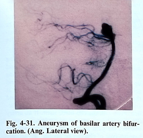
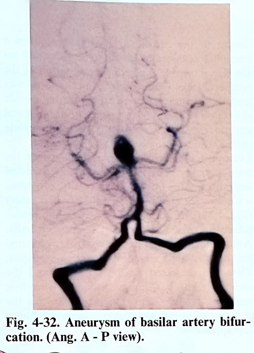
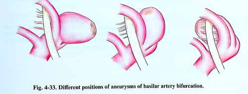
Basilar bifurcation is one of the commonest places of posterior circulation aneurysms. The aneurysms are directed anteriorly, superiorly or posteriorly, and special attention is paid on its relation to perforating arteries, usually originating on its posterior wall (Figs. 4-31; 4-32; 4-33). Direct clipping is preferred, as endo-vascular attempts are at times associated with incomplete thrombosis or propagation of the thrombus, occlusion of perforators, and even intraoperative rupture. The endovascular treatment, however, is considered always when direct clipping is not possible.
Collateral circulation has to be studied preoperatively in details. The ability of posterior communicating arteries to compensate the flow in the posterior part of the Willisian circle is essential for predicting the consequences of aneurysm trapping. Opposite to that, the Alcock test (vertebrobasilar angiographic imaging with carotid compression), reveals the role which posterior circulation may play for the supply of other vascular territories.
The aneurysms of the basilar artery, irrespective of the level of their location can be reached conveniently through a pterional or temporobasal craniotomy. The choice of approach for clipping a basilar bifurcation aneurysm depends on the position of the neck regarding the tentorial edge and the direction of the aneurysm.
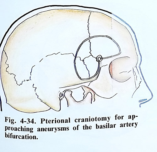
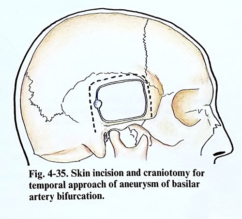
High position of the basilar bifurcation requires extensive subtemporal retraction avoided in transylvian route. This route is gained through a pterional craniotomy (Fig. 4-34). An additional orbitozigomatic flap can provide better angle of observation to highly positioned lesions. Aneurysms directed anteriorly or not high in the interpeduncular fossa are better approached subtemporally (Fig. 4-35). Most difficult to clip are aneurysms directed posteriorly, as very often that can compromise perforants. Broad view of more difficult lesions, even combining pterional and subtemporal approaches can provide safer dissection and clipping. The subtemporal approach can be extended by anterior petrosectomy. Anteriorly directed lesions are reached subtemporally, remembering that their dome can be adherent to dorsum sellae. Low position of the basilar bifurcation can require tentorial incision, always caring for the trochlear nerve under the tentorial edge.
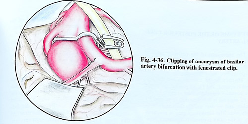
As a general rule the interpeduncular cistern in subtemporal approach is reached by tracing the superior cerebellar artery and finding its origin from the basilar artery and the Pl segment of the posterior cerebral artery. In this penetration the surgeon retracts the IIIrd cranial nerve in the same direction as the parahyppocampal gyrus is displaced. It is better to penetrate first anteriorly to the basilar artery as there are no perforators originating from the arterial wall. In aneurysms pointing anteriorly and superiorly the neck will be the next to reach. Greater difficulties are encountered in the dissection of the posterior aneurysmal wall. Sometimes the peduncle has to be slightly moved posteriorly in order to reveal the posterior surface of the posterior cerebral artery, the basilar artery and the aneurysmal sac. The fine perforating arteries are seen better with this slight retraction and away from the aneurysmal neck. Having found these vessels, a blunt probe or hook must be inserted behind the aneurysmal sac to displace it slightly in anterior direction. In this manipulation of the aneurysmal sac the origin of the opposite posterior cerebral artery must also be seen (Fig. 4-36).
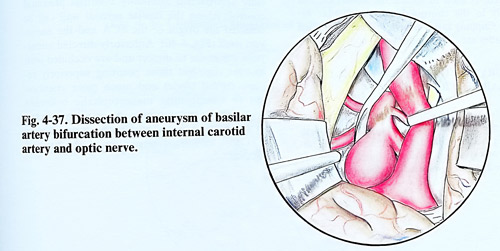
The basilar aneurysms whose neck is located superiorly to the level of the postrior clinoid process are reached conveniently through the pterional approach and a corridor between the carotid and the optic nerve or lateral to the carotid artery. That requires a pterional craniotomy, with some extension on the temporal side for better mobilisation of the temporal lobe. Dura is opened in the usual way and the carotid artery is exposed after a wide dissection of the Sylvian fissure. Both optic and carotid cisterns are dissected to the complete mobilisation of the artery and the nerve. The Lilliequist membrane is opened to expose the basilar artery. The carotid artery is retracted laterally and the optic nerve medially, providing a window for manipulation on the basilar artery (Fig. 4-37). This direction of view on the basilar bifurcation visualises both Pl segments usually. A place for proximal basilar control is prepared and it is just above the posterior clinoid process. Then are dissected both Pl origins and the aneurysmal neck. Clipping through this route is requiring often bayonet and thin blade clips, as the problem of obstructed vision is frequent during clip application.
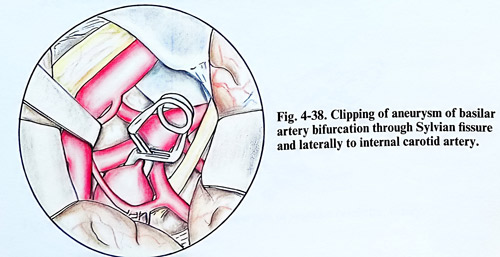
When the space between the optic nerve and internal carotid artery is narrow, the aneurysms of the basilar bifurcation above the level of the posterior clinoid process can be approached laterally to the internal carotid artery. After opening of the carotid cistern and Lilliequist membrane, the internal carotid artery is retracted medially to expose the posterior communicating artery and its perforating branches, Pl segment, and aneurysm neck. The posterior communicating artery is retracted medially. The dissection of the aneurysm neck and the ipsilateral proximal Pl segment completes the path for clip application. (Fig. 4-38).
There are some particular tips for clipping according to the direction of the aneurysm and the approach to it. Those aneurysms projecting up and forward don't allow the opposite P1 and some perforators there to be seen. Therefore at the time of clip blade passing, the neck and aneurysmal wall require gentle minimal retraction, even with the clip blade itself. If an artery (Pl segment for instance) or oculomotor nerve obstruct the path to the neck, a fenestrated or bayonet type of clip can be applied. Posteriorly directed aneurysms present difficulties with preservation of perforating arteries. Paying maximum attention to that avoids ischaemic complications of the brain stem.
Some aneurysms after complete dissection can result unclippable as is the case when haemodinamically important Pl or multiple perforators originate from its dome. In such a case the only solution remains with wrapping, a technique with uncertain effect.
ANEURYSMS OF THE POSTERIOR CEREBRAL ARTERY
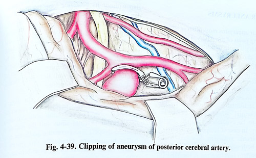
The aneurysms of the posterior cerebral artery are most often originating from the P2 and P3 segments of the artery. These segments are approached most conveniently subtemporally. Medially to the tentorial notch the arachnoid is opened, and P1, P2 point at the origin of the posterior communicating artery is identified. P2 segment is followed distally to find P3 branches, and by that usually the aneurysm is already seen. The aneurysmal neck is prepared caring of the small perforators appearing from that segment (Fig. 4-39). Temporary clipping, if required, is used to isolate the aneurysm, but distally to the choroidal and peduncular arteries. Clipping rarely produces complications if the blades are closed under visual control.
ANEURYSMS OF THE SUPERIOR CEREBELLAR ARTERY
The aneurysms of the trunk of the basilar artery are situated most often in its upper third, at the place of separation of the superior cerebellar artery or in its lower third, where the inferior anterior cerebellar arteries come out.
The aneurysms of the upper third of the basilar artery arise most often from the distal angle between this artery and the superior cerebellar and have a well formed neck. They are situated anteriorly or laterally in the subarachnoid space, are not closely attached to the perforating arteries and may form a bed in the peduncles.
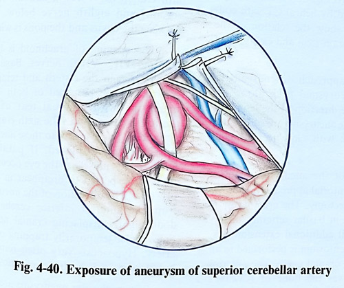
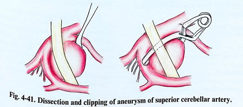
The approach to the aneurysms in this location is always subtemporal. After the mesencephalic cistern is opened and the trochlear nerve is preserved, the tentorial edge is retracted with a stitch, and the arachnoid is divided, anteriorly through the Lilliequist membrane. At this stage P1, and P2 arterial segments are clearly identifiable and the basilar trunk is carefully approached intending eventual proximal control. Along the basilar arterial wall can be detected the origin of the SCA and the aneurysm itself (Fig. 4-40). Anteriorly directed aneurysms are easier to dissect and are occluded with straight clip, but those pointing superiorly require curved blades and much more attention not to compromise mesencephalic branches (Fig. 4-41). Oculomotor nerve is often affected by the dome, can suffer damage during dissection and may need retraction at the time of clipping.
MIDBASILAR ANEURYSMS
Midbasilar aneurysms most often originate at the emerging point of the AICA. This part of the basilar artery is more difficult for access and very selective choice of supra-, infra- or combined transtentorial approaches do exist, demanding very appropriate decision-making.
The difficulties and risks faced with open surgery give preference to endovascular techniques in those cases where perforators or the AICA itself are not too close to the aneurysmal neck and can be accidentally compromised by the occlusion. Potential collaterals of the basilar artery have to be evaluated by angiography before surgery. In case of temporary clipping of the basilar trunk, sufficient flow has to be provided on both sides of the trapped segment.
Another important investigation is the MRI (in some cases substituted by helical CT with contrast) and it has to show the relation of the lesion to clivus, the brain stem and emerging of cranial nerves.
The choice of approach depends on the position of the aneurysmal neck along the basilar trunk and its direction. In this regard midbasilar aneurysms are subdivided into two subgroups: with "low" and "high" position.
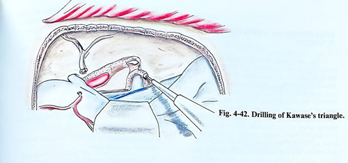
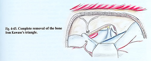
High position of the aneurysms requires subtemporal, transtentorial approach preferably with anterior petrosectomy. The craniotomy is similar to that used in basilar top aneurysms, but with small additional extension posteriorly. Section of tentorium is performed in all cases, preserving IV cranial nerve. The anterior petrosectomy removes the bone of Kawase's triangle in the medial petrous apex and provides space for approaching the petroclival region of the upper posterior fossa, and to the upper half of the basilar trunk (Figs. 4-42; 4-43).
Anterior petrosectomy technique includes selective removal of part of the petrous bone. After separation of the dura from the temporal fossa, the exposed bony surface reveals a number of important structures and middle meningeal artery is divided at its exit from foramen spinosum, the greater superficial petrosal nerve is sacrificed and the bone toward its origin at the geniculate ganglion is drilled out. The horizontal part of the ICA is exposed also without its posterior loop because of the danger of bleeding. The remaining bone medially and anteriorly to the geniculate ganglion and the superior semicircular canal (arcuate eminence) can be drilled out safely. The dural sleeve of the internal acoustic meatus has to be exposed, and if the drilling is always medially to the Miller's line (connecting intersection of carotid and trigeminus with Bill's bar), there is no danger of damaging cochlea.
In the subtemporal route, the dura is opened and a spatula is placed to retract the temporal lobe. The tentorium is divided along the superior petrosal sinus and the spatula is repositioned deeper, also retracting gently the cerebellar hemisphere. Gradually this spatula is moved, until it exposes the pons between the fifth cranial nerve above and the roots of the seventh and eighth nerve below. The space between the clivus and the pons is widened.
The opening of arachnoid is between the Vth and VII - VIIIth nerves. Basilar artery is usually seen in the cistern and temporary clipping places are prepared of both sides of the aneurysm, with minimal tear of adhesions around the aneurysm. The sixth cranial nerve is situated in close vicinity to these aneurysms, it is dissected and displaced. If paralysis of the lateral rectus muscle of the eye occurs, it is most probably expected to disappear within few days or weeks. The dissection of the aneurysmal neck from the anterior inferior cerebellar arteries done gently displacing cranial nerves and at times under temporarily trapping of the basilar artery. If occurring, hypoacusis very often remains lasting, but facial palsy has a better prognosis.
Low midbasilar aneurysms are approached through lateral suboccipital craniectomy, at times enlarged with petrosectomy.
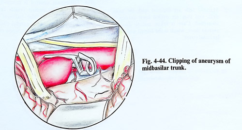
In the lateral suboccipital approach, after retraction of the cerebellar hemisphere, arachnoid medially to floculus is opened and all the space between the VII - VIIIth and IX - Xth nerves is used for the approach. The dissection further on follows the same principle as in the previous approach. Very often have to be used angulated clips to avoid basilar artery stenosis (Fig 4-44).
ANEURYSMS OF VERTEBROBASILAR JUNCTION
The aneurysms of vertebrobasilar junction create significant problems for treatment. Located in one of the most difficult to approach places, they very often have complex configuration and poorly defined neck, thence straight forward decision for the method of treatment cannot be taken with ease. Options include endovascular and direct techniques, but preference is given to the first one. When the aneurysm doesn't have arterial branches arising very close to its neck and its shape and neck after thrombosis will not compromise the flow in the parent artery, is proceeded with interventional neuroradiology. It is common however, to find a situation when the direct attack is the only choice. In these cases is considered the low lateral suboccipital approach, which can be extended with posterior petrosectomy and temporal flap, or if the lesion is closer to foramen magnum, removal of the medial third of the occipital condyle.
The posterior petrosectomy is an adjunct to the confined supra- and infratentorial approaches (posterior temporal and lateral suboccipital). After these craniotomies are completed, several further steps are followed to gain the presigmoid route. These steps in brief are as follows:
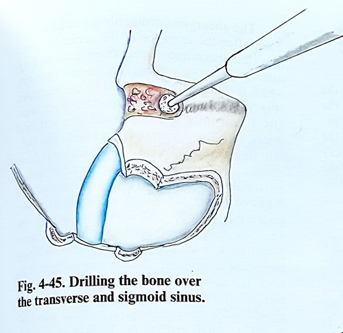
1. The bone over the transverse and sigmoid sinuses is removed completely with the high-speed drill (Fig. 4- 45).
2. Mastoid cells are exposed with the same drill, and they are drilled until antrum is identified. Care is taken not to damage antrum floor, which is the lateral semicircular canal cover.
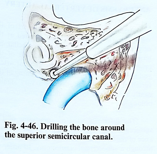
3. The sino-dural angle bone is removed with the diamond drill to the posterior semicircular canal, together with the remaining bone around the superior semicircular canal to expose the superior petrous sinus (Fig. 4-46).
4. Mastoid tip cells are removed also to the digastric ridge (close to the stylomastoid foramen). The Fallopian canal is followed superiorly at the same depth as the posterior semicircular canal.
5. Dura is opened superiorly and inferiorly to the superior petrous sinus. The sinus is clipped (ligated) and divided.
6. The tentorium is incised parallel to the superior petrous sinus.
This approach preserves all structures, except the superior petrous sinus. It mobilises the transverse to sigmoid junction and the entry point of the vein of Labbe. All these steps, however, require skills, which can be mastered only with sufficient additional training with the otological surgeon. Dura is opened from foramen magnum upward to the most superior part of the exposure. CSF is evacuated from cisterna magna.
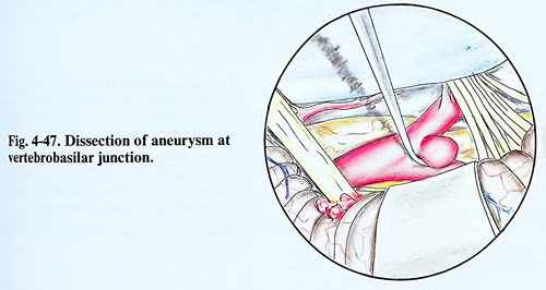
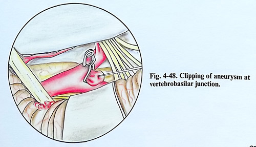
Floculus is retracted and the lower cranial nerves are exposed. Under the nerves is identified the vertebral artery, and it is followed distally. After the visualisation of the PICA origin, the vertebral trunk is exposed between the VII - VIIIth and IX - X - XIth nerves, reaching the junction and the aneurysmal neck (Fig. 4-47). The dissection cares for the perforators and the anterior spinal artery origin in this area. Temporary control is extremely difficult, but the accessible parent vessels can by controlled with endovascular balloons navigated there. Clipping requires careful choice of the shape of blades to avoid distortion of the junction (Fig. 4-48).
ANEURYSMS OF VERTEBRAL ARTERY
Aneurysm of the intracranial part of the vertebral artery are most often situated at the origin of the posterior inferior cerebellar arteries. As a result to the variable locations of the origin of the posterior inferior cerebellar artery and the variety of shape and lumen of the vertebral artery, these aneurysms vary significantly in their relations to midline and medulla oblongata.
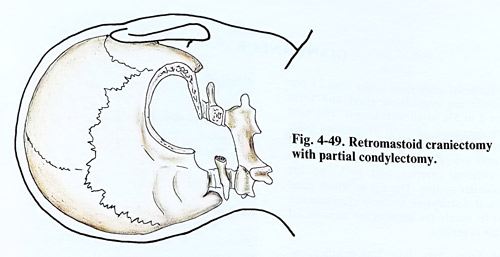
The inferiorly located aneurysms of the vertebral artery, especially those at the appearance of the PICA are most conveniently reached through a suboccipital craniectomy. In order to expose the space anteriorly to the pons and the medulla oblongata better, the craniectomy is widened on account of the resection of the posterior lamina of the atlas reaching the atlanto-occipital joint (Fig. 4-49). The vertebral artery entry through the atlanto-occipital membrane is reached and the posterior part of the occipital condyle is drilled out to the hypoglossal canal. The posteromedial one third of the condyle and the lateral mass of atlas can be removed without biomechanical compromise. This removal can be time consuming and not necessary in many cases of more distally located lesions.
Dural incision begins at Cl and extends vertically through foramen magnum to proceed laterally to the superolateral apex of the craniotomy and cisterna magna is opened.
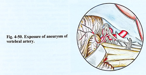
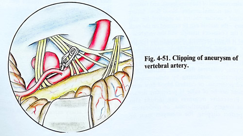
Aneurysms usually are situated 1 or 2 cm after the entrance of the vertebral artery into the subarachnoid space from foramen magnum and anteriorly to the first dentate ligament, which is thinned and distended by the aneurysm (Fig. 4-50). In order to achieve some additional space laterally to medulla and to dissect the vertebral artery with the aneurysmal neck well, this dentate ligament is divided. Additional bone removal allows control of the vertebral artery proximally for clipping. The vertebral artery is followed under the spinal accessory and next under the IX - X - XI nerves to identify the anatomy of the aneurysm. Sometimes the aneurysmal neck is deeper to the posterior inferior cerebellar artery and towards the midline. The neck is, however, quite always at the angle formed by the origin of PICA. The dissection should safely assure clipping without affection of PICA, small arteries or the flow in the VA (Fig. 4-51).
At present postoperative care in addition to the routine can include some additional important measures, also applicable to ruptured aneurysms with higher risks in other locations:
1. The triple H therapy: Hypervolemia, hyper-tension, haemodilution, with invasively monitored parameters of hemodynamics (preferably with Swan Ganz catheter).
2. Transcranial Doppler monitoring of vasospasm.