INTRACEREBRAL HAEMATOMA
Inracerebral haematomas, except those of traumatic origin, are due usually to hypertension and have predilected locations. About 50% of them are in the basal ganglia, 15% in thalamus, 10% in the cerebellum and 10 to 20% in the white matter of the hemispheres. In patients without hypertensive disease, the cause of haematoma is most frequently rupture of an aneurysm, arteriovenous malformation orcoagulopathy.
The basic principles of surgical treatment of intracerebral haematomas are completely different from those in other parts of the human body. In case of haemorrhage outside of the intracranial cavity the aim of surgery is to stop the bleeding as early as possible, to treat the resulting anaemia, and save patient's live. In intracerebral haemorrhage blood is of limited amount and does not affect circulation, rapidly provokes destruction of the brain and raises the intracranial pressure. The neurological disturbances resulting from the bleeding are due more to destruction of the brain tissue then to the blood volume as an expanding lesion.
This concept explains the controversy existing in the choice of method of surgical treatment in intracerebral haemorrhage. Till now there are differences regarding the indications and the potential benefit of the surgical treatment.
SELECTION OF PATIENTS FOR SURGICAL TREATMENT
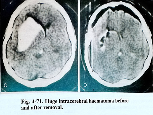
The surgical indications depend on the location and the volume of the haematoma, considering the general and neurological conditions of the patent. The haematomas located in the thalamus, the brain stem and intraventricular hemorrhages after evacuation do usually not improve patient's condition. The haematomas in the brain hemisphere, those located laterally to the internal capsule in the basal ganglia and in the cerebellum are considered more suitable for evacuation (Fig. 4-71). Better results from surgical treatment are obtained in patients with slow progressive clinical deterioration for hours or days. The surgical evacuation does not change evidently the clinical evolution when clinically there is acute onset and bad initial clinical condition. Improvement can be expected in haematomas, when CT scan images show displacement of midline brain structures.
The haematomas in the cerebellum need special attention, because their evolution cannot be predicted, and frequently there is sudden deterioration. In cases of cerebellar haematomas with data for brain stem compression there is an indication for urgent evacuation. Even in a patient in stable condition, the operation is indicated in big haematomas. Intraventricular bleeding with the formation of clots can deteriorate additionally patients condition due to obstruction of CSF outflow, and if obviously this is the cause of deterioration, an urgent ventricular drainage can be effective.
The general condition of the patient is essential in the selection for surgical treatment. Deeply comatose patients, with significant brainstem disfunction do not improve even with clearly decompressive effect of haematoma removal.
SURGICAL TECHNIQUE
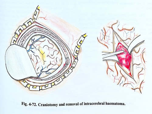
The choice of surgical technique for evacuation of an intracerebral haematoma depends on the volume and the location of the haematoma. The worse the condition of the patient, the less surgical trauma he will be able to sustain. A craniotomy, and even craniectomy up to 3 - 4 cm in diameter is enough to provide save access to the haematoma, if done at the most appropriate place following the clinical and CT scan data. After opening the dura, the brain tissue is found under increased pressure and with the tendency to protrude through the craniectomy. Sometimes a yellowish coloured cortex can be seen, and that indicates the most superficial part of the haematoma. The brain is punctured with a ventricular cannula and when the haematoma is reached the maximum possible amount of blood is evacuated. Direct penetration into the haematoma cavity and complete removal of the blood is recommendable (Fig. 4-72).
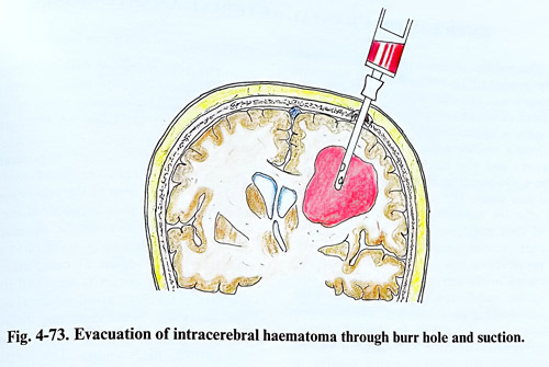
In case of huge haematomas, however, can be evacuated mainly the fluid portion, if such exists. This is enough efficient in reducing intracranial pressure and brain tissue compression, and safer regarding postoperative rebleeding (Fig. 4-73).
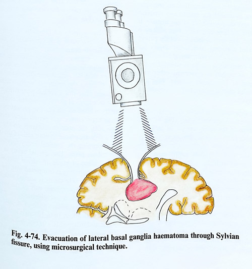
The direct penetration, described above, refers to haematomas whose cavities are very close to the surface, quite under pia. That is not the same in deeply seated lesions, where to reach the cavity has to be penetrated eloquent brain, apparently functionally intact. To avoid additional damage, the approach has to be selected avoiding functionally important areas, gaining penetration to the depth through sulci or the anatomically important fissures, especially the Sylvian and the interhemispheric. Dissection of a deep sulcus requires the surgical microscope. The same is valid for exposing insular and opercular cortical surfaces through the Sylvian fissure. After such dissection vascular damage and pial incisions are minimal, but sufficient access is provided to the haematoma (Fig. 4-74).
In the great majority of cases, the bleeding vessel has thrombosed immediately after rupture and it is not necessary to do haemostasis. However, when the operation is performed in the first hours after the onset of the haemorrhage, a bleeding vessel can be discovered and the haemostasis can be performed by clipping, bipolar coagulation or oxydased cellulose. In the event of uncertain complete haemostasis, the haematoma cavity is drained for 24 hours.
With patients in good general condition a sufficient in size craniotomy is preferable. This offers the possibility to inspect well the haematoma cavity, and if there is a suspicion of pathological tissue presence, a small specimen is taken for biopsy.
If an aneurysmal rupture is the cause of the haematoma, the surgeon has to make an effort to dissect the aneurysm and exclude it from the circulation. When the haemorrhage is due to an arteriovenous malformation, the operation is much more complicated. In such cases it is better to remove big part of the haemato-ma, if there are vital indications. The dissection of the malformation must be left for the next stage, after the patient has completely recovered. Later, in a relatively good general condition of the patient, the malformation is investigated properly and can be removed without causing an additional trauma to the brain.