GIANT ANEURYSMS
Intracranial aneurysms larger than 25 mm in diameter are considered giant and these comprise 2 to 5% of all such lesions. The most common locations of giant aneurysms differ from those smaller in size. About 40% are seen on the carotid artery, 25% on the anterior and middle cerebral arteries, and 30 % on the vertebrobasilar system. The age at which giant intracranial aneurysms become symptomatic seems to be nearly the same as for intracranial aneurysms in general.
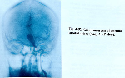
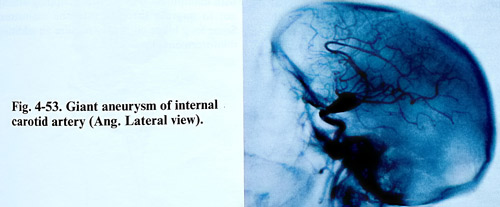
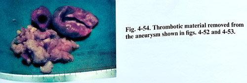
Plan X-rays may show 'ring calcification' which is pathognomic for these lesions, or bony erosion due to chronic compression. The CT scan is usually positive showing round mass with a ring enhancement around the aneurysmal wall, nonenhanced thrombi and enhanced central region due to blood flow. MRI demonstrates the presence of giant aneurysms quite well by the flow inside as a signal void area and differentiating old clots from recent ones. "CT angiography" with spiral scanning techniques can add a lot about the shape and location of the lesion. Cerebral angiography is still the most important investigation for surgery of giant aneurysms (Figs. 4-52 - 4-54). It should clarify the exact anatomy around the neck of the aneurysm and determine the collateral flow. Functional assessment of the collateral flow, including balloon occlusion tests can be needed preoperatively, as long lasting parent artery occlusion may take place during surgery.
The treatment goal for giant intracranial aneurysms is not only prevention of rupture, but also removal of the mass lesion, and restoration of the normal cerebral blood flow. Secure proximal vascular control is required for safe surgery - the carotid can be exposed on the neck or at the Glasscock triangle and the vertebral - where it is just emerging intracranially. For the same purpose also balloons can be navigated in appropriate locations. The treatment strategy has to be planned, depending aneurysm location, collateral flow, and patient's age.
Intraoperative monitoring by motor and/or somatosensory evoked potentials, or cerebral blood flow measurement can be of value in preventing ischaemia. Preventive measures can include also bypass surgery, hypothermia, or medication.
For precise clipping, the aneurysm must be slackened by suction decompression, thrombectomy, or cardiac standstill. The surgeon should select the best fitting clips and check the result by intraoperative angiography. Endovascular treatment can prevent rupture, but does not remove the existing mass lesion and its effects.
DIRECT SURGICAL TECHNIQUES
Giant carotid aneurysms. They are classified according to their projection into four groups: anterolateral, anteromedial, posteromedial and posterior.
With the patient in supine position the cervical carotid artery is exposed for temporary trapping and a pterional craniotomy is made.
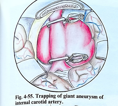
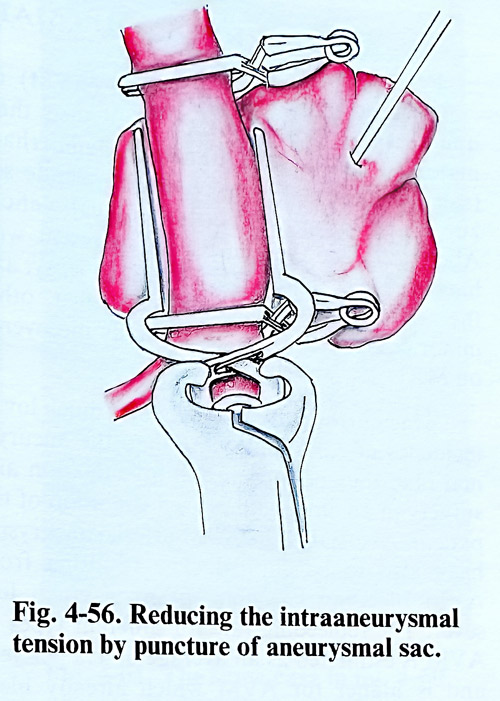
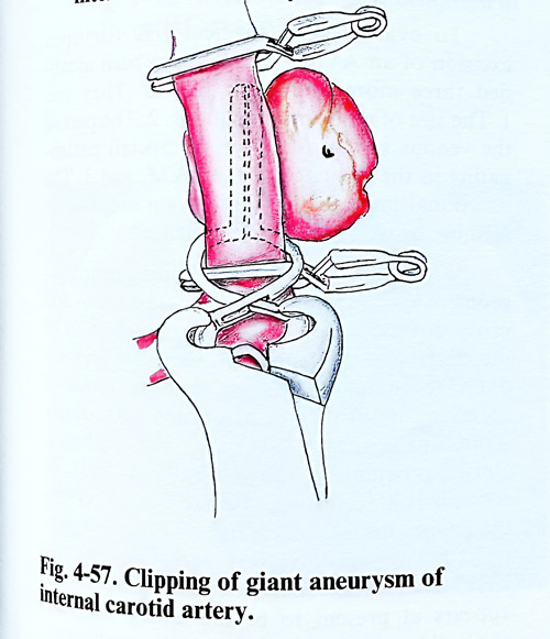
The frontal branch of the superficial temporal artery is preserved as a possible donor for an extracranial-intracranial bypass. Before approaching the aneurysm, bone in paraclinoid region should be drilled, optic canal and distal dural ring opened widely to release the optic nerve and C3 portion of the carotid artery. A more intense bleeding from the cavernous sinus can be controlled by elevation of the patient's head and meticulous packing with oxidised cellulose. The body of the aneurysm is dissected and the neck is isolated. At least two-thirds of the major circumference of the aneurysm must be dissected from adjacent tissues for satisfactory clipping. When the intraaneurysmal pressure is too high to isolate the aneurysm, clipping on the neck under temporary trapping of the parent artery can be suggested. Thrombi and calcified plaques at the neck can be avoided or removed after trapping of the parent artery. A good choice of with the optimal in fitting to the neck clips is essential (Figs. 4-55 - 4-57).
Surgery for giant aneurysms of the middle cerebral artery is generally easier than surgery in other locations because the operative field is wider and trapping is easier. The most important goal in these cases is to avoid stenosis and occlusion of distal branches of the middle cerebral artery and lenticulostriate arteries. Sometimes after using multiple clips in a proper combination, remaining portion of the aneurysm may need wrapping. In case of stenosis of the parent artery extra-intracranial arterial bypass is performed.
To clip a giant aneurysm of the anterior cerebral- anterior communicating artery complex is more appropriate to use the interhemispheric approach because it better exposes the Al and
A2 segments simultaneously on both sides. The lower intra-aneurysmal pressure in the cases of this location is an advantage. In addition, this area seems to tolerate temporary occlusion better than others. However, the anatomy of these aneurysms is comparatively more complicated as Al, A2 and the anterior communicating artery must be preserved. In some cases, side-to-side or end to side anastomosis to A2 must be performed as a solution to restore the flow.
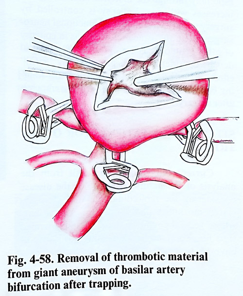
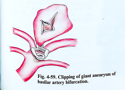
The natural course of a giant basilar bifurcation aneurysm is poor because of frequent fatal bleedings. The pterional or frontoorbital approach with additional removal of the lateral orbital wall is quite useful in widening the operative field. In some cases a temporobasal craniotomy with opening of tentorial notch gives also good access to the lesion. With increased use of temporary basilar artery clip-
ping, the results of aneurysm surgery in this location have become more successful (Figs. 4-58; 4-59).
Most of the giant vertebral artery aneurysms are partially thrombosed and their clinical presentation is brain stem affection with long tract signs, lower cranial nerve palsies and respiratory distress. The treatment of choice may be intravascular occlusion of the vertebral artery if the patient can tolerate the occlusion test and has a good collaterization from the opposite side. If the patient's condition doesn't improve after occlusion, trapping with excision may be necessary. Long lasting deficit does not improve with decompression.
Partially thrombosed aneurysms make at times clip blades closure impossible. The clip usually slides to the parent artery and constrict its lumen. In such cases aneurysm thrombectomy is necessary. Great care should be taken with manipulation or multiple clipping attempts not to migrate thrombi into the mainstream of the parent artery.Once the aneurysm clipped, some long blade clips may require an additional reinforcement clip.