Legacy: "Atlast of Neurosurgery" / L.Karaguiosov, A. Ramadan, K.Karaguiosov / Kiwait/ 1998
Read Previous: 3.Tumors - Chapter 2
From the variety of vascular diseases of the brain, surgical treatment is most often applied in cases of aneurysms, arteriovenous malformation, carotid cavernous fistulae, intracerebral haematomas, and sometimes in cases of extra-or intracranial cerebral arterial occlusion. The types of methods that are currently used include: open surgery through a craniotomy, endovascular techniques in interventional neuroradiology and external high energy beams and particles. In big number of cases these methods are complementing each other.
INTRACRANIAL ANEURYSMS: GENERAL PRINCIPLES OF MANAGEMENT
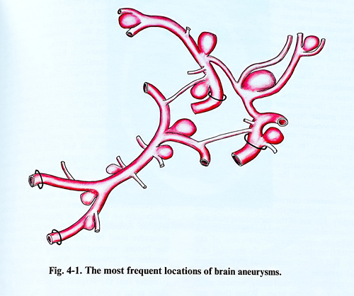
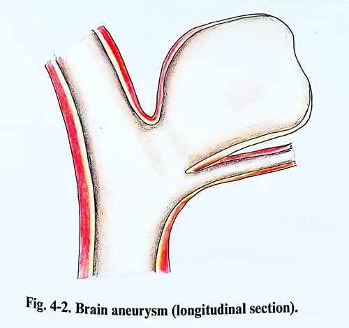
The aim of the surgical treatment of intracranial aneurysms is prophylactic: to prevent new rupture of the aneurysmal sac, and only in some cases to evacuate an intracerebral haematoma, which endangers the patient's life (Figs.4-1; 4-2).
GENERAL PRINCIPLES OF ANEURYSM SURGERY
Complete angiographic study is required of the four main vessels with multiple views. Collateral circulation is evaluated on static images or by dynamic tests as temporary occlusion with balloon. These last tests are compulsory when temporary occlusion of major arteries or 'trapping' can be expected.
The direct surgical procedure with exclusion of the aneurysmal sac from the brain circulation is universally aimed. Alternative methods are trapping of the aneurysm, by proximal and distal occlusion of the parent artery, wrapping of the aneurysmal sac, proximal endovascular occlusion, or intra aneurysmal endovascular occlusion, with induction of thrombosis inside the sac.
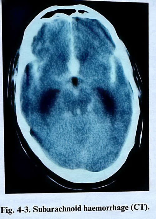
When multiple aneurysms are discovered, the ruptured one can be identified by localisation of the haemorrhage on CT, assessment of haemosiderin deposition on MRI, aneurysm dome configuration on the angiography, or clinical neurological findings (for example sudden IlIth nerve palsy) (Fig. 4-3). If more then one aneurysm should be treated in one surgical session, the first should be the ruptured one.
During the surgical approach, brain relaxation is achieved by appropriate use of hyperventilation, osmotic agents, diuretics, and evacuation of CSF (preoperative placed lumbar drainage or aspiration after the craniotomy). The surgeon should be prepared to delay the operation if relaxation is not sufficient.
Proximal vascular control must be established on the parent artery if possible, if not proximal vascular supply on the neck has to be isolated and occasionally occluded or a balloon is inserted in the internal carotid artery. Giant aneurysms of the basilar artery during the surgery are best controlled by temporary intra-vascular occlusion with balloon catheter.
The sequence of vascular exposure is the proximal parent vessel, then the distal parent vessel, and finally the aneurysmal neck. Sharp dissection is preferable to blunt one, intending to reduce traction forces applied to the aneurysm. Once the aneurysmal neck is isolated, critical vessels adjacent to the aneurysm are defined and dissected away when possible. This dissection can be often postponed until the aneurysm is collapsed after clip application.
Local hypotension by trapping with low closing force temporary clips is preferable to systemic arterial hypotension. Temporary trapping should by used to avoid aneurysmal rupture anticipating the complication when the anatomical conditions create difficulties. If vascular control cannot be achieved within the operative exposure, a temporary balloon can be navigated into the appropriate location.
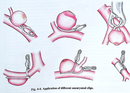
The clipping axis is preferable to be along the axis of the parent artery. Clipping across the axis of the parent artery increases the risk of stenosis and bending of the artery, or tearing close to the aneurysmal neck. Application of multiple clips is necessary either to enforce the single already applied clip or in case of a complex aneurysm (Fig. 4-4). Once the permanent clip is placed and temporary clips are removed, the aneurysm is punctured with a fine needle and aspirated to check for residual filling. In case of large or complex aneurysms may be required temporary trapping, aspiration of the sac before placing the final clip, and only than the dissection is completed. Residual filling of the aneurysm requires repositioning of the clip, multiple clips, or a reinforcing clip to increase the closing pressure. Patency of the parent artery is evaluated by assessing the vessel diameter, colour and pulsation, intraoperative doppler study or intraopreative angiography.
SELECTION OF PATIENTS FOR OPERATION
The proper selection of patients with intracranial aneurysms for surgical treatment is essential for the safe course of the operation and the operative results. In considering the indications for surgical treatment it is necessary to assess the patient's general and neurological condition, localisation of the aneurysm and its anatomical peculiarities, conditions of the cerebral arteries, patient's age and the presence of intracranial haematoma.
The patient's neurological and general condition with intracranial aneurysms plays a decisive part in determining the indications for operative treatment. Different scales for grading the patient's condition after subarachnoid haemorrhage have been introduced. All of them intend to select those patients, whose condition is not to much affected to tolerate without additional deterioration and complications the preventive surgery proposed. Such is the scale of Hunt and Hess subsequently modified on different occasions:
Grade I (minimal bleeding). The patient is alert without neurological deficit. Grade II (mild bleeding). The patient is awake but with headache, neck rigidity or minimal neurological deficit, like paresis of the third cranial nerve. Grade III (moderate bleeding). The patient is drowsy or confused with or without focal signs. Grade IV (moderate or severe bleeding). The patient is semicomatous with or without neurological focal signs. Grade V (severe bleeding). The patient is in coma and decerebrate, failing vital signs. At any moment every patient with a SAH can be evaluated and scored according to one of the existing scales. When the patient is over 50 years of age, the gravity of the condition is assessed with one grade higher than the observed signs would indicate. Usually over the age of 65 operative treatment is not recommended, but in many countries with achievements in population longevity these limits have been moved forward. If the patient has serious heart, lung, kidney or liver diseases, one more grade is added too, as these conditions represent an additional risk for operative treatment. The best surgical results are achieved with patients in grade I and II.
The condition of the cerebral arteries is of great importance in the assessment of the indications for operative treatment. Arterial atherosclerotic plaques may discredit the technical performance of the operation. They oblige the surgeon to be more careful when working on the vessels and especially when he has to clip or ligate, as at this time the delicate and sometimes rigid arterial wall may be torn.
The collateral circulation has to be taken into consideration, when the indications for operative treatment of an aneurysm are evaluated. All segments of the circle of Willis have to be studied as size and direction of flow. The pre-operative planning should assess the option of collateral supply if the parent vessel is occluded. For instance, when aneurysms are in the region of the anterior communicating artery, the surgeon must carefully establish the patency of the anterior cerebral arteries and what is the direction of flow.
Arterial spasm, following subarachnoid haemorrhage, is one of the major complications that has to be carefully evaluated in considering the indications for operative treatment. Practice shows, that when an operation is performed in patients with severe arterial spasm, the results are poor and the mortality is increased. In such cases it is advisable to postpone the operation until the spasm resolves. In the majority of patients with clinically and angiographically significant diffuse spasm, their condition improves after a period of three weeks.
The localisation of the aneurysm also influences the selection of patients for operation.
From surgical point of view the aneurysms of the middle cerebral artery are the most accessible, because they are superficially situated in the Sylvian fissure. Aneurysms of the internal carotid artery require greater retraction of the frontal and temporal lobes, and the dissection of arteries and the aneurysmal sac need more time and attention. When it is a case of an anterior communicating artery aneurysm, the operative approach is even more difficult, due to the close relation of the aneurysm to important vessels and the greater depth of its localisation. The favourable localisation of the aneurysm on middle cerebral artery makes surgery to be done earlier than in the cases of internal carotid artery aneurysms, while in aneurysms of the anterior communicating artery and the vertebrobasilar circulation the delay is usually the longest.
The anatomic characteristics of the aneurysm are also important for patients’ selection for surgery. The size of the aneurysmal neck and its anatomical features influence the selection of the technical approach for isolation of the aneurysm from the circulation. In the event of a narrow, well formed aneurysmal neck, without arterial branches in its vicinity, the dissection and clipping are performed easily, without additional trauma on the brain. When there are large blood vessels around the aneurysmal sac and its neck, then the dissection is much more difficult and requires greater retraction of brain surface. If the neck of the aneurysm is wide, the clipping becomes more difficult, even impossible without narrowing the parent artery. With large aneurysms (giant aneurysms) it is very often necessary to interrupt temporary the circulation in the parent artery, and at the end it may be necessary to make microsutures of the walls.
The more complex the anatomical topography around the aneurysmal neck, the more indications for operation have to be carefully considered. The finding of subdural, intracerebral or intraventricular hematoma in cerebral aneurysms rupture has to be taken thoroughly into consideration in the indications for operation. When the patient's condition obviously is deteriorated and that is due to the intracranial haematoma as a mass lesion effect, there are indications of urgent surgery and the operation has the task not only to exclude the aneurysm from the circulation, but to evacuate the haematoma and save the life of the patient.
The rupture of an aneurysm during pregnancy sets special requirements in deciding in favour of operative management. There are no contraindications on the part of pregnancy for angiographic investigation of the brain. Preventive measures must be taken against irradiation of the foetus, especially during the first three months of pregnancy. When an aneurysm is established in a pregnant patient, many authors consider that the surgical decision has to be the same as in a non pregnant, i.e. have operative treatment performed irrespective of the pregnancy. During the last weeks of pregnancy, the surgery can be anticipated by a Caesarean section and the operative treatment performed immediately afterwards.
ANEURYSMS OF ANTERIOR CIRCULATION
ANEURYSMS OF INTERNAL CAROTID ARTERY
Internal carotid artery aneurysms present different technical problems according to the segment of the artery they involve. The following segments are currently considered: intracavernous, ophthalmic, posterior communicating and anterior choriodal, and internal carotid bifurcation.
The relation of the aneurysmal neck to the optic nerve, anterior clinoid process and the distal dural ring determine the principles of the technique to be applied also and the expected complications. Proximal arterial control can be dificult in aneurysms of the cavernous and ophthalmic segments and preoperative precise location of the aneurysmal neck, can allow the surgeon to choose the safest technique (at the neck, in the Glasscock triangle or with endovascular balloon). To assess the consequences of elective or emergency trapping of a part of the internal carotid has to be investigated the functional effect of a temporary balloon occlusion.
ANEURYSMS OF THE INTRA-CAVERNOUS CAROTID ARTERY
The options for the treatment of intra-cavernous aneurysms include endovascular intra-aneurysmal placement of a balloon or thrombogenic substances, trapping procedures by direct or endovascular technique, and direct intracavernous aneurysmal clipping. The direct clipping of cavernous internal carotid artery aneurysm is a suitable when the aneurysm is paraclinoid or intracavernous, expands the distal dural ring and enters the intradural space.
The intracavernous aneurysms not amenable to indirect technique can be also managed directly.
Fortunately, most intracavernous carotid aneurysms arise from the C3 - C4 segment (proximal anterior loop) and can be clipped directly after opening the distal dural ring, opening the roof of the cavernous sinus, and partially mobilising the lateral wall of the cavernous sinus.
DIRECT CLIPPING OF INTRA-CAVERNOUS ANEURYSMS
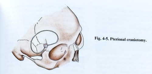
The patient is placed in a supine position on the operating table with the head rotated 30 degrees to the opposite side. The pterional craniotomy is the most suitable, but has to be extended anteriorly to the midpoint of the orbital entrance at the level of orbital roof. Laterally temporal fossa is opened to its floor (Fig. 4-5).
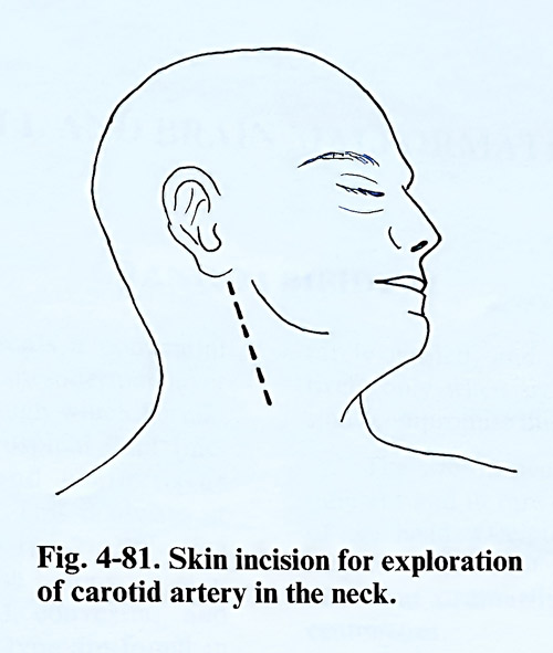
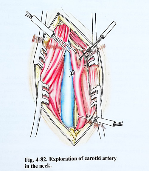
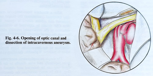
Carotid bifurcation is exposed in the neck after skin incision along the skin creases. Internal and external carotid arteries are dissected and isolated with vessel loops to establish proximal vascular control. The internal carotid artery is also available for catheterization, balloon occlusion and angiography. An alternative option exists either by using endovascular technique and balloon occlusion, or by exposure of the artery in the Glasscock's triangle. CSF is aspirated through the previously placed lumbar catheter or the exposed cisterns. The dura is retracted to expose the orbital roof, which is entered by high speed drill. The posterior half of the orbital roof is removed by bone nibbler. The superior orbital fissure is exposed removing additional bone from the lesser wing of sphenoid. A high speed diamond drill is used to remove the anterior clinoid process and the roof of the optic canal. Care is needed to avoid entering the ethmoidal sinus, which is medial to the optic canal and can produce postoperative CS fistula and infection. The bone lateral to the superior orbital fissure and medial to the foramen rotundum, is thinned with the drill and fractured laterally to expose fully the superior orbital fissure. An incision of the dura is made medial by and parallel to the superior orbital fissure. The dural incision is extended medially and laterally to provide access to the floor of both anterior and middle cranial fossae (fig. 4-6)
The distal dural ring that encircles the internal carotid is incised sharply, using an arachnoid knife in direction away of the arterial wall. Beginning at the oculomotor foramen, the lateral wall of the cavernous sinus is incised along the axis of the optic nerve and the mid-portion of the oculomotor triangle. The next dissection reflects the dura propia of the lateral wall to expose cranial nerves III and IV and the first division of V. The inner membranous layer formed by the perineural sheaths (of cranial nerves) when preserved allows mobilisation of dura propia without disruption of the venous channels of the cavernous sinus. Beginning at the falciform ligament, the optic nerve sheath is incised along the course of the nerve and the dura on the medial side is exposed.
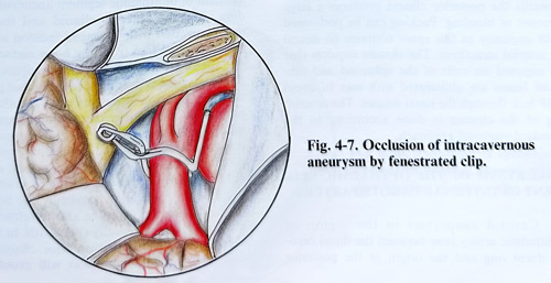
The optic nerve is retracted medially to expose the proximal dural ring and it is opened along the medial edge of the oculomotor nerve, retracting the nerve laterally to expose the fundus and the aneurysmal neck. Using the dissector, the relationship of the aneurysmal neck to the carotid artery and the distal dural ring can by established. A right-angle fenestrated clip is applied along the axis of the carotid artery preserving its patency (Fig. 4-7). In some circumstances aneurysms of the carotid cavernous region may require collapse of the aneurysmal sac for effective dissection and clipping. The collapse of the aneurysm can be obtained by temporary occlusion of the internal carotid artery in the neck and distally to the intracranial dissection site. Aspiration of the sac will permit dissection and precise placement of the clip. Intraoperative angiography is desirable in aneurysms in this area because direct inspection for completeness of clipping is often difficult.
At the completion of the intradural part of the procedure, haemostasis should be complete. Small pieces oxidised cellulose may be inserted in the anterior triangle between the internal carotid and the optic nerve, around the carotid siphon tip, and pressing against the medial wall of the superior orbital fissure dura. Oxidised cellulose must be modestly used to avoid excessive pressure on the oculomotor nerve that is running closely. Opening of the medial triangle towards the posterior clinoid produces a large amount of bleeding. Packing can be performed with impunity as this space contains no neural or arterial structures. The closure requires that all exposed air cells of the sphenoid and ethmoid bones are obliterated with wax to avoid CSF leak through the nasal sinuses. The remaining of the closure is done according to the standard pterional approach.
ANEURYSMS OF THE OPTHALMIC SEGMENT OF INTERNAL CAROTID ARTERY
Carotid aneurysms in the region of ophthalmic artery arise between the distal carotid dural ring and the origin of the posterior communicating artery. The aneurysmal neck can be located: next to the ophthalmic artery origin, lateral to the ophthalmic artery or juxta-ophthalmic near the origin of the superior hypophyseal artery. Superiorly and medially projecting aneurysms are under the optic nerve and anterior clinoid process, leading early to visual field defect. Aneurysms at the origin of the superior hypophyseal artery usually project posteriorly or laterally. Endovascular intra-aneurysmal occlusion must be considered as alternative treatment to direct open surgery. If occlusion of the carotid artery is anticipated, the patient must pass a preoperative test by tolerating internal carotid occlusion for 10 to 30 minutes with a quantitative study of the cerebral blood flow. Failure to tolerate occlusion shows the need for preoperative extracranial - intracranial arterial bypass.
After a standard frontotemporal craniotomy (pterional craniotomy) the frontal lobe is retracted and sylvian cistern is opened. Sufficient CSF should be aspirated to enhance the reduction of the intracranial pressure obtained through the use of osmotic diuretics. Arachnoid around the internal carotid artery and the optic nerve is opened widely to enhance further retraction of the frontal lobe. Care should be taken to preserve, if possible, the olfactory tract.
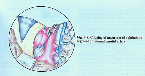
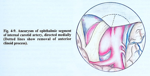
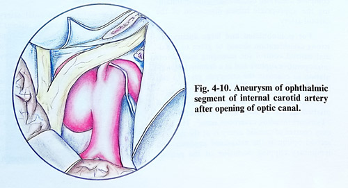
The simple paraophthalmic aneurysms that point superiorly and arise distally to the distal dural ring can be dissected and clipped easily (Fig. 4-8). In many occasions the dissection of the aneurysmal neck needs removal of the anterior clinoid process. This removal is one of the most critical procedures to the successful management of ophthalmic segment aneurysms. The dura over the anterior clinoid and the optic canal is incised with T-shaped incision and two flaps are separated from the bone surface (Fig. 4-9). Before that careful dissector palpation over the optic canal is needed to identify that portion that may be covered only by dura and therefore may allow inadvertent thermal or compression injury to the optic nerve. It is advantageous to remove the entire clinoid process to expose the proximal cavernous sinus and the distal carotid dural ring. It is also important to unroof the optic nerve completely, especially the bone on the medial border, so to be able to retract optic nerve medially or laterally (Fig. 4-10). In case of excessively pneumatised anterior clinoid pro-cess, packing with bone wax will avoid post-operative CSF rhinorrhoea. If sphenoid sinus is opened and, especially when decompressing the medial aspect of the optic canal and mucosa is opened, exenteration of all mucosa, packing with piece of muscle and sealing with glue are mandatory.
Opening of the inner layer of dura (dura propia) along the lateral aspect if the optic nerve is necessary. This exposes the carotid artery more proximaly and allows better exposure of the origin of the ophthalmic artery. If the neck of the aneurysm extends further proximal then the opening of the distal carotid ring, dissection of superior part of cavernous sinus will be necessary. Once the appropriate segment of carotid artery has been exposed, the extent of the aneurysm neck should be identified and dissection begins. The ophthalmic artery should be dissected free of the aneurysmal neck.
Superior hypophyseal branches as well as the branches to the optic chasm should be identified and preserved at the time of clipping. Intermittent retraction of the optic nerve may be necessary. Depending on the size of the aneurysm, the origin of the posterior communicating artery and anterior choroidal artery may need to be dissected. Prior to clip application, a fine dissector should be passed completely behind the neck of the aneurysm to ensure an easy passing of both aneurysm clip blades. Any apparent resistance should be immediately followed by further dissection technique. Following complete mobi-lisation and dissection of the aneurysm, the decision is made regarding clip selection. Except in aneurysms with very narrow necks, the clip should be placed with the blades along the axis of the parent vessel to avoid kink or tearing of the neck. Sometimes, several sizes or configurations of clips may need to be tried in an effort to obtain complete aneurysm obliteration while maintaining the appropriate lumen dimension of the carotid artery.
For polilobulated and large aneurysms, strong consideration should be given to proximal carotid control for managing inadvertent intraoperative rupture and as a definitive manipulation to evacuate the aneurysm prior to clip placement (Figs. 4-11; 4-12).
Proximal control however, can be accomplished at three sites as was described in "Tumours invading cavernous sinus". The easiest control of the carotid artery is at the neck. A second option is the extradural exposure and temporary clipping occlusion of the petrous portion of the carotid artery (Glasscock triangle). A third and more distal site for carotid control is the short segment of carotid between the two carotid rings. It is important to leave a small amount of aneurysmal neck between the clip blade and the parent vessel to avoid constriction of the carotid artery. In cases of bilateral carotid ophthalmic aneurysms the contralateral aneurysm occasionally can be clipped through the same craniotomy. This is easyly accomplished, when the contralateral aneurysm is reasonably small and projects medially or posteriorly from the carotid artery.
ANEURYSMS OF THE POSTERIOR COMMUNUCATING ARTERY AND ANTERIOR
CHOROIDAL ARTERY REGION
Aneurysms that arise at the junction of the carotid and posterior communicating arteries can be round, elongated, polilobular in shape, varying in size, although aneurysms grater than 2,5 cm in diameter are rarely seen. The most of the aneurysms with this location (86%) project posterolaterally and involve the III cranial nerve in about one third of the cases (Fig. 4-13). In a small group of these patients the aneurysm extends above the tentorial edge and the aneurysmal sac may adhere to the temporal lobe.
Medially directed aneurysms arising at the internal carotid artery-posterior communicating artery junction occur in only 4%. They tend to extend beneath the optic nerve and produce visual symptoms.
The relationship of aneurysmal sac with anterior clinoid process should be determined on the angiography to decide the need of drilling this process.
The anterior choroidal artery aneurysms are rare and arise from the inferior aspect of the internal carotid artery 3 - 6 mm proximally to the carotid bifurcation. They project laterally, while the anterior choroidal artery runs medially.
Surgical technique. With the patient in supine position and the head rotated at 45 degrees to the opposite side, the standard pterional craniotomy with a half-and-half exposure of the frontal and temporal lobes is performed. After opening the dura, a self-retaining retractor is placed under the orbital surface of the frontal lobe and the surgeon follows the edge of the lesser wing of the sphenoid. We prefer initially to open the sylvian cistern, and by retracting the frontoorbital cortex to continue opening the sylvian fissure. The temporal lobe is not retracted at this stage. By reaching the proximal Ml segment and the carotid bifurcation, the carotid cistern is opened and the frontal lobe is freely separated medially to the chiasmatic cistern, allowing additional space for manipulation.
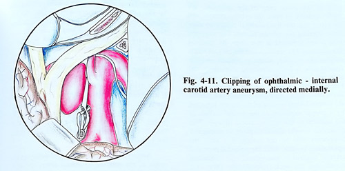
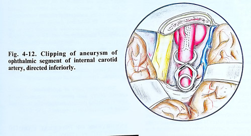
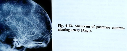
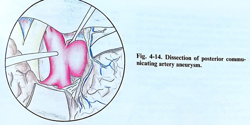
Considering the position of the aneurysmal neck and dome, initially is dissected the antero-lateral surface of the parent artery, followed by a proper exposure of the medial surface of C2 segment in the optico-carotid triangle. This dissection usually allows to place an emergency temporary clip on the carotid just after the ophthalmic branch. After the potential proximal arterial control is guaranteed, the dissection approaches the neck from distally (M1 - C1 segments) and if the aneurysm is not in contact with the medial temporal lobe, uncus can be retracted. The dissection of the neck is completed to allow free passage of the clip blades (Fig. 4-14).
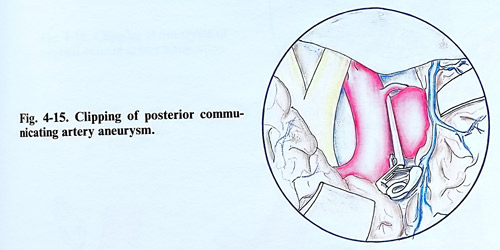
At times a shorter C2 segment or a bigger dural fold laterally to the clinoid may require incision or coagulation of dura to produce safe space for dissection and the proximal clip blade. Clip is positioned preferably along the artery, not stretching the walls of the carotid and not obliterating the posterior communicating artery (Fig. 4-15).
In cases of posterior communicating artery aneurysms, the establishment of the size of this artery and the direction of the blood flow is important for the dissection and clipping of the aneurysm. In cases of a small artery with insignificant blood flow, it can be clipped together with the aneurysmal neck. When the posterior communicating artery is big and gives the origin of the posterior cerebral artery (“fetal” type) or the blood flow is from the vertebrobasilar system to the internal carotid artery (on the Alcock test), it needs very careful dissection and any stenosis of this artery during the clipping should be avoided. In case of third nerve palsy before the operation, the aneurysm is punctured and its contents evacuated after clipping. No attempt is made to dissect the aneurysm from the nerve. Third nerve palsy can be expected to resolve within a few months.
Rupture at an early stage of dissection presents serious threat for the favourable outcome of surgery. If other techniques are ineffective, temporary "trapping" of the area may be needed. Temporary clips are placed just distally to the ophthalmic artery and distally to the aneurysmal neck area, as often retrograde flow is significant. Even then posterior communicating artery can sometimes produce persisting bleeding, but permanent clipping should be done better under these circumstances. Then the temporary ICA clips are removed, starting from the distal one. Ruptures close or at the aneurysmal neck require always temporary trapping.
If a clip cannot be placed effectively on the neck, the artery is clipped by a specially designed fenestrated clip or is wrapped preserving its patency as suture can be time consuming. Permanent carotid occlusion at the neck is the last remedy.
Aneurysms of internal carotid artery at the origin of the anterior choroidal artery are situated a little higher and at 3 - 6 mm from the internal carotid artery bifurcation. That allows bigger space for dissection on the proximal side of the neck. Anterior choroid artery is situated medially to the aneurysmal sac and sometimes is difficult to be identified during dissection. This artery should be carefully dissected free of the aneurysmal sac and preserved. In about 15% of patients, sacrifice of the anterior choroidal artery results in the infarction of the internal capsule. This artery can arise from the aneurysmal sac and its clipping may provoke acute neurological deficit as hemiplegia, hemianaesthesia, even impairment of consciousness.
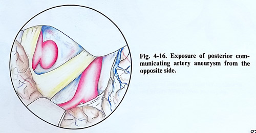
Some aneurysms of ophthalmic - internal carotid artery and posterior communicating artery, pointing postero-medially can be clipped easily by contralateral approach (Fig. 4-16).
ANEURYSMS OF INTERNAL CAROTID ARTERY BIFURCATION
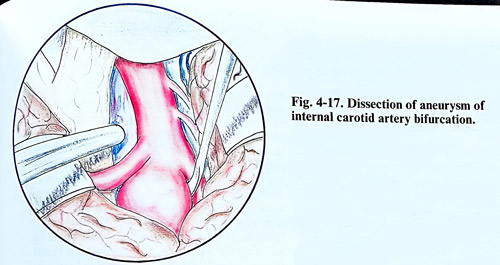
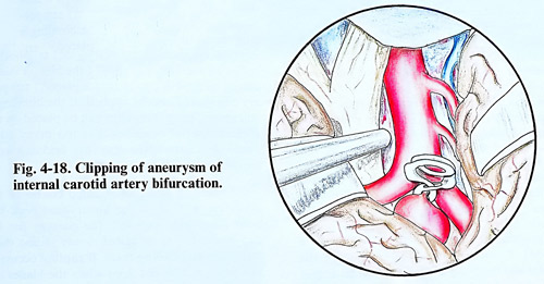
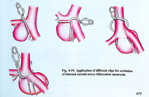
They represent 5% of all intracranial aneurysms and are located between the anterior and middle cerebral arteries. Most often project superiorly into the anterior perforate substance or posteriorly, distorting the optic tract. Frequently these aneurysms excavate bed in the anterior perforate substance. Sometimes they are direct continuation of internal carotid trunk or have a broad-based neck that incorporates the junction of the internal carotid artery together with part of the anterior cerebral or middle cerebral arteries trunks. These anatomical peculiarities demand fine and cautious dissection of the aneurysmal neck to avoid any damage of the central arteries, arising from the proximal segments (Al and M1) of anterior and middle cerebral arteries. The dissection should be sufficient to insert and then inspect the clip for inclusion of any perforators prior to its final placement (Figs.4-17; 4-18; 4-19).
Further inspection must confirm that the origin of anterior and middle cerebral arteries is not compromised by clip placement and that the aneurysmal neck is completely obliterated. The majority of surgical complications are attributed to the occlusion of perforating arteries that arise from the proximal segment of the anterior and middle cerebral arteries or adherence of anterior choriodal or the recurrent artery of Heubner to the aneurysm.
Tearing or rupturing of the aneurysmal neck should be avoided by all means because there are no effective options for management of this complication. An encircling clip will occlude the perforators. The only option might be trapping the aneurysm, which deprives circulation to the hemisphere and requires an emergency bypass. Intraoperative angiography is advisable for complex aneurysms of the region because of the difficulty in accurately visualising the anatomical details.
ANEURYSMS OF THE MIDDLE CEREBRAL ARTERY
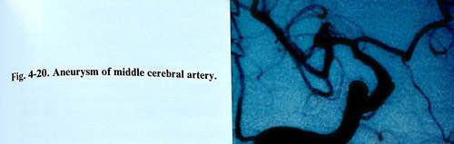
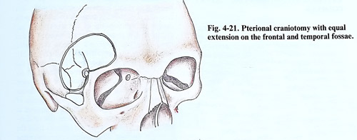
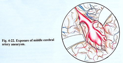
Commonly middle cerebral artery aneurysms arise at the bifurcation or trifurcation of this artery and are often partially embraced by the distal insular branches (Fig. 4-20). In a few cases the aneurysms arise from the more proximal part of the middle cerebral artery, often at the origin of the anterior temporal branch. The frontotemporal or "pterional" craniotomy with equal extension on the frontal and temporal fossae is the most suitable to reach middle cerebral artery aneurysms (Fig. 4-21). The dura is opened with a flat half-circle shaped line based anteriorly. The next step is to elevate the frontal lobe and identify the ipsilateral optic nerve, using gentle retraction. The chiasmatic and carotid arachnoid cisterns are opened, and CSF is removed from the basal cisterns, as this provides yet more brain relaxation prior to sylvian dissection. Beginning from the level of internal carotid artery, using a sharp arachnoid knife, the arachnoid bands connecting both lobes are divided. Continued opposing traction of the frontal and temporal lobes facilitates splitting of the Sylvian fissure. The small veins bridging the fissure can be coagulated and divided, and the main venous trunk (Sylvian vein) is usually retracted together with the temporal lobe (Fig. 4-22).
The more common distal bifurcation aneurysms can be approached after directly exposing the Ml segment if it is long enough or following carotid bifurcation if it is short. During the dissection blood clots should be washed out from within Sylvian fissure in an effort to minimise postoperative vasospasm.
When a large temporal haematoma is present, a cortical incision is made where the clot is close to the surface, usually through the superior temporal gyrus, until the haematoma is entered. The haematoma can be removed completely until the aneurysm is reached. This manipulation should be very cautious to prevent aneurysm rupture, or the haematoma is removed partially enough to slacken the brain and then the surgeon turns to a transsylvian exposure of the aneurysm.
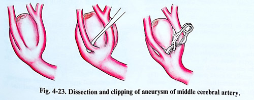
Once the aneurysm is reached the anatomy of the area should be exposed in a systematic way. Since the Ml segment at the level of the aneurysm bifurcates or trifurcates, all branches need to be identified. It is not possible absolutely to predict from the angiograms how many branches arise from the main divisions of the middle cerebral artery (Fig. 4-23). When these vessels are adherent and complete dissection of the aneurysm needed, or when the aneurysm dome is very thin walled, Ml temporary occlusion may be carried out for repeated short intervals as required to facilitate dissection by softening the aneurysm. If the aneurysm bleeds during its final dissection, proximal Ml temporary clip should also be applied. If the bleeding is still severe, temporary clips can be placed on the efferent arterial branches, but too many additional clips narrow the operative field and disturb further dissection. Otherwise in uncomplicated course of the operation and when the aneurysmal neck has been adequately exposed, the passage for the clip blades has to be tested slowly sliding them into place and closing them. If rupture occurs at this point and it stops when the blades are filly closed, it means that the hole was distal to the clip and the blades completely occluded the neck of the aneurysm. If bleeding originating proximal to the clip continues, the surgeon resorts to temporary clipping. The place of rupture is found and the clip positioned to isolate the aneurysm and rupture site.
Some large MCA aneurysms which project anteriorly from the bifurcation can almost reach the surface of the Sylvian fissure. Such aneurysms should be suspected from the angiograms, and because of the blood in the subarachnoid space, the red aneurysmal dome can be difficult to identify during dissection and can be damaged inadvertently. Large aneurysms of this type can block proximal Ml exposure in some cases and this relation may need the aneurysm to be approached from the distal part of the Sylvian fissure. If during the dissection of the Sylvian fissure the aneurysm ruptures prior to its identification, a temporary clip should be placed upon Ml and arterial blood pressure maintained in normal or slightly hypertensive values. Dissection must be quick, but safe in exposing M1, and an area for tentative clipping at a more distally should be considered as early as possible through the dissection. After clipping, the entire aneurysm and clip should be visualised from all sides, ensuring complete obliteration and absence of any branches included within the clip blades, with clip repositioning (definitive clipping) if necessary. On certain very rare occasions a small residual part of the aneurysm can be left unobliterated in order to preserve some major vessel origin patent. In such a case the residual part can be only wrapped.
ANEURYSMS OF ANTERIOR CEREBRAL - ANTERIOR COMMUNICATING ARTERY
COMPLEX
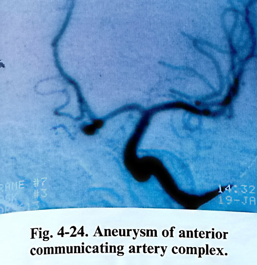
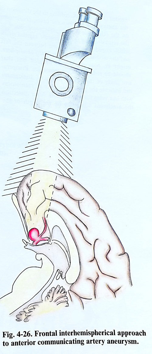
The aneurysms of the complex of the anterior cerebral and anterior communicating arteries are reached through a pterional craniotomy. For some aneurysms pointing anteriorly and adherent to the chiasm and tuberculum sellae dura can be used an interhemispherial approach or frontoorbital craniotomy (Figs. 4-24; 4-26).
The dissection in a pterional approach begins from the internal carotid artery, continues with its bifurcation, and anterior cerebral artery without uncovering completely or interrupting any branches. At opening the internal carotid cistern the arachnoid is additionally divided distally to the sylvian fissure, providing larger field and access to the anterior communicating artery area with less retraction pressure applied to the cortex. Frontal lobe retraction in anteriorly directed aneurysms should be cau-tious, as adherence of the dome to the basal dura can provoke premature rupture. Aneurysms with more complex anatomy requiring broader dissection area may need an additional orbitozigomatic osteotomy to reduce retraction.
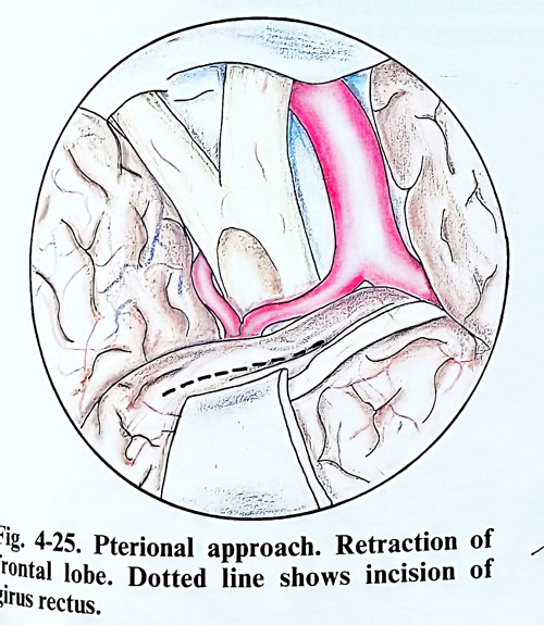
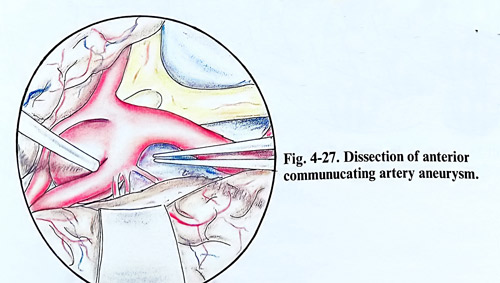
Once carotid is fully exposed, the surgeon follows Al segment and carefully separates gyrus rectus and olfactory tracts from the chiasm to gain approach to the opposite Al, and by that complete proximal vascular control. For broader exposure of the anterior communicating artery complex aneurysms the ipsilateral gyrus rectus can be partially removed under the pia outside the interhemispherical fissure, usually to avoid premature rupture (Fig.4-25). In that way, the interhemispherical fissure, usually very tight after SAH, becomes easier to dissect. The interhemispherial space, however should be opened following the Al and anterior communicating artery, and than the initial part of A2. This sequence of structures to expose should give access to the neck of the aneurysm before the fundus, with secured proximal control (Fig. 4-27).
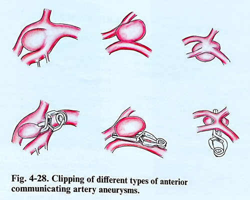
As in other locations of cerebral aneurysms, a great variety is observed in respect to the size, shape and interrelation to large arteries. Most often segments A2 arise from only one Al segment. The aneurysm is most frequently found at the bifurcation of Al and is usually directed upward. During the dissection of the aneurysm the surgeon must be guided by the angiography. From the angiographic data however, is not always possible to establish with details the relationship of the aneurysmal sac and its neck in respect to the main vessels and in these cases the surgeon's actions are guided primarily by the operative findings (Fig. 4-28).
Great attention should be paid in dissecting the proximal portions of the anterior cerebral arteries not to approach too near the bifurcation of the internal carotid artery, as some perforating arteries may be injured. The appearance of a recurrent (Heubner) artery should be noticed in advance. At clipping care is taken not to compromise the hypothalamic perforators from the posterior surface of the anterior communicating artery complex.
Large aneurysms of the anterior communicating artery aneurysm and those with intraoperative rupture on the neck, may require special technique of occlusion: special clips, trapping, even suture. For this purpose the dissection of both distal and proximal parts of the cerebral arteries and their temporary occlusion is absolutely necessary.
ANEURYSMS OF THE PERIPHERAL PART OF THE ANTERIOR CEREBRAL ARTERY
The aneurysms of the peripheral part of the anterior cerebral artery are most frequently found at the origin of the callosomarginal artery. There are also aneurysms at the origin of the frontopolar artery, in the region or branching of the callosomarginal artery and in the distal part of the pericallosal artery.
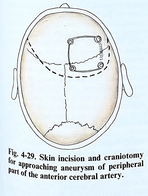
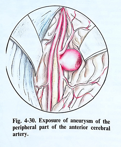
For surgical approach of these aneurysms, craniotomy close to the midsagittal line is needed (Fig. 4-29). According to the direction of the aneurysmal sac, in some cases, craniotomy on the other side is more convenient. Some of the veins draining in the superior sagittal sinus, which impede the penetration between the falx and the medial surface of the brain hemisphere, might be coagulated and divided but that should be reduced to the minimum. Preoperative study of the venous phases of angiograms can indicate the preferable place of craniotomy with less veins for interruption. After dissection the arachnoid adhesions on the depth of the interhemispherial fissure, one or both pericallosal arteries and the aneurysm are reached (Fig.4-30).
Very often these aneurysms do not lie freely in the subarachnoid space, but are buried into the medial surface of the frontal lobe or in some cases into the opposite hemisphere. There can be an anastomosis at the level of the aneurysm between the pericallosal arteries similar to the anterior communicating artery. In such cases the surgeon should protect it.
ANEURYSMS OF POSTERIOR CIRCULATION
The history of surgical treatment of aneurysms of the vertebro-basilar system is much shorter than that of the carotid, probably due to the difficulties of the greater surgical inaccessibility to these arteries.
In view of operative technique, the aneurysms of the vertebro-basilar system can be divided into six groups: at the bifurcation of the basilar artery, at the posterior cerebral artery, at the superior cerebellar artery, at the midbasilar trunk, at the vertebrobasilar junction, and at the vertebral artery.
ANEURYSMS OF THE BASILAR ARTERY BIFURCATION
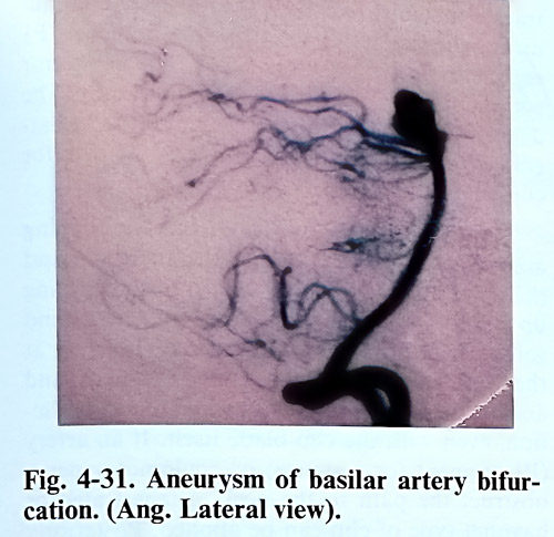
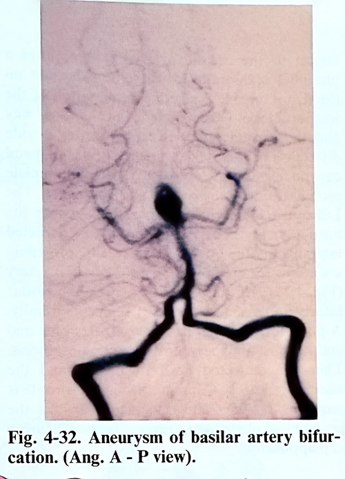
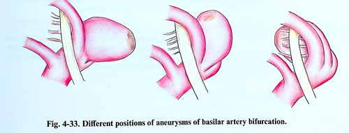
Basilar bifurcation is one of the commonest places of posterior circulation aneurysms. The aneurysms are directed anteriorly, superiorly or posteriorly, and special attention is paid on its relation to perforating arteries, usually originating on its posterior wall (Figs. 4-31; 4-32; 4-33). Direct clipping is preferred, as endo-vascular attempts are at times associated with incomplete thrombosis or propagation of the thrombus, occlusion of perforators, and even intraoperative rupture. The endovascular treatment, however, is considered always when direct clipping is not possible.
Collateral circulation has to be studied preoperatively in details. The ability of posterior communicating arteries to compensate the flow in the posterior part of the Willisian circle is essential for predicting the consequences of aneurysm trapping. Opposite to that, the Alcock test (vertebrobasilar angiographic imaging with carotid compression), reveals the role which posterior circulation may play for the supply of other vascular territories.
The aneurysms of the basilar artery, irrespective of the level of their location can be reached conveniently through a pterional or temporobasal craniotomy. The choice of approach for clipping a basilar bifurcation aneurysm depends on the position of the neck regarding the tentorial edge and the direction of the aneurysm.
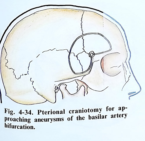
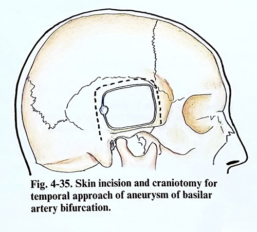
High position of the basilar bifurcation requires extensive subtemporal retraction avoided in transylvian route. This route is gained through a pterional craniotomy (Fig. 4-34). An additional orbitozigomatic flap can provide better angle of observation to highly positioned lesions. Aneurysms directed anteriorly or not high in the interpeduncular fossa are better approached subtemporally (Fig. 4-35). Most difficult to clip are aneurysms directed posteriorly, as very often that can compromise perforants. Broad view of more difficult lesions, even combining pterional and subtemporal approaches can provide safer dissection and clipping. The subtemporal approach can be extended by anterior petrosectomy. Anteriorly directed lesions are reached subtemporally, remembering that their dome can be adherent to dorsum sellae. Low position of the basilar bifurcation can require tentorial incision, always caring for the trochlear nerve under the tentorial edge.
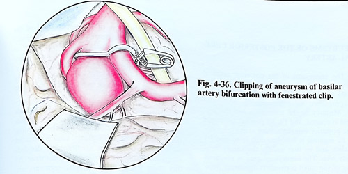
As a general rule the interpeduncular cistern in subtemporal approach is reached by tracing the superior cerebellar artery and finding its origin from the basilar artery and the Pl segment of the posterior cerebral artery. In this penetration the surgeon retracts the IIIrd cranial nerve in the same direction as the parahyppocampal gyrus is displaced. It is better to penetrate first anteriorly to the basilar artery as there are no perforators originating from the arterial wall. In aneurysms pointing anteriorly and superiorly the neck will be the next to reach. Greater difficulties are encountered in the dissection of the posterior aneurysmal wall. Sometimes the peduncle has to be slightly moved posteriorly in order to reveal the posterior surface of the posterior cerebral artery, the basilar artery and the aneurysmal sac. The fine perforating arteries are seen better with this slight retraction and away from the aneurysmal neck. Having found these vessels, a blunt probe or hook must be inserted behind the aneurysmal sac to displace it slightly in anterior direction. In this manipulation of the aneurysmal sac the origin of the opposite posterior cerebral artery must also be seen (Fig. 4-36).
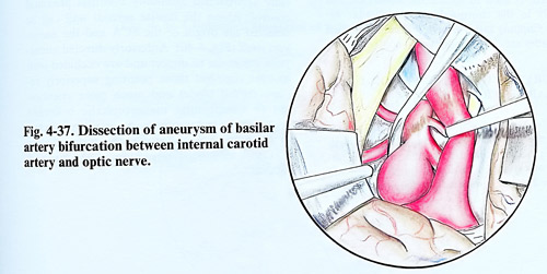
The basilar aneurysms whose neck is located superiorly to the level of the postrior clinoid process are reached conveniently through the pterional approach and a corridor between the carotid and the optic nerve or lateral to the carotid artery. That requires a pterional craniotomy, with some extension on the temporal side for better mobilisation of the temporal lobe. Dura is opened in the usual way and the carotid artery is exposed after a wide dissection of the Sylvian fissure. Both optic and carotid cisterns are dissected to the complete mobilisation of the artery and the nerve. The Lilliequist membrane is opened to expose the basilar artery. The carotid artery is retracted laterally and the optic nerve medially, providing a window for manipulation on the basilar artery (Fig. 4-37). This direction of view on the basilar bifurcation visualises both Pl segments usually. A place for proximal basilar control is prepared and it is just above the posterior clinoid process. Then are dissected both Pl origins and the aneurysmal neck. Clipping through this route is requiring often bayonet and thin blade clips, as the problem of obstructed vision is frequent during clip application.
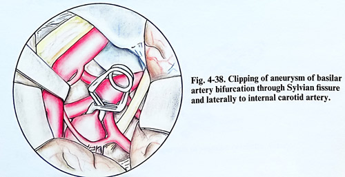
When the space between the optic nerve and internal carotid artery is narrow, the aneurysms of the basilar bifurcation above the level of the posterior clinoid process can be approached laterally to the internal carotid artery. After opening of the carotid cistern and Lilliequist membrane, the internal carotid artery is retracted medially to expose the posterior communicating artery and its perforating branches, Pl segment, and aneurysm neck. The posterior communicating artery is retracted medially. The dissection of the aneurysm neck and the ipsilateral proximal Pl segment completes the path for clip application. (Fig. 4-38).
There are some particular tips for clipping according to the direction of the aneurysm and the approach to it. Those aneurysms projecting up and forward don't allow the opposite P1 and some perforators there to be seen. Therefore at the time of clip blade passing, the neck and aneurysmal wall require gentle minimal retraction, even with the clip blade itself. If an artery (Pl segment for instance) or oculomotor nerve obstruct the path to the neck, a fenestrated or bayonet type of clip can be applied. Posteriorly directed aneurysms present difficulties with preservation of perforating arteries. Paying maximum attention to that avoids ischaemic complications of the brain stem.
Some aneurysms after complete dissection can result unclippable as is the case when haemodinamically important Pl or multiple perforators originate from its dome. In such a case the only solution remains with wrapping, a technique with uncertain effect.
ANEURYSMS OF THE POSTERIOR CEREBRAL ARTERY
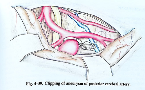
The aneurysms of the posterior cerebral artery are most often originating from the P2 and P3 segments of the artery. These segments are approached most conveniently subtemporally. Medially to the tentorial notch the arachnoid is opened, and P1, P2 point at the origin of the posterior communicating artery is identified. P2 segment is followed distally to find P3 branches, and by that usually the aneurysm is already seen. The aneurysmal neck is prepared caring of the small perforators appearing from that segment (Fig. 4-39). Temporary clipping, if required, is used to isolate the aneurysm, but distally to the choroidal and peduncular arteries. Clipping rarely produces complications if the blades are closed under visual control.
ANEURYSMS OF THE SUPERIOR CEREBELLAR ARTERY
The aneurysms of the trunk of the basilar artery are situated most often in its upper third, at the place of separation of the superior cerebellar artery or in its lower third, where the inferior anterior cerebellar arteries come out.
The aneurysms of the upper third of the basilar artery arise most often from the distal angle between this artery and the superior cerebellar and have a well formed neck. They are situated anteriorly or laterally in the subarachnoid space, are not closely attached to the perforating arteries and may form a bed in the peduncles.
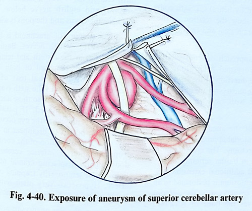
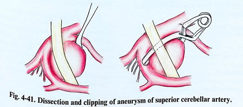
The approach to the aneurysms in this location is always subtemporal. After the mesencephalic cistern is opened and the trochlear nerve is preserved, the tentorial edge is retracted with a stitch, and the arachnoid is divided, anteriorly through the Lilliequist membrane. At this stage P1, and P2 arterial segments are clearly identifiable and the basilar trunk is carefully approached intending eventual proximal control. Along the basilar arterial wall can be detected the origin of the SCA and the aneurysm itself (Fig. 4-40). Anteriorly directed aneurysms are easier to dissect and are occluded with straight clip, but those pointing superiorly require curved blades and much more attention not to compromise mesencephalic branches (Fig. 4-41). Oculomotor nerve is often affected by the dome, can suffer damage during dissection and may need retraction at the time of clipping.
MIDBASILAR ANEURYSMS
Midbasilar aneurysms most often originate at the emerging point of the AICA. This part of the basilar artery is more difficult for access and very selective choice of supra-, infra- or combined transtentorial approaches do exist, demanding very appropriate decision-making.
The difficulties and risks faced with open surgery give preference to endovascular techniques in those cases where perforators or the AICA itself are not too close to the aneurysmal neck and can be accidentally compromised by the occlusion. Potential collaterals of the basilar artery have to be evaluated by angiography before surgery. In case of temporary clipping of the basilar trunk, sufficient flow has to be provided on both sides of the trapped segment.
Another important investigation is the MRI (in some cases substituted by helical CT with contrast) and it has to show the relation of the lesion to clivus, the brain stem and emerging of cranial nerves.
The choice of approach depends on the position of the aneurysmal neck along the basilar trunk and its direction. In this regard midbasilar aneurysms are subdivided into two subgroups: with "low" and "high" position.
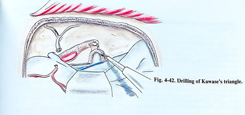
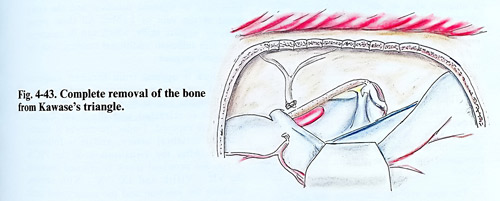
High position of the aneurysms requires subtemporal, transtentorial approach preferably with anterior petrosectomy. The craniotomy is similar to that used in basilar top aneurysms, but with small additional extension posteriorly. Section of tentorium is performed in all cases, preserving IV cranial nerve. The anterior petrosectomy removes the bone of Kawase's triangle in the medial petrous apex and provides space for approaching the petroclival region of the upper posterior fossa, and to the upper half of the basilar trunk (Figs. 4-42; 4-43).
Anterior petrosectomy technique includes selective removal of part of the petrous bone. After separation of the dura from the temporal fossa, the exposed bony surface reveals a number of important structures and middle meningeal artery is divided at its exit from foramen spinosum, the greater superficial petrosal nerve is sacrificed and the bone toward its origin at the geniculate ganglion is drilled out. The horizontal part of the ICA is exposed also without its posterior loop because of the danger of bleeding. The remaining bone medially and anteriorly to the geniculate ganglion and the superior semicircular canal (arcuate eminence) can be drilled out safely. The dural sleeve of the internal acoustic meatus has to be exposed, and if the drilling is always medially to the Miller's line (connecting intersection of carotid and trigeminus with Bill's bar), there is no danger of damaging cochlea.
In the subtemporal route, the dura is opened and a spatula is placed to retract the temporal lobe. The tentorium is divided along the superior petrosal sinus and the spatula is repositioned deeper, also retracting gently the cerebellar hemisphere. Gradually this spatula is moved, until it exposes the pons between the fifth cranial nerve above and the roots of the seventh and eighth nerve below. The space between the clivus and the pons is widened.
The opening of arachnoid is between the Vth and VII - VIIIth nerves. Basilar artery is usually seen in the cistern and temporary clipping places are prepared of both sides of the aneurysm, with minimal tear of adhesions around the aneurysm. The sixth cranial nerve is situated in close vicinity to these aneurysms, it is dissected and displaced. If paralysis of the lateral rectus muscle of the eye occurs, it is most probably expected to disappear within few days or weeks. The dissection of the aneurysmal neck from the anterior inferior cerebellar arteries done gently displacing cranial nerves and at times under temporarily trapping of the basilar artery. If occurring, hypoacusis very often remains lasting, but facial palsy has a better prognosis.
Low midbasilar aneurysms are approached through lateral suboccipital craniectomy, at times enlarged with petrosectomy.
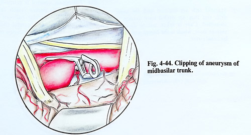
In the lateral suboccipital approach, after retraction of the cerebellar hemisphere, arachnoid medially to floculus is opened and all the space between the VII - VIIIth and IX - Xth nerves is used for the approach. The dissection further on follows the same principle as in the previous approach. Very often have to be used angulated clips to avoid basilar artery stenosis (Fig 4-44).
ANEURYSMS OF VERTEBROBASILAR JUNCTION
The aneurysms of vertebrobasilar junction create significant problems for treatment. Located in one of the most difficult to approach places, they very often have complex configuration and poorly defined neck, thence straight forward decision for the method of treatment cannot be taken with ease. Options include endovascular and direct techniques, but preference is given to the first one. When the aneurysm doesn't have arterial branches arising very close to its neck and its shape and neck after thrombosis will not compromise the flow in the parent artery, is proceeded with interventional neuroradiology. It is common however, to find a situation when the direct attack is the only choice. In these cases is considered the low lateral suboccipital approach, which can be extended with posterior petrosectomy and temporal flap, or if the lesion is closer to foramen magnum, removal of the medial third of the occipital condyle.
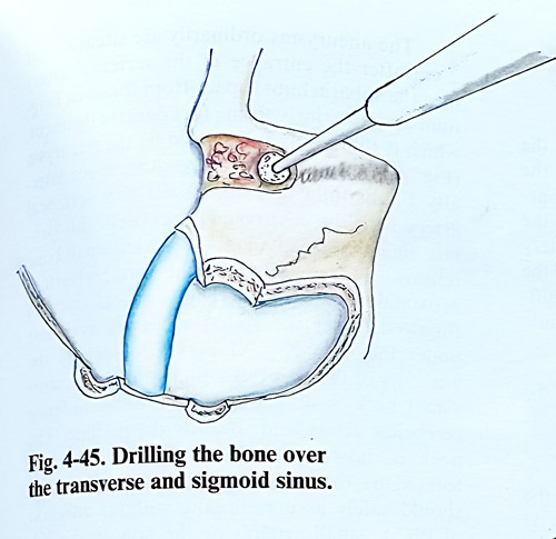
The posterior petrosectomy is an adjunct to the confined supra- and infratentorial approaches (posterior temporal and lateral suboccipital). After these craniotomies are completed, several further steps are followed to gain the presigmoid route. These steps in brief are as follows:
1. The bone over the transverse and sigmoid sinuses is removed completely with the high-speed drill (Fig. 4- 45).
2. Mastoid cells are exposed with the same drill, and they are drilled until antrum is identified. Care is taken not to damage antrum floor, which is the lateral semicircular canal cover.
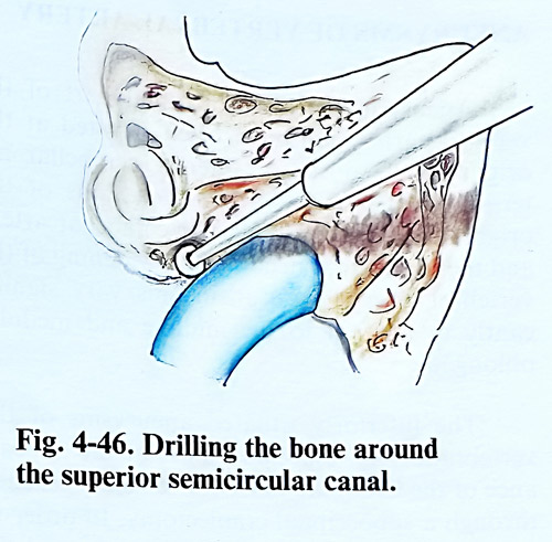
3. The sino-dural angle bone is removed with the diamond drill to the posterior semicircular canal, together with the remaining bone around the superior semicircular canal to expose the superior petrous sinus (Fig. 4-46).
4. Mastoid tip cells are removed also to the digastric ridge (close to the stylomastoid foramen). The Fallopian canal is followed superiorly at the same depth as the posterior semicircular canal.
5. Dura is opened superiorly and inferiorly to the superior petrous sinus. The sinus is clipped (ligated) and divided.
6. The tentorium is incised parallel to the superior petrous sinus.
This approach preserves all structures, except the superior petrous sinus. It mobilises the transverse to sigmoid junction and the entry point of the vein of Labbe. All these steps, however, require skills, which can be mastered only with sufficient additional training with the otological surgeon. Dura is opened from foramen magnum upward to the most superior part of the exposure. CSF is evacuated from cisterna magna.
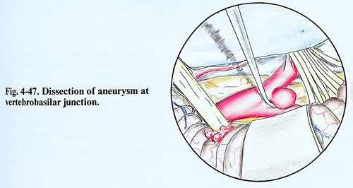
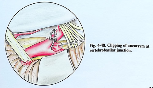
Floculus is retracted and the lower cranial nerves are exposed. Under the nerves is identified the vertebral artery, and it is followed distally. After the visualisation of the PICA origin, the vertebral trunk is exposed between the VII - VIIIth and IX - X - XIth nerves, reaching the junction and the aneurysmal neck (Fig. 4-47). The dissection cares for the perforators and the anterior spinal artery origin in this area. Temporary control is extremely difficult, but the accessible parent vessels can by controlled with endovascular balloons navigated there. Clipping requires careful choice of the shape of blades to avoid distortion of the junction (Fig. 4-48).
ANEURYSMS OF VERTEBRAL ARTERY
Aneurysm of the intracranial part of the vertebral artery are most often situated at the origin of the posterior inferior cerebellar arteries. As a result to the variable locations of the origin of the posterior inferior cerebellar artery and the variety of shape and lumen of the vertebral artery, these aneurysms vary significantly in their relations to midline and medulla oblongata.
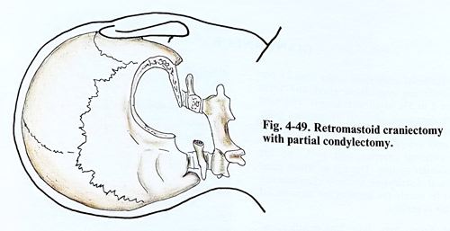
The inferiorly located aneurysms of the vertebral artery, especially those at the appearance of the PICA are most conveniently reached through a suboccipital craniectomy. In order to expose the space anteriorly to the pons and the medulla oblongata better, the craniectomy is widened on account of the resection of the posterior lamina of the atlas reaching the atlanto-occipital joint (Fig. 4-49). The vertebral artery entry through the atlanto-occipital membrane is reached and the posterior part of the occipital condyle is drilled out to the hypoglossal canal. The posteromedial one third of the condyle and the lateral mass of atlas can be removed without biomechanical compromise. This removal can be time consuming and not necessary in many cases of more distally located lesions.
Dural incision begins at Cl and extends vertically through foramen magnum to proceed laterally to the superolateral apex of the craniotomy and cisterna magna is opened.
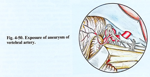
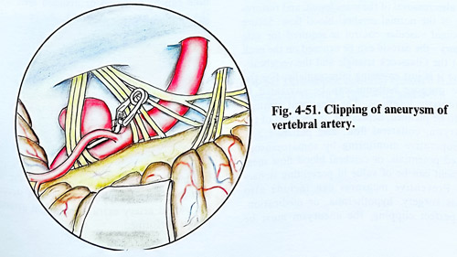
Aneurysms usually are situated 1 or 2 cm after the entrance of the vertebral artery into the subarachnoid space from foramen magnum and anteriorly to the first dentate ligament, which is thinned and distended by the aneurysm (Fig. 4-50). In order to achieve some additional space laterally to medulla and to dissect the vertebral artery with the aneurysmal neck well, this dentate ligament is divided. Additional bone removal allows control of the vertebral artery proximally for clipping. The vertebral artery is followed under the spinal accessory and next under the IX - X - XI nerves to identify the anatomy of the aneurysm. Sometimes the aneurysmal neck is deeper to the posterior inferior cerebellar artery and towards the midline. The neck is, however, quite always at the angle formed by the origin of PICA. The dissection should safely assure clipping without affection of PICA, small arteries or the flow in the VA (Fig. 4-51).
At present postoperative care in addition to the routine can include some additional important measures, also applicable to ruptured aneurysms with higher risks in other locations:
1. The triple H therapy: Hypervolemia, hyper-tension, haemodilution, with invasively monitored parameters of hemodynamics (preferably with Swan Ganz catheter).
2. Transcranial Doppler monitoring of vasospasm.
GIANT ANEURYSMS
Intracranial aneurysms larger than 25 mm in diameter are considered giant and these comprise 2 to 5% of all such lesions. The most common locations of giant aneurysms differ from those smaller in size. About 40% are seen on the carotid artery, 25% on the anterior and middle cerebral arteries, and 30 % on the vertebrobasilar system. The age at which giant intracranial aneurysms become symptomatic seems to be nearly the same as for intracranial aneurysms in general.
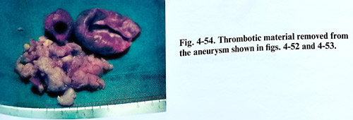
Plan X-rays may show 'ring calcification' which is pathognomic for these lesions, or bony erosion due to chronic compression. The CT scan is usually positive showing round mass with a ring enhancement around the aneurysmal wall, nonenhanced thrombi and enhanced central region due to blood flow. MRI demonstrates the presence of giant aneurysms quite well by the flow inside as a signal void area and differentiating old clots from recent ones. "CT angiography" with spiral scanning techniques can add a lot about the shape and location of the lesion. Cerebral angiography is still the most important investigation for surgery of giant aneurysms (Figs. 4-52 - 4-54). It should clarify the exact anatomy around the neck of the aneurysm and determine the collateral flow. Functional assessment of the collateral flow, including balloon occlusion tests can be needed preoperatively, as long lasting parent artery occlusion may take place during surgery.
The treatment goal for giant intracranial aneurysms is not only prevention of rupture, but also removal of the mass lesion, and restoration of the normal cerebral blood flow. Secure proximal vascular control is required for safe surgery - the carotid can be exposed on the neck or at the Glasscock triangle and the vertebral - where it is just emerging intracranially. For the same purpose also balloons can be navigated in appropriate locations. The treatment strategy has to be planned, depending aneurysm location, collateral flow, and patient's age.
Intraoperative monitoring by motor and/or somatosensory evoked potentials, or cerebral blood flow measurement can be of value in preventing ischaemia. Preventive measures can include also bypass surgery, hypothermia, or medication.
For precise clipping, the aneurysm must be slackened by suction decompression, thrombectomy, or cardiac standstill. The surgeon should select the best fitting clips and check the result by intraoperative angiography. Endovascular treatment can prevent rupture, but does not remove the existing mass lesion and its effects.
DIRECT SURGICAL TECHNIQUES
Giant carotid aneurysms. They are classified according to their projection into four groups: anterolateral, anteromedial, posteromedial and posterior.
With the patient in supine position the cervical carotid artery is exposed for temporary trapping and a pterional craniotomy is made.
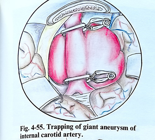
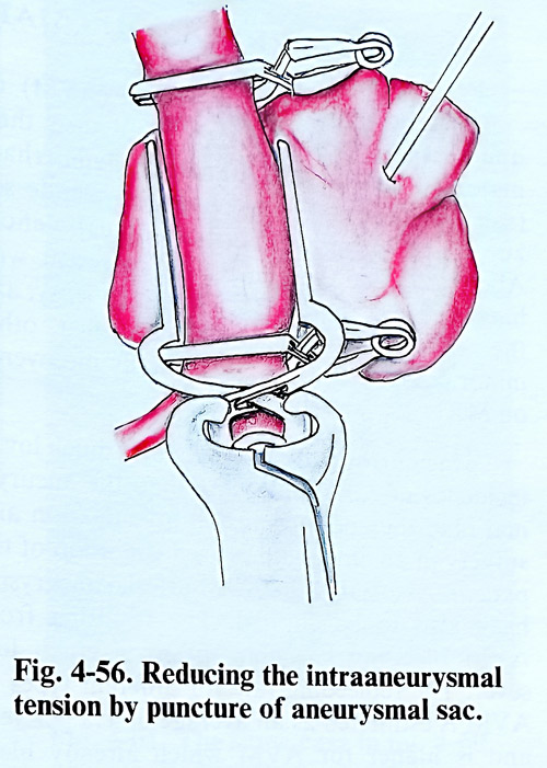
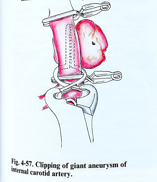
The frontal branch of the superficial temporal artery is preserved as a possible donor for an extracranial-intracranial bypass. Before approaching the aneurysm, bone in paraclinoid region should be drilled, optic canal and distal dural ring opened widely to release the optic nerve and C3 portion of the carotid artery. A more intense bleeding from the cavernous sinus can be controlled by elevation of the patient's head and meticulous packing with oxidised cellulose. The body of the aneurysm is dissected and the neck is isolated. At least two-thirds of the major circumference of the aneurysm must be dissected from adjacent tissues for satisfactory clipping. When the intraaneurysmal pressure is too high to isolate the aneurysm, clipping on the neck under temporary trapping of the parent artery can be suggested. Thrombi and calcified plaques at the neck can be avoided or removed after trapping of the parent artery. A good choice of with the optimal in fitting to the neck clips is essential (Figs. 4-55 - 4-57).
Surgery for giant aneurysms of the middle cerebral artery is generally easier than surgery in other locations because the operative field is wider and trapping is easier. The most important goal in these cases is to avoid stenosis and occlusion of distal branches of the middle cerebral artery and lenticulostriate arteries. Sometimes after using multiple clips in a proper combination, remaining portion of the aneurysm may need wrapping. In case of stenosis of the parent artery extra-intracranial arterial bypass is performed.
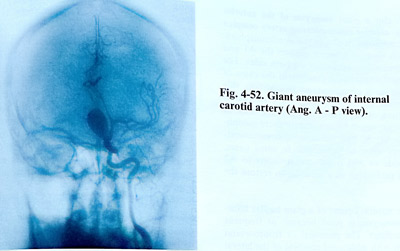
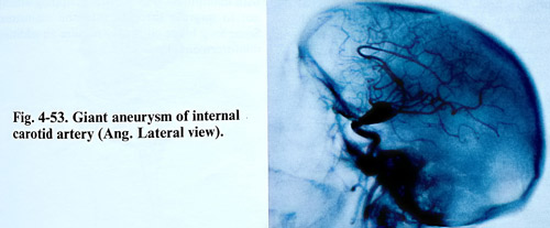
To clip a giant aneurysm of the anterior cerebral- anterior communicating artery complex is more appropriate to use the interhemispheric approach because it better exposes the Al and
A2 segments simultaneously on both sides. The lower intra-aneurysmal pressure in the cases of this location is an advantage. In addition, this area seems to tolerate temporary occlusion better than others. However, the anatomy of these aneurysms is comparatively more complicated as Al, A2 and the anterior communicating artery must be preserved. In some cases, side-to-side or end to side anastomosis to A2 must be performed as a solution to restore the flow.
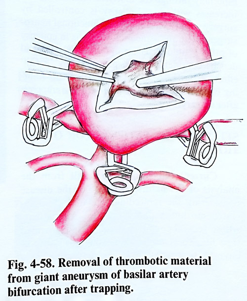
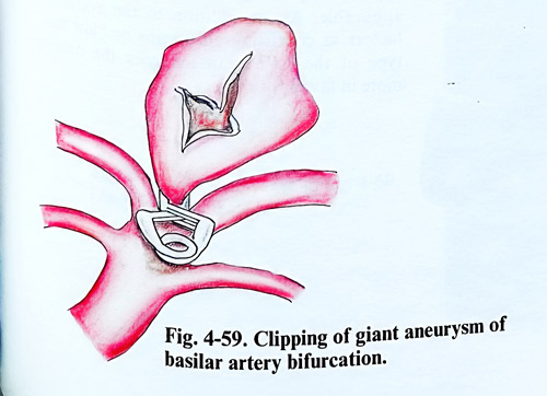
The natural course of a giant basilar bifurcation aneurysm is poor because of frequent fatal bleedings. The pterional or frontoorbital approach with additional removal of the lateral orbital wall is quite useful in widening the operative field. In some cases a temporobasal craniotomy with opening of tentorial notch gives also good access to the lesion. With increased use of temporary basilar artery clip-
ping, the results of aneurysm surgery in this location have become more successful (Figs. 4-58; 4-59).
Most of the giant vertebral artery aneurysms are partially thrombosed and their clinical presentation is brain stem affection with long tract signs, lower cranial nerve palsies and respiratory distress. The treatment of choice may be intravascular occlusion of the vertebral artery if the patient can tolerate the occlusion test and has a good collaterization from the opposite side. If the patient's condition doesn't improve after occlusion, trapping with excision may be necessary. Long lasting deficit does not improve with decompression.
Partially thrombosed aneurysms make at times clip blades closure impossible. The clip usually slides to the parent artery and constrict its lumen. In such cases aneurysm thrombectomy is necessary. Great care should be taken with manipulation or multiple clipping attempts not to migrate thrombi into the mainstream of the parent artery.Once the aneurysm clipped, some long blade clips may require an additional reinforcement clip.
ARTERIOVENOUS MALFORMATIONS OF THE BRAIN
Arteriovenous malformations (AVM) require surgical treatment because of their three major clinical manifestations: haemorrhage (subarachnoid and intracerebral), epileptic seizures and progressive neurological deficit.
About 50% of the clinical cases present with haemorrhage, another 25% - with epilepsy, and the rest (25%) with neurological deficit, other minor complains or are accidentally discovered on MRI.
Haemorrhage due to AVM has much lower incidence of early rebleeding than the aneurysmal, thus permitting full investigation and surgery in an already improved condition of the patient. Related to the presence of AVM arterial aneurysms have also to be clipped. The resulting from AVM bleeding vascular spasm is much less severe. The rebleeding rate for different types of AVM is estimated as an average of 4% per year and is higher for AVMs which already bled, especially through the first year after bleeding, and increases with age. Epilepsy is controlled by surgery with the techniques of functional neurosurgery, with the same indications as in epilepsy of other aetiology. The progressive neurological deficit, considered to be provoked by "steal" effect of the AVM circulation on the surrounding brain tissue is expected to improve after surgery.
SELECTION OF PATIENTS FOR OPERATION
Current indications are the result from the analysis of the natural outcome of the disease considering rebleeding rate with its associated morbidity and mortality. All patients whose general condition permits (and provided their life expectancy is long enough to justify it) have to be evaluated for surgery. The same refers to all patients with epilepsy or other complains, taking into account their natural prognosis. The risk of treating them conservatively has to be compared to the surgical risk of total excision of the malformation by craniotomy.
At present from existing three methods of surgery: "open" surgical excision through cra-niotomy, endovascular obliteration and radio-surgery (stereotactic radiosurgery), only the first can provide high degree of certainty for radicalism. Therefore, we consider the other two as a second line or adjuvant to the surgical excision.
For the evaluation of surgical risk for open excision of an AVM, recently have been identified three more important factors. They are: 1. The size of malformation nidus. 2. The part of the venous system (deep or superficial) participating in the drainage of the AVM, and 3. The functional importance of the brain areas occupied by the nidus, particularly its functional importance or "eloquence". With the contribution of leading neurosurgeons, AVM surgical cases, evaluated retro- and prospectively, have shown that the affection of the deep venous system by the high pressure drainage, by big AVMs in "eloquent" areas of cortex or brainstem, is associated with clearly significant increase of mortality and morbidity.
Only by balancing the natural risk with the surgical risk according to these factors can be achieved the proper decision for surgery. For these purposes have been developed several grading systems, from which that of Spetzler and Martin appears at present to be the most universally applicable. As an addition to the evaluation, factors as coexisting aneurysms or "low flow" type of the AVM can influence the decision more in favour of surgery.
EXCISION OF THE ARTERIOVENOUS MALFORMATIONS
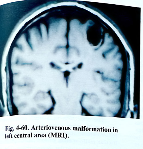
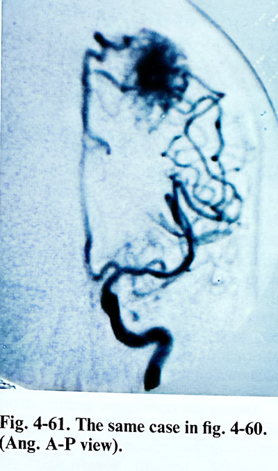
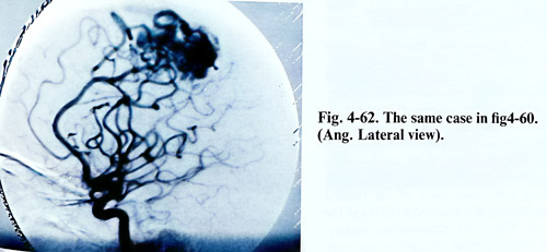
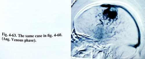
When planning the AVM surgery it has to be decided if the feeding can be reduced by endo-vascular procedures in advance. That procedures have to precede shortly the day of surgery, usually at the time of superselective angiographic study. On this last study the position of all major feeders and drainers has to be identified in relation to bony, dural and cortical reference points. Although the angiography can be very precise, additional space for hidden feeding vessels and free manipulation upon them has to be provided, so larger craniotomies with generous distances to the nidus are recommended (Figs.4-60 - 4-63). Very often during the operation the malformation proves to be of larger size than that shown on the angiography.
During the craniotomy greater attention is paid to haemostasis as in many cases vessels from of the external circulation communicate freely with the malformation. In such a case, the opening of the dura is made with an incision that gets around such communications. Frequently after opening of the dura only enlarged arterialized veins are seen in the cortical surface.
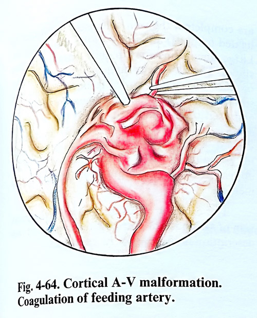
The arachnoid around these veins is opened. The dissection of the malformation begins sub-pial and if the feeding arteries are not seen on the surface, should be searched carefully for them considering the angiographic data (Fig. 4-64).
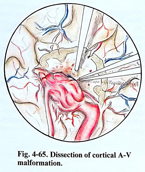
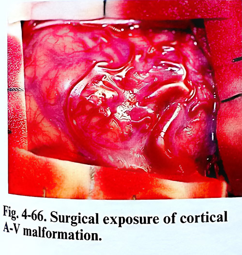
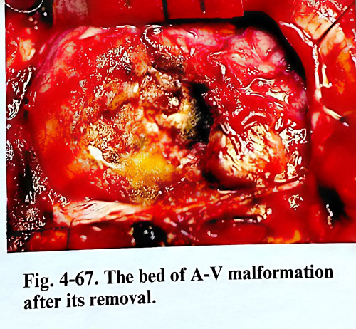
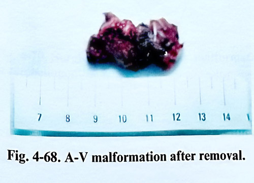
After clipping and coagulation of the feeding arteries, they are divided near to the nidus of the malformation. The interruption of the supplying arteries causes partial collapse of the malformation and this facilitates the dissection to follow (Fig. 4-65). In case of perinidal haematoma, the residual cavity around the malformation makes the dissection easier. If there are difficulties to follow the plane of cleavage around the malformation, the thin layer of gliosis around the nidus is a good indicator. When it is impossible to find a feeding artery near the malformation and there is a risk of injury of apparently normal brain tissue, temporary clipping of some branches of the circle of Willis more proximally is an option. After interruption of the feeding arteries and dissection of the nidus, the draining veins are clipped and divided last. The malformation bed is inspected carefully for residual pathological vessels and final haemostasis is achieved. If arterial hypotension is applied, the blood pressure should be raised to normal before deciding on safety of haemostasis (Figs. 4-66; 4-68).
Rupture during the dissection causes at times dramatic bleeding and in these circumstances manipulation may cause destruction of brain tissue or considerable loss of blood with abrupt decrease of the systolic blood pressure. In such cases one of the malformation walls must be dissected very fast and a broad clip be applied on the bleeding area until bleeding is controlled and dissection continues in the usual meticulous way.
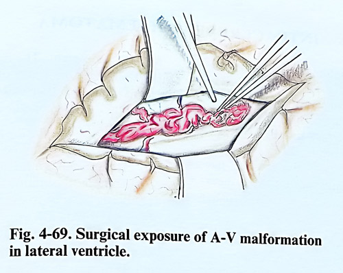
The dissection of a deep arteriovenous malformation has some particular points for consideration. The removal of a para- and intraventricular malformation carries risk of producing severe intraventricular haemorrhage. The feeders in completely intraventricular AVMs originate from the anterior or posterior choroidal arteries, and can be accessible in the ventricular cavity (Fig. 4-69).
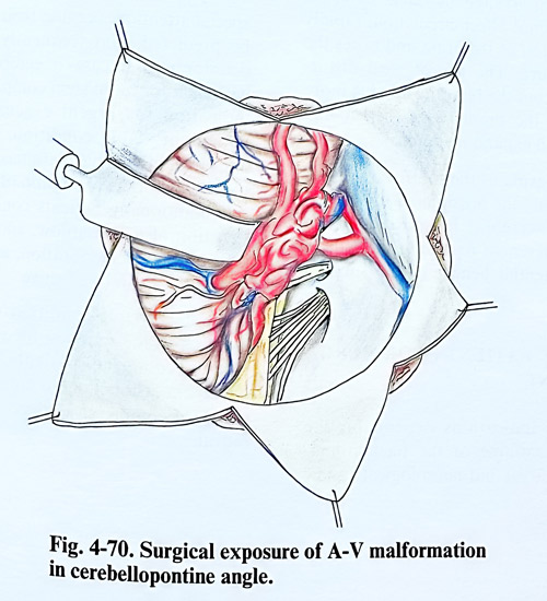
The excision of an arteriovenous malformation from the cerebellum is requiring the clipping of some cerebellar arteries and when it is situated in the cerebellar hemisphere, partial lobectomy can safely be performed, without causing any cerebellar symptoms (Fig. 4-70). The removal of an arteriovenous malformation from the vermis usually does not present any special difficulties.
The malformations of the great vein of Galen are particular type of AVM. In spite of the fact that they are rare, the decision for surgery has been always a major point of controversy. In this type of malformations due to high intravenous pressure, the great vein is dilated to the shape of a sphere or ellipsoid with diameter 3 or more centimetres. The feeding vessels are branches of the anterior and middle cerebral arteries and enter directly into the vein uni- or bilaterally.
This type of arteriovenous malformations can be treated in very rare occasions by direct attack despite the existing controversies. That is not the first choice at present, and endovascular procedures have to be attempted after complete selective angiographic studies. On the basis of these last investigations and the assessment of the general condition (often impaired due to compromise of the cardiac function, resulting from the AV shunt), is taken the final decision for the procedure to be performed. The approach is done through parietal and parietotemporal craniotomy close to the midline, depending on the location of the feeders. When the parietal approach is used, the surgeon penetrates between the medial surface of the hemisphere and the falx, followed by division of corpus callosum. The approach is facilitated a lot by lateral ventricle puncture. The aneurysmal wall (the widened great vein of Galen) is carefully dissected and all arteries, which according to the angiographic data penetrate into it are divided. When these divisions are completed, the vein collapses, and it can be ligated (clipped) anteriorly to the straight sinus.
Large aneurysms may require surgery in two stages. The first stage undertakes a craniotomy on the side of the wider feeders. During the second stage the same is made on the other side and excision is completed. When the supplying arteries are located more posteriorly, more convenient is the parietooccipital craniotomy with or without incision of the tentorium. This approach does not require corpus callosum incision.
The excision of an aneurysm of the great vein of Galen is not a routine technique and this description indicates only its guidelines.
INTRACEREBRAL HAEMATOMA
Inracerebral haematomas, except those of traumatic origin, are due usually to hypertension and have predilected locations. About 50% of them are in the basal ganglia, 15% in thalamus, 10% in the cerebellum and 10 to 20% in the white matter of the hemispheres. In patients without hypertensive disease, the cause of haematoma is most frequently rupture of an aneurysm, arteriovenous malformation orcoagulopathy.
The basic principles of surgical treatment of intracerebral haematomas are completely different from those in other parts of the human body. In case of haemorrhage outside of the intracranial cavity the aim of surgery is to stop the bleeding as early as possible, to treat the resulting anaemia, and save patient's live. In intracerebral haemorrhage blood is of limited amount and does not affect circulation, rapidly provokes destruction of the brain and raises the intracranial pressure. The neurological disturbances resulting from the bleeding are due more to destruction of the brain tissue then to the blood volume as an expanding lesion.
This concept explains the controversy existing in the choice of method of surgical treatment in intracerebral haemorrhage. Till now there are differences regarding the indications and the potential benefit of the surgical treatment.
SELECTION OF PATIENTS FOR SURGICAL TREATMENT
The surgical indications depend on the location and the volume of the haematoma, considering the general and neurological conditions of the patent. The haematomas located in the thalamus, the brain stem and intraventricular hemorrhages after evacuation do usually not improve patient's condition. The haematomas in the brain hemisphere, those located laterally to the internal capsule in the basal ganglia and in the cerebellum are considered more suitable for evacuation (Fig. 4-71). Better results from surgical treatment are obtained in patients with slow progressive clinical deterioration for hours or days. The surgical evacuation does not change evidently the clinical evolution when clinically there is acute onset and bad initial clinical condition. Improvement can be expected in haematomas, when CT scan images show displacement of midline brain structures.
The haematomas in the cerebellum need special attention, because their evolution cannot be predicted, and frequently there is sudden deterioration. In cases of cerebellar haematomas with data for brain stem compression there is an indication for urgent evacuation. Even in a patient in stable condition, the operation is indicated in big haematomas. Intraventricular bleeding with the formation of clots can deteriorate additionally patients condition due to obstruction of CSF outflow, and if obviously this is the cause of deterioration, an urgent ventricular drainage can be effective.
The general condition of the patient is essential in the selection for surgical treatment. Deeply comatose patients, with significant brainstem disfunction do not improve even with clearly decompressive effect of haematoma removal.
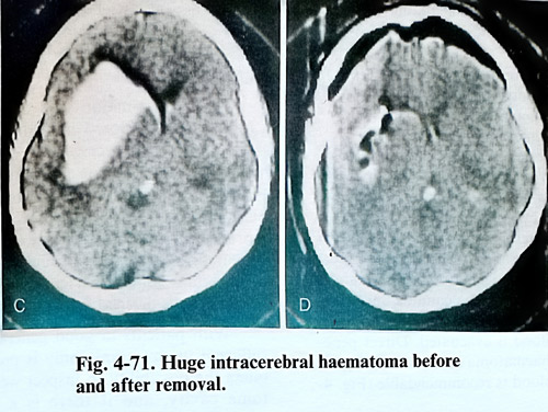
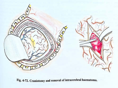
SURGICAL TECHNIQUE
The choice of surgical technique for evacuation of an intracerebral haematoma depends on the volume and the location of the haematoma. The worse the condition of the patient, the less surgical trauma he will be able to sustain. A craniotomy, and even craniectomy up to 3 - 4 cm in diameter is enough to provide save access to the haematoma, if done at the most appropriate place following the clinical and CT scan data. After opening the dura, the brain tissue is found under increased pressure and with the tendency to protrude through the craniectomy. Sometimes a yellowish coloured cortex can be seen, and that indicates the most superficial part of the haematoma. The brain is punctured with a ventricular cannula and when the haematoma is reached the maximum possible amount of blood is evacuated. Direct penetration into the haematoma cavity and complete removal of the blood is recommendable (Fig. 4-72).
In case of huge haematomas, however, can be evacuated mainly the fluid portion, if such exists. This is enough efficient in reducing intracranial pressure and brain tissue compression, and safer regarding postoperative rebleeding (Fig. 4-73).
The direct penetration, described above, refers to haematomas whose cavities are very close to the surface, quite under pia. That is not the same in deeply seated lesions, where to reach the cavity has to be penetrated eloquent brain, apparently functionally intact. To avoid additional damage, the approach has to be selected avoiding functionally important areas, gaining penetration to the depth through sulci or the anatomically important fissures, especially the Sylvian and the interhemispheric. Dissection of a deep sulcus requires the surgical microscope. The same is valid for exposing insular and opercular cortical surfaces through the Sylvian fissure. After such dissection vascular damage and pial incisions are minimal, but sufficient access is provided to the haematoma (Fig. 4-74).
In the great majority of cases, the bleeding vessel has thrombosed immediately after rupture and it is not necessary to do haemostasis. However, when the operation is performed in the first hours after the onset of the haemorrhage, a bleeding vessel can be discovered and the haemostasis can be performed by clipping, bipolar coagulation or oxydased cellulose. In the event of uncertain complete haemostasis, the haematoma cavity is drained for 24 hours.
With patients in good general condition a sufficient in size craniotomy is preferable. This offers the possibility to inspect well the haematoma cavity, and if there is a suspicion of pathological tissue presence, a small specimen is taken for biopsy.
If an aneurysmal rupture is the cause of the haematoma, the surgeon has to make an effort to dissect the aneurysm and exclude it from the circulation. When the haemorrhage is due to an arteriovenous malformation, the operation is much more complicated. In such cases it is better to remove big part of the haemato-ma, if there are vital indications. The dissection of the malformation must be left for the next stage, after the patient has completely recovered. Later, in a relatively good general condition of the patient, the malformation is investigated properly and can be removed without causing an additional trauma to the brain.
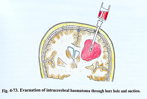
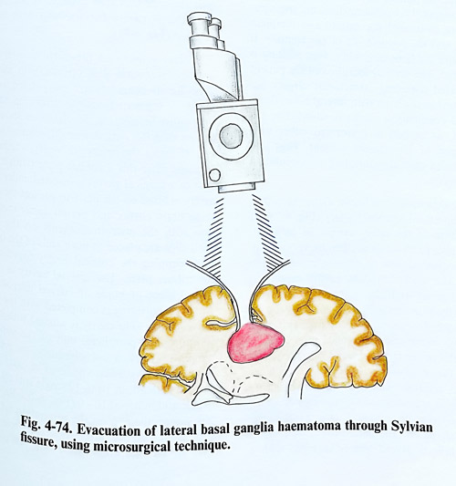
EXTRA-INTRACRANIAL ARTERIAL ANASTOMOSIS
The results of extra-intracranial bypass study published in 1985 indicated that this technique was not effective in reducing stroke incidence or stroke related death. Currently the operation is applied rarely in selected patients who have a vascular lesion directly related their symptoms and who have decreased blood flow in the corresponding hemisphere on cerebral blood flow study. The same patients should not have effective collateral circulation on evaluation of their angiograms and functional tests.
Extra-intracranial anastomosis is applied also with prophylactic aim when an ischaemic lesion is expected in the territory of the internal carotid or the middle cerebral arteries before the removal of big basal tumours, which envelop the internal carotid artery. In view of all this, cerebral revascularization with different EC -IC techniques is not any more an accepted routine in cerebral ischemia.
Once the patient has been decided to be in need of cerebral revascularization, his angiographies are evaluated for donor and recipient arteries and the possible site of anastomosis. If donors are deficient in quality, vein grafting is considered. The general condition of the patient is evaluated regarding intracranial surgery under general anaesthesia with several hours duration and coagulation profile is adjusted to the required, as surgery is performed either with normalised or heparinized patient. That will also demand careful monitoring of coagulation after surgery.
The skin incision is linear, along the course of the superficial temporal artery (Fig. 4-75). If the position of the artery cannot be palpated, the portable Doppler device can by very useful. The dissection should be done in a way not to damage the small branches or the superficial temporal artery itself. Its anatomical location is between the skin and galea, so once it is exposed distally, the dissection proceeds following it at the above mentioned anatomical plane. Either the parietal or the frontal branch of the artery is selected for anastomosis (Fig. 4-76). Together with the arterial branches are also running veins, which are recognised by the lack of pulsations. The temporal muscle is incised in a linear or " T " shape and the bone surface is exposed. A burr hole is placed and is enlarged with a craniotomy as a small free flap.
The dura is opened with a stellate incision and the tips of the flaps are lifted with traction sutures. A suitable recipient artery (about 1 mm in diameter) is identified on the cortex. The artery may be any branch of the middle cerebral artery, but a temporal one is preferable. If a suitable branch cannot be found or it appears very close to the edge of the craniotomy, small additional nibbling is required. If the cortical surface is tending to protrude, the arachnoid is opened. Seldom has to be resorted to a lumbar puncture. More often at completion of the anastomosis, the cortex is separated from dura and care is taken to avoid subdural collections postoperatively. When dissecting the donor, the periarterial tissue is left around it, coagulating and cutting all branches. The length of the dissected artery must be about 5 cm. The adventitia is removed only from the isolated end of the superficial temporal artery and its proximal part is clipped. A thin catether is inserted into the arterial cavity for washing with heparin solution (Fig. 4-77).
Next is isolated the cortical artery and the arachnoid around it is opened with microscisors. Small branches are coagulated and divided. A segment from the artery up to 1 cm long must be isolated. Its diameter should not be less than 0.8 mm. A fine dissector or hook is passed under the artery, to verify whether it is free and after this a rubber plate (with a thickness of a surgical glove material) is inserted under it. The artery is lifted on the plate to protect the underlying brain cortex and permit easier manipulation with the microinstruments on the artery.
Two clips are placed at both ends of the isolated part, clipping the cortical artery together with the rubber plate. The arterial cavity is opened with a small blade or curved microscissors. An elliptic opening is completed with the curved microscissors. The cavity is washed with heparin solution. The peripheral end of the superficial temporal artery is cut obliquely, to get wider aperture to the anastomosis. The later is performed with about 10 to 12 single sutures with a needle and material 10 - 0 (Fig. 4-78). Before the last suture, the clip on the proximal part of the superficial temporal artery is slightly released so that air bubbles are pushed out. All clips are removed, starting with those on the cortical artery and after that on the superficial temporal artery. The place of the anastomosis is slightly pressed with cottonoid and oxidise cellulose, as very often minor bleeding occurs at the place of the sutures, which stops in a few minutes only with this technique. If the bleeding continues, clips are applied anew and an additional suture is placed (Figs. 4-79; 4-80).
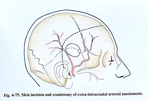
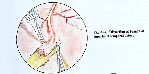
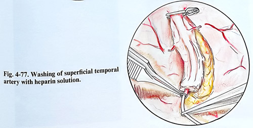
The dural flaps are turned back on the cortical surface and closed with single sutures.
The temporal muscle and its fascia are closed separately, taking care not to strangulate the donor artery. The skin is sutured, applying fine drainage in the epidural space.
Operative variants. A double anastomosis can be performed, using both branches of the superficial temporal artery and each of them is connected with a separate cortical branch. No obvious benefit has been shown yet from this technique.
The occipital artery can be used for anastomosis with a branch of the middle cerebral artery, when the superficial temporal artery and its branches are inadequate.
Another option for the high-flow requirements compensation of the middle cerebral artery circulation after carotid occlusion are the vein grafts. Two types have been used: "short" - between the proximal superficial temporal artery and a cortical branch, and "long" - between the carotid artery in the neck and the middle cerebral artery branches. The main characteristic property of vein graft is their diameter and consequently, the higher flow through them. "Short" grafts are substitute of inadequate for anastomosis superficial temporal artery branches, thus providing sufficient diameter and length of the donor. "Long" grafts intend to provide the benefits of the proximal high flow and pressure in the carotid, delivering them directly to the middle cerebral artery circulation. Technically the graft is anastomosed on both ends with microsutures and passed through a tunnel. "Short" grafts are positioned in a similar way through the tissues as the standard STA - MCA bypass donor artery. "Long" venous grafts however, require tunelization between the wound at the neck and the craniotomy usually positioning them retro- or pre-auricularly. Grafts are heparinized after harvesting and are sutured with continuous or interrupted monofilaments under magnification. Postoperative angiographies after these techniques show very often excellent filling through the graft.
There are options for revascularization of the posterior circulation and the donor (most often occipital artery) is sutured to PICA branch. Another approach uses STA to reach the superior cerebellar artery, via tentorial incision. Few centres have developed and are performing these techniques.
Postoperatively the wound care present occasionally problems with healing. It is a result from the devascularized wound edges at surgery. That however rarely requires any special treatment and is resolved within the usual period of time. Another important point to remember is the position of the donor or graft under the soft epicranial tissue. It permits easy Doppler control of the flow, but can be pressed or injured there much easier.
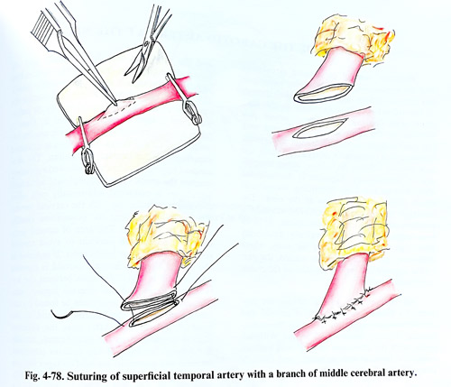
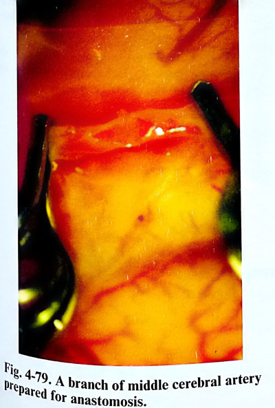
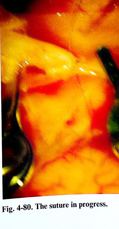
EXPOSURE OF THE CAROTID ARTERY AT THE NECK
Proximal control on carotid circulation and the application of endovascular techniques has demanded from neurosurgeons the exposure of carotid artery and it branches on the neck more often than any other major vessel. Not satisfied by the complications associated with the carotid ligation in intracranial aneurysms, neurosurgical community rarely resorts to it and now prefers balloon occlusions, either temporary or permanently. Ligation is discontinued also, due to the huge ascending thrombosis which follows the occlusion at the neck. The isolation of the carotid, however, remains a solution when unclippable aneurysms, or skull base tumours are treated surgically, preferably after high flow bypass. As in the cases, where temporary proximal control is needed, the carotid is exposed just before the start of the major intracranial operation.
All interventions on the carotid are preceded by detailed investigation of carotid and vertebrobasilar circulation assessing the contribution of every main vessel to the Willisian circle, with the alteration of CBF with balloon occlusion (clinical test included) and the collateralization that exists.
The incision has to be done along the anterior border of the sternocleidomastoid muscle or horizontally at thyroid cartilage level if only the bifurcation has to be exposed (Fig. 4-81). Platisma is incised on the same plane, with transverse cervical nerve sacrificed only if necessary. The medial edge of internal jugular vein is identified and the inflowing branches medially that obstruct dissection are divided. The descendent hypoglossal branch is retracted medially. Medially to the internal jugular vein is dissected the common carotid artery, recognised by its pulsation, and a tape is passed around it. Care is taken to separate vagus nerve, which usually lies posteriorly to the artery. Dissection is extended superiorly to the carotid bifurcation. The descendent hypoglossal branch, followed superiorly reaches the trunk of the hypoglossal nerve and sometimes is retracted additionally only after dividing its connection with the cervical nerves.
The lymphnodes over the bifurcation usually need to be removed. The area of carotid sinus can be blocked with lidocaine to avoid carotid reflex hypotension or bradicardia. If included in the aim of surgery, internal carotid is exposed additionally in distal direction between the hypoglossal nerve and the internal jugular vein.
The superior thyroid artery can be found on the medial surface of the external carotid at a level just above the bifurcation. After that the external carotid is exposed to the next branch, usually the lingual artery. All big arteries (external, internal and common are held on tapes and are ready for any further intervention (Fig. 4-82).
The flow can be arrested in any of them in an emergency situation, but away from the bifurcation to avoid detachment of atheromatous debris.
Complications are rare and in quite all cases are related to ischaemia produced by carotid occlusion or emboli.