SKULL CONVEXITY TUMOURS
Tumours arising from the cranial bones can be benign or malignant. Those of them which have a clear tendency to recur and to do so with significant growth must be excised totally, as are those that are symptomatic. Complete removal with both tables leaves a skull defect after surgery. If growth is extremely slow and insignificant they can be excised, reshaping skull convexity. Nonrecurring tumours in diploe can be submitted to currettage only, preserving the inner table. Surgery should provide histopathological proof in all cases and in some secondary skull lesions a biopsy of the skull lesion can establish the diagnosis. Multiple malignancies are rarely removed by surgery.
Skull tumours arise from the several types of tissue present in the convex part of the skull. They can originate from osseous, cartilaginous, connective, vascular and haemopoietic tissue, as well as some other common type of tumour or tumour-like lesions can be found. Not always very distinctive, the difference between benign and malignant types of tumours in these groups is an essential factor determining the surgical technique.
BENIGN TUMOURS are common and they can be osteomas, osteoid-osteomas, osteo-blastomas, chondromas, chondroblastomas, chondromyxoid fibromas, intradiploic meningiomas, benign nonossifying fibromas, giant cell tumours, lipomas, haemangiomas, dermoids and epidermoids.
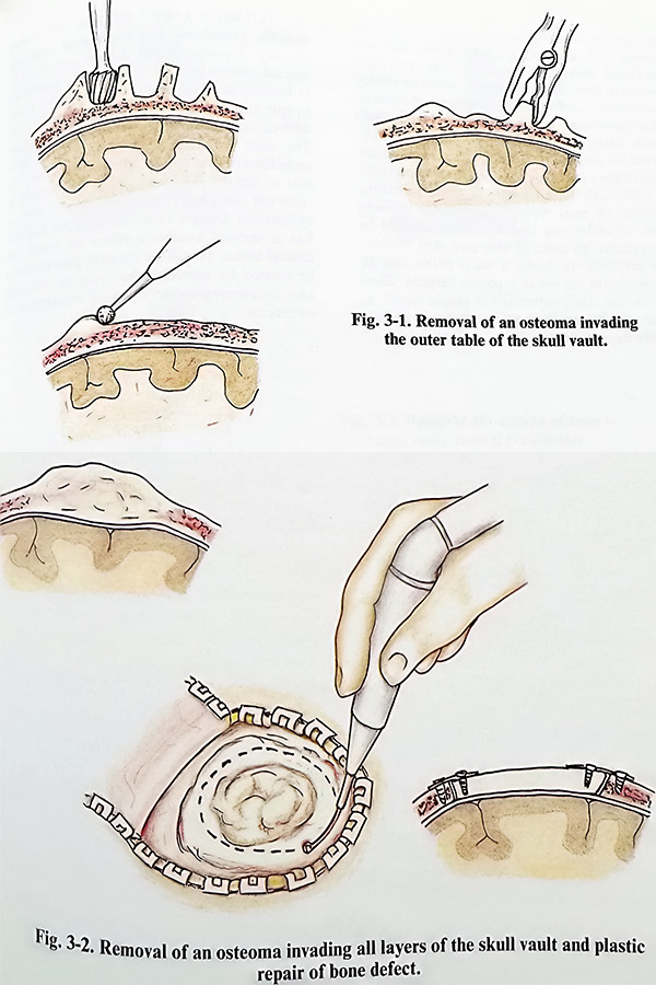
Osteomas are the most common benign neoplasms (in middle-aged patients, in women more often than men) growing from the cranial bones. They appear as circumscribed, slowly growing tumours from the inner or outer table of the cranial vault (extra- and/or intra-cranial growth). Growth from the outer table is removed by drilling out the bony tissue and reshaping the convexity of the skull, preserving the inner table. Osteomas growing inside the skull cavity can be approached only by craniot-omy. Care is taken at lifting the flap with the osteoma, as its surface can be strongly adherent to the dura. Flaps are not convenient when the osteoma involves areas of dural venous sinuses or aerated cavities. The osteoma can be drilled out from the flap and its outer table preserved.
Returning and fixing the flap avoids cranioplasty. A more difficult preservation task is faced in osteomas affecting both tables and diploe. Very often these tumours are nibbled or drilled out completely, leaving a bone defect. If no doubts exist on the type of tumour, the defect can be repaired with autologous bone at the same session (Figs. 3-1; Fig. 3-2).
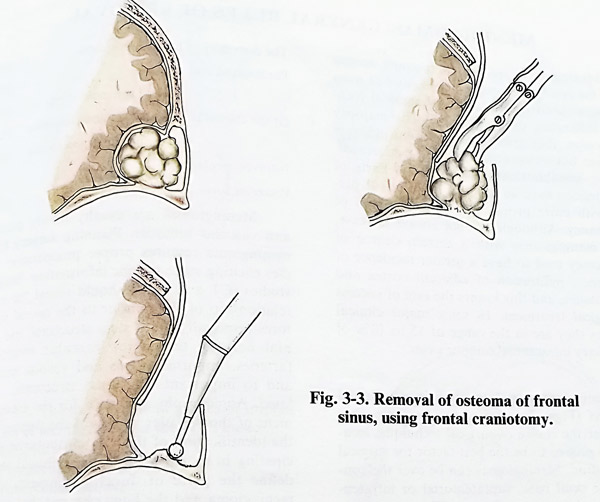
Frontal sinus osteomas need neurosurgical care if growing inside the cranial cavity from the posterior wall of the sinus. They are approached by a craniotomy to the edge of the sinus, which permits careful separation of the dura and complete drilling of the osteoma. If the sinus cavity is opened, the mucosa is stripped. Dural tears, if provoked, should be closed meticulously before flap repositioning and suture of the soft flap (Fig. 3-3).
Haemangiomas account for about 10% of all benign tumours of the skull, more often seen in women than in men. There are two basic types of these tumours: cavernous and capillary. The cavernous type of haemangioma is more frequent and usually grows in the frontal or parietal convex region. They involve all layers of skull bone, and their removal results in a bone defect. The removal is achieved by including them in a craniotomy flap or nibbling. Bleeding is controlled by waxing the edges of the bone cutting. The confirmed histopathology permits cranioplasty on the same session.
The epidermoids and dermoids are found in the parietal and frontal region and sometimes they penetrate to the orbit. These tumours are soft and not vascular. Their curettage to normal tissue is easy and at the end the capsule should be removed. As they arise from diploe, the removal also leads to a limited defect, which after being smoothed, is repaired immediately.
Fibromas, giant cell tumours, lipomas and teratomas are more rare lesions to the cranial bones. Their curettage is the treatment of choice.
The most frequent primary MALIGNANT CRANIAL TUMOURS are osteosarcomas, chondrosarcomas, and fibrosarcomas, but mesenchymal chondrosarcomas, angiosarcomas,
solitary myelomas (plasmacytomas), Ewing sarcomas and non-Hodgkin lymphomas also can be found. Metastatic tumours of the skull vault are often from lung carcinoma, breast or kidney carcinoma, and multiple myeloma. Advanced skin carcinoma invades the underlaying skull bone. Malignant tumours of the skull are indicated for surgical removal, if the general condition of the patient and his or her life expectancy is sufficiently long enough. Surgical technique consists of total removal, sometimes excising over- or underlying anatomic layers affected by the tumour. In cases of ulcerated skin carcinoma invading the bone, a scalp defect can be covered with the aid of a flap. Skull defects are rarely submitted to plastic repair, as local recurrences and life expectancy do not justify the procedure.
TUMOUR LIKE LESIONS such as eosinophilic granuloma and aneurysmal bone cyst usually develop in the diploe and must be treated surgically. The eosinophilic granuloma (a histiocytic type lesion) can be found in young people and causes a characteristic type of bone destruction. Removal is achieved by curettage. The inner table can be preserved. The surgical technique of extirpation on an aneurysmal bone cyst is similar. The wall of the cyst should be removed completely. Recurrences after such an extirpation are not observed. Fibrous dysplasia has a serious cosmetic effect on facial and cranial bones. Its diffuse invasive growth cannot be treated by resection, but only by reshaping and decompression in cases of entrapment of structures.