Legacy: "Atlast of Neurosurgery" / L.Karaguiosov, A. Ramadan, K.Karaguiosov / Kiwait/ 1998
Read NEXT: 3. TUMORS - Chapter 2
3.TUMORS
Surgery for cranial and intracranial neo-plastic lesions aims at the achievement of several goals. The first is to reduce or abolish the effects of the lesion as a mass, with its focal and general efects on the intracranial contents and the second is to eradicate the neoplastic tissue to control systemic affections due to tumour cell function and prevent recurrence. Additionally, there can be cosmetic problems to solve with skull tumours or general diagnostic problems in biopsy of a systemic disease with intracranial involvement. However, all goals are attained by a certain amount of removal, always with the goal of radicality at minimal or no functional cost for the patient.
The type of surgery is always related to the type of neoplastic affection. The grade of malignancy, the stage of development and the general and neurological condition of the patient with an intracranial neoplasm influence the decision to undertake surgical treatment. Surgery should be approached with a well preplanned technique, reserving emergency treatment for the cases with already uncontrollable precipitous progression of deterioration. In the latter case, results are definitely worse. Therefore, surgical treatment of intracranial tumours should be undertaken as early as possible, but not as an emergency. Emergency surgery is reserved for the cases of increased intracranial pressure or optic nerve compression. those presenting with imminent brain shifts and herniation or significant progressing visual damage or neurological deficit.
Surgical planning in intracranial tumour surgery should include imaging and functional studies to define the precise location of the lesion and its relation to adjacent normal structures. The analysis of this information permits planning of the approach and the expected extent of surgery. Decision making involves considering whether the lesion is a primary or secondary one, deciding on the expected grade of malignancy, and finally determining how skull and thecal layers may interfere with the approach. Secondary lesions are considered with respect to possible multiplicity. Malignancies are treated in the full context of the need of possible adjuvant treatment according to histopathology, and therefore frozen sections intra operatively are of great value. Surgical planning in tumours with skull base involvement should preview the possible need of combined approaches with maxillofacial or ENT techniques. All intracranial work is best accomplished with the aid of the surgical microscope and, if avail-able, of an ultrasonic aspirator.
During the application of surgical techniques in different types of tumours the above-mentioned general rules are always taken into consideration. All methods for particular types of tumors are in full accordance with them.
SKULL CONVEXITY TUMOURS
Tumours arising from the cranial bones can be benign or malignant. Those of them which have a clear tendency to recur and to do so with significant growth must be excised totally, as are those that are symptomatic. Complete removal with both tables leaves a skull defect after surgery. If growth is extremely slow and insignificant they can be excised, reshaping skull convexity. Nonrecurring tumours in diploe can be submitted to currettage only, preserving the inner table. Surgery should provide histopathological proof in all cases and in some secondary skull lesions a biopsy of the skull lesion can establish the diagnosis. Multiple malignancies are rarely removed by surgery.
Skull tumours arise from the several types of tissue present in the convex part of the skull. They can originate from osseous, cartilaginous, connective, vascular and haemopoietic tissue, as well as some other common type of tumour or tumour-like lesions can be found. Not always very distinctive, the difference between benign and malignant types of tumours in these groups is an essential factor determining the surgical technique.
BENIGN TUMOURS are common and they can be osteomas, osteoid-osteomas, osteo-blastomas, chondromas, chondroblastomas, chondromyxoid fibromas, intradiploic meningiomas, benign nonossifying fibromas, giant cell tumours, lipomas, haemangiomas, dermoids and epidermoids.
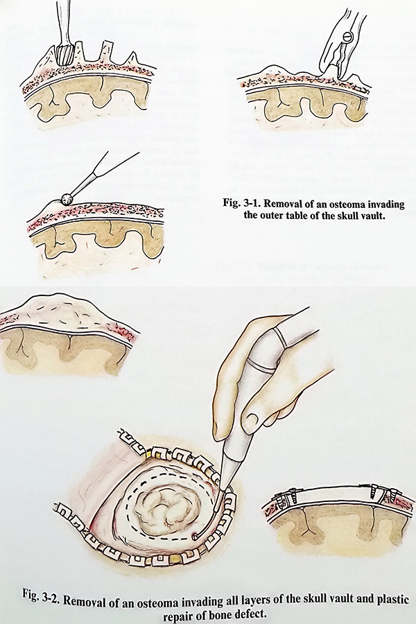
Osteomas are the most common benign neoplasms (in middle-aged patients, in women more often than men) growing from the cranial bones. They appear as circumscribed, slowly growing tumours from the inner or outer table of the cranial vault (extra- and/or intra-cranial growth). Growth from the outer table is removed by drilling out the bony tissue and reshaping the convexity of the skull, preserving the inner table. Osteomas growing inside the skull cavity can be approached only by craniot-omy. Care is taken at lifting the flap with the osteoma, as its surface can be strongly adherent to the dura. Flaps are not convenient when the osteoma involves areas of dural venous sinuses or aerated cavities. The osteoma can be drilled out from the flap and its outer table preserved.
Returning and fixing the flap avoids cranioplasty. A more difficult preservation task is faced in osteomas affecting both tables and diploe. Very often these tumours are nibbled or drilled out completely, leaving a bone defect. If no doubts exist on the type of tumour, the defect can be repaired with autologous bone at the same session (Figs. 3-1; Fig. 3-2).
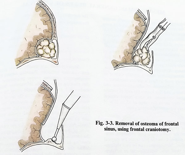
Frontal sinus osteomas need neurosurgical care if growing inside the cranial cavity from the posterior wall of the sinus. They are approached by a craniotomy to the edge of the sinus, which permits careful separation of the dura and complete drilling of the osteoma. If the sinus cavity is opened, the mucosa is stripped. Dural tears, if provoked, should be closed meticulously before flap repositioning and suture of the soft flap (Fig. 3-3).
Haemangiomas account for about 10% of all benign tumours of the skull, more often seen in women than in men. There are two basic types of these tumours: cavernous and capillary. The cavernous type of haemangioma is more frequent and usually grows in the frontal or parietal convex region. They involve all layers of skull bone, and their removal results in a bone defect. The removal is achieved by including them in a craniotomy flap or nibbling. Bleeding is controlled by waxing the edges of the bone cutting. The confirmed histopathology permits cranioplasty on the same session.
The epidermoids and dermoids are found in the parietal and frontal region and sometimes they penetrate to the orbit. These tumours are soft and not vascular. Their curettage to normal tissue is easy and at the end the capsule should be removed. As they arise from diploe, the removal also leads to a limited defect, which after being smoothed, is repaired immediately.
Fibromas, giant cell tumours, lipomas and teratomas are more rare lesions to the cranial bones. Their curettage is the treatment of choice.
The most frequent primary MALIGNANT CRANIAL TUMOURS are osteosarcomas, chondrosarcomas, and fibrosarcomas, but mesenchymal chondrosarcomas, angiosarcomas,
solitary myelomas (plasmacytomas), Ewing sarcomas and non-Hodgkin lymphomas also can be found. Metastatic tumours of the skull vault are often from lung carcinoma, breast or kidney carcinoma, and multiple myeloma. Advanced skin carcinoma invades the underlaying skull bone. Malignant tumours of the skull are indicated for surgical removal, if the general condition of the patient and his or her life expectancy is sufficiently long enough. Surgical technique consists of total removal, sometimes excising over- or underlying anatomic layers affected by the tumour. In cases of ulcerated skin carcinoma invading the bone, a scalp defect can be covered with the aid of a flap. Skull defects are rarely submitted to plastic repair, as local recurrences and life expectancy do not justify the procedure.
TUMOUR LIKE LESIONS such as eosinophilic granuloma and aneurysmal bone cyst usually develop in the diploe and must be treated surgically. The eosinophilic granuloma (a histiocytic type lesion) can be found in young people and causes a characteristic type of bone destruction. Removal is achieved by curettage. The inner table can be preserved. The surgical technique of extirpation on an aneurysmal bone cyst is similar. The wall of the cyst should be removed completely. Recurrences after such an extirpation are not observed. Fibrous dysplasia has a serious cosmetic effect on facial and cranial bones. Its diffuse invasive growth cannot be treated by resection, but only by reshaping and decompression in cases of entrapment of structures.
MENINGIOMAS: GENERAL RULES OF REMOVAL
Meningiomas are benign tumours arising from the arachnoid layer which expand as mass lesions with an extracerebral intracranial location, infiltrating dura and bone. The majority are benign, they grow slowly, do not infiltrate adjacent brain tissue, and are well demarcated, making possible their total removal. Parts of these lesions have an atypical hystological pattern with more pronounced characteristics of malignancy. Although it is not always the case, those meningiomas with a certain degree of malignancy tend to have a greater incidence of recurrence, infiltration of adjacent cortex and other tissues, and this lowers the rate of success of surgical treatment. In some major clinical statistics they are in the range of 13 to 18% of all primary intracranial tumour cases.
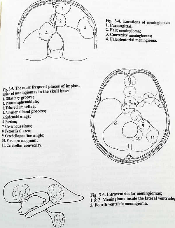
Meningiomas tend to grow in predilected locations (Figs. 3-4: 3-5; 3-6). As these sites determine the choice of surgical technique, location has proved to be the best factor for surgical classification. Meningiomas can be over the convexity or skull base, supratentorial or infratentorial, and intraventricular. The tumour is described according its place of attachment (initial growth) at the dura.
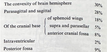
Convexity meningiomas are truly “convex”, parasagittal and falx. In their most posterior location as parasagittal they also involve also the confluens - torcular meningiomas, or falx and tentorium - falcotentorial meningiomas. Occasionally they can be attached only to the free edge of the tentorium - tentorial notch meningiomas. Skull base locations involve the midline of the anterior fossa - olfactory groove, tuberculum and diaphragma sellae, sphenoid wings - from pterion to the clinoid, the cavernous sinus, superior petrous surface and petroclival area, posterior petrous surface, cerebellar convexity and foramen magnum. After this classification, a rough approximation of the distribution in some major clinical statistics shows:
Meningiomas are usually slowly growing and vascular tumours. Planning surgery for a meningioma requires proper preoperative studies eliciting some specific information. Imaging studies (CT and MRI) should reveal the exact relationship of the tumour to the neural structures (especially brain stem structures and cranial nerves), the major vascular structures (arteries, important veins and venous sinuses) and to important skull base structures, if related. Angiography is needed for the establishment of the vascular supply of the tumour and the identification of the major tributaries participating in it. All investigations should clearly define the place of dural attachment of the meningioma and the bony changes that coexist (infiltration of bone and enostosis).
The basic steps of meningioma removal include: 1. Epicranial and bone flaps. 2. Dural opening. 3. Approach to the attachment of the tumour and interruption of arterial feeders. 4. Debulking of the tumour. 5. Dissection of the tumour from adjacent structures. 6. Removal of infiltrated structures and enostosis. 7. Repair of defects at attachment. 8. Closure of craniotomy.
These rules can be approximately the same and valid in all meningioma cases, except those which are intraventricular. Surgery is done under general anaesthesia, with intracranial hypotension by osmotic agents and diuretics, and with well preplanned blood loss estimation, (providing the necessary number of blood units plus one or two).
Epicranial and bone flaps have to comply with some important requirements. The most essential are that the exposed area of craniotomy has to give broad and comfortable access to the attachment place with sufficient space of manipulation around it. In the case of a meningioma on the convexity, the task is easier - between the attachment and border of the craniotomy there should be enough distance to allow easy work upon the dura (it is usually 1-2 cm at least). When the tumour is attached to a dural fold (falx, tentorium), or on the skull base, the craniotomy edge should widely expose the accessible border of the structure of attachment in such a way, that after retraction of the hemisphere there will be access from both sides of the attachment (Fig. 3-7). Although these rules are followed strictly, exceptions are possible, for instance, in a case where wider access has to be gained to important structures around the attachment, especially venous sinuses.
It should always be kept in mind that craniotomies in meningioma cases can lead to substantial blood loss because of pathologically hypervascular bone and dura. Some practical tips can be used. For instance, burr or entry hole bleeding is controlled easily by wax, bipolar coagulation and packing, but bone cutting lines are not. That suggests that the surgeon should do in advance as many holes as are needed, but slightly away from the hypervascular bone. The moment of major uncontrolled bleeding is between the bone cutting and the lifting of the flap, so the anaesthesia team should be alert for that. This also requires shortening the time between bone cutting and lifting of the flap, as after elevation there is much better access to the bleeders. The most vascular bone cutting is left last. The turning of the flap is followed by fast and efficient control of all bleeding, first by covering the entire surface with cottonoids and surgicel, wet gauzes, and gradually uncovering small areas of dura for controlling bleeders with coagulation.
Epicranial flaps are planned in a standard way. Attention, however, must be paid to cases of meningiomas penetrating the bone. These lead to hypervascular epicranial tissues, and therefore to flaps with altered vascular pattern and that increases blood loss during surgery.
Dural opening is adjusted to the place of attachment. It is done after application of dehydrating agents for intracranial hypotension. The rule is to open the dura at the border between affected and unaffected dura, starting with the interruption of major arterial feeders of the tumour. Affected and unaffected dura is held on traction sutures. In cases where the attachment is not seen after the craniotomy, the dura is opened according to the plan of access intracranially.
Approach to the attachment place and interruption of arterial feeders will provide the proper conditions for a removal with less bleeding and injury to the neural structures. In the case of a convexital meningioma the goal is achieved with the craniotomy. More important and time consuming is the step in meningiomas with attachment locations deep and far from the most convenient craniotomy site. Approaches have always been very much dependent on this step. The attachment site is optimally approached both intradurally and extradurally.
However, the extradural approach is not always possible. In tumours attached to falx and tentorium it cannot be performed. The extradural approach, if planned, has to follow craniotomy and haemostasis. Now skull base attachment places are approached with techniques additional to those of specific skull base approach. Skull base exposure aims early, extracranial control of the arterial supply to the lesion and prevention of damage of important structures within the skull base during detachment of the tumour. At the end of surgery it permits radical removal of the lesion. In more medially located tumours which have invaded the skull base, that is a very difficult task and requires high neurosurgical skills. The intradural part of the approach (routinely done under magnification) consists of brain retraction until the attachment place is seen and further separation of the lesion with bipolar coagulation and micro-instrumentation follows. Important structures in relation to the attachment are dissected and separated. Removal of a small part of the tumour may be needed to provide space for manipulation and access. Care should be taken at this stage not to separate the dura from the bone, as the bleeding provoked is from bony vascular channels, which cannot be coagulated and have to be waxed (not an easy task in the depth of soft abnormal tissue). Intradural separation is much easier, if it has been anticipated, by extradural control of arterial supply. At present, with the advent of endovascular techniques (interventional neuroradiology), a session of feeder obliteration can take place a day before surgery.
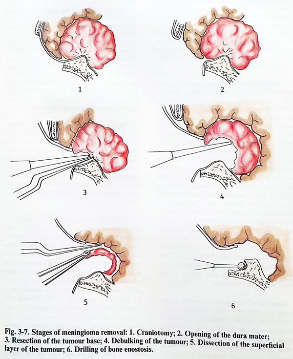
Debulking of the tumour should be carried out while preventing manipulation of the mass lesion intracranially, minimising injury to the neural structures in its vicinity. The already less vascular tumour must be reduced in volume by removing its internal part without applying force and provoking movements. There are several techniques to do this, the classical being use of the loop of the monopolar coagulator.
The cutting loop removes slices, but can give spread of currents (dangerous when close to the brain stem and cranial nerves especially). The laser is now preferred. It evaporates tissue in a touchless way. Cavitational destruction (ultrasonic aspiration) is more frequently used, but is not effective against fibrous tissue (contained in large amounts by some meningiomas). Debulking should leave a thin and increasingly flexible layer of the tumour, allowing easier dissection from neural structures. However, debulking should not risk penetration beyond tumour limits and injury of normal structure. The extent of debulking requires thorough judgement on how far to go, especially in a region of big arteries overlaying the tumour.
Dissection of the tumour is possible in the majority of cases only after debulking, except for very small nodules, which are removed in small pieces. Actually, the dissected and spared structure is the surrounding normal tissue, and the thin layer of remaining tumour is freely manipulated, pulled and torn with the aim of protecting the important neural and vascular structures. There are several important points on the dissection of meningiomas which should be emphasised. The first of these is the location of the tumour tissue regarding arachnoid layers.
The completely benign tumour which is noninvasive of the adjacent tissues should be covered by a single arachnoid layer. This should be the natural plane of dissection. Another important point is to identify and follow from normal anatomical sites all main important intracranial anatomical structures (especially arteries and nerves) with attention when they are involved on the surface of the tumour (there will be significant distortion).
Classical examples are the optic nerve and the internal carotid artery at the anterior clinoid process. The aim is to preserve and mobilise some structures and this can also allow work in the subarachnoid space. This dissection should be considered the most difficult microsurgical work, as the degree of postoperative complications and morbidity depend very much upon it.
Removal of infiltrated tissues and enostosis has a direct relationship to the recurrence rate of meningiomas. To achieve the lowest rate (about 5%), all infiltrated dura, bone and other tissues should be meticulously resected. The infiltrated dura is excised and any bony enostosis, commonly found at the place of attachment must be drilled out. In cases of deeply seated meningioma the most suitable tool for that is the micro-drill.
However, radical excision can lead to a defect of dura and bone, whose repair at the place of attachment is important to prevent postoperative complications. Basal dura has to be patched if there is no intact bony surface remaining instead and there will be no communication with the epidural space. In cases where communication is possible with an open aerated sinus, muscle packing and dural plastic repair are absolutely necessary. Defects of falx and tentorium are not necessarily repaired. In me-ningiomas of convexity the plastic repair is the only way to get a watertight closure. Dural layers can be split and the upper one turned to cover the defect. Otherwise it is fascia or allograft that can be used. We do not prefer allografts, unless the options to use other materials are completely limited.
If a bone defect remains because of removal of infiltrated bony tissue, the plastic repair can be done at the same session or at a later stage. The decision depends on the expectation and control of perifocal and postoperative brain oedema.
The closure of craniotomy is done in the routine way.
CONVEXITY MENINGIOMAS
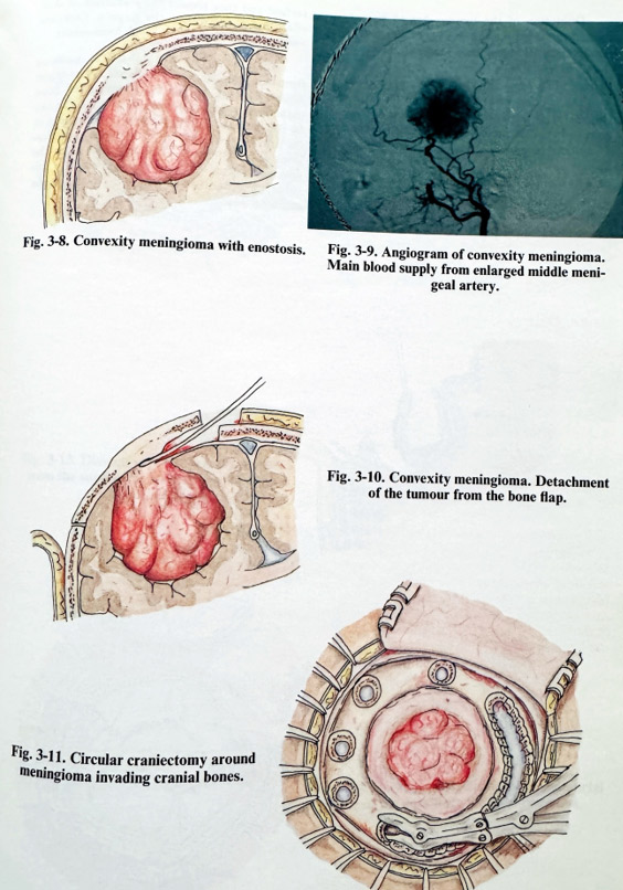
These meningiomas are attached on the dura of the convex surface of cerebral hemispheres and they do not invade any of the dural sinuses. Their shape is spherical or polylobu-lated and they excavate a bed in the cortical surface. They are found more often in the central area, then in the frontal and last in the parietal or temporal areas. At the place of attachment there is often an exostosis or enostosis (Fig. 3-8). Main arterial supply is provided by enlarged meningeal arteries (mainly the middle meningeal artery), but epicranial arteries can contribute in the case of exostoses or skull bone infiltration. Cortical arteries can also give off branches to the tumour (Fig. 3-9).
The craniotomy is planned to expose the tumour attachment area in the centre of the exposed dura. An exostosis or infiltration should be included in the preplanned flap. Bone cutting lines should be within normal bone, outside the tumour involvement. Sufficient space for manipulation of 1-2 cm should be provided between the attachment area and the bone edge.
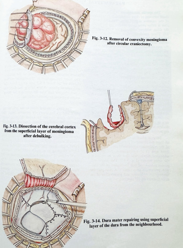
Craniotomy is performed in the standard way, including special precautions to avoid excessive bleeding, and there can be more than the usual number of burr holes. The flap is lifted after careful separation of the infiltrated dura and tumour from the inner flap surface. A fine periosteal elevator or dissector is used to scrape the inner table to avoid pulling of the tumour (and in so doing, also the important adjacent neural and vascular structures) (Fig. 3-10). Haemostasis after the flap lift is the most important step of the craniotomy; it requires skilful handling by the surgeon and should prevent excessive bloodloss at this early stage of the operation. Bleeding dural arteries are coagulated with bipolar coagulation, vascular bony channels are waxed and bleeding under the craniotomy edges is packed with oxidised cellulose. All abnormal bone must be removed. In some cases of extensive meningioma infiltration and growth inside the cranial bone, lifting a flap is rather risky, as severe haemorrhage and brain injury may occur. These tumours are approached with a circular craniectomy done with the high speed drill or with multiple burr holes and nibbling, interrupted periodically for haemostasis by waxing. After the lesion is encircled, it can be dissected at the dural level. If epidural dissection is inefficient, the dura is opened in a circular way close to the affected bone, the procedure followed being that for routine removal (sometimes with all the tumour "en block") (Figs. 3-11; 3-12).
The dura is opened in the standard way for a meningioma: first at a point which is non-adherent to the underlying cortex area, and then as close as possible to the attachment. Dural edges are held on traction sutures. If an enlargement is needed for safe manipulation around the tumour, short radial additional cuts can be added.
Debulking is started through the attachment place after biopsy sample taking. Marginal parts of the tumour are preferred for biopsy. In debulking a convexity meningioma additional bleeding appears from cortical branches to the tumour when penetrating into the depth of the tumour. They are carefully stopped with bipolar coagulation, especially when over a thin remaining layer of tumour. Debulking continues until the remaining tumour layer becomes flexible.
Dissection of the tumour has to be performed under magnification and, as a rule, with preservation of arachnoid. Manipulation is at the sacrifice of the tumour, protecting the cortical surface. The tumour is retracted more easily with the traction sutures of the dura on its side (Fig. 3-13). If the subarachnoid space is opened, arterial branches are followed until they enter the tumour before being coagulated and divided.
Special attention, as in parasagittal tumours, is paid to cortical venous drainers, whose damage is often the cause of postoperative focal deficit.
Some tumours are more adherent to the cortical surface and they require the maximum of skill, effort and patience from the surgeon. Complete removal is followed by thorough haemostasis.
There is always a dural defect resulting from this removal, and it is closed in one of the accepted methods (Fig. 3-14). Infiltrated bone tissue is removed. A normal part of the flap can be retained and the remaining defect closed at the same surgical session if there are no indications for significant brain oedema after the removal (steroids have to be considered for this surgery). All other layers are closed in the common way.
PARASAGITTAL MENINGIOMAS
This specific meningioma location refers to those tumours in close relationship to the edge of the superior sagittal sinus. The attachment area is either close to or reaches the border of the sinus, without infiltrating its walls (Fig. 3-15). These commonly seen meningiomas are round-shaped lesions excavating the cerebral cortex near the midline, and originating from the area of arachnoidal villae (Pachionian granulations) found in vicinity of the superior sagittal sinus. The tumour is mainly supplied by pathologically enlarged branches of the middle meningeal artery. Additional arterial supply is provided by cortical, falcine and epicranial branches. The lesions often involve cranial bones and can seriously affect the venous drainage, changing the pattern of venous flow through parasagittal, cortical anastomotic and diploic veins. Not infrequently, without really reaching the midline, the area of attachment is next to broad venous lacunae collecting cortical and tumour venous drainers and widely opening into the superior sagittal sinus. Preoperative imaging should reveal all these morphological relationships and angiography, which is necessary in all, except very small tumours, should disclose not only the feeders, but also the draining pattern (Fig. 3-16).
Surgical planning takes into consideration all these morphological particularities. In general, the craniotomy area should cover the attachment area as in meningiomas, but with the additional requirement that there be access to the sinus border (Fig. 3-17). This access will permit radical and safe finalization of the excision, preventing sinus rupture. A clear plan for interruption of feeders and preservation of venous drainers should anticipate surgery.
Patient positioning can be supine, oblique, lateral, prone, or semi-sitting, depending on the location of the lesion. The routine measures to avoid venous congestion are essential in this location of the meningioma. Epicranial incisions are designed in the common way, although they can be extended to the contralateral side for functional or cosmetic reasons. Bone flaps are performed in the manner used in convexity meningiomas with particular attention to the medial border of the craniotomy. In this place any dural injury can provoke massive venous bleeding. Therefore, some technical tips are essential. Certain identification of the skull sagittal midline (sagittal suture) should be done before placing burr holes close to the midline.
The bone cutting line to the sagittal midline should be at least 1.5-2 cm away from the midsagittal plane. After lifting the flap, the sagittal border can also be removed for better access with drill or careful nibbling.
The dura is incised close to the attachment place and as far away as possible from the sagittal sinus border to avoid draining veins crossing the subdural space. Opening continues along the attachment border, sparing intact dura. The most medial part of opening is left last to avoid opening venous dural lacunae. It is possible also to find an enlarged sphenoparietal dural sinus near the posterior frontal area, whose bleeding is stopped only after bending dural edges. If venous bleeding appears at the most medial part of the dural incision, it is better to stop it with all methods for control of sinus bleeding, although the already opened dura permits easy and immediate control by sutures or clips.
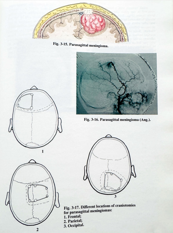
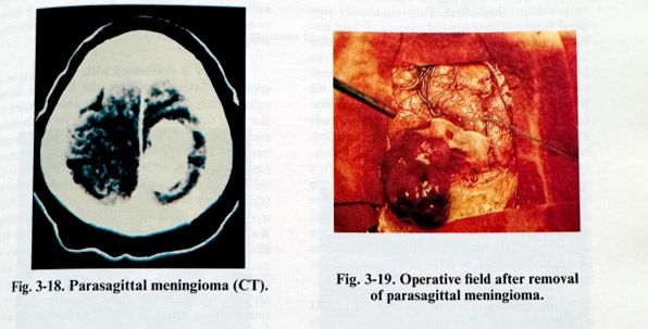
Small tumours with a broad base can be removed in one piece without damaging the brain cortex (Figs. 3-18; 3-19). Big tumours should be removed by debulking in the common way after which dissection of the residual layer of tumour follows, starting laterally and moving medially. Some of the venous collectors on the cortical surface are found very stretched and adherent to the tumour. If followed most medially, just before emerging epidurally, they will be found separated from the tumour. There can be found the proper plane of dissection to be followed. If the laser has to be the tool of removal, veins are cautiously and completely covered with soaked cottonoids. Special attention is paid to the most medial dural involvement, where, because of bleeding, small residual areas infiltrated with tumour can be left and cause early recurrence. It is necessary to perform at least the steps of residual tumour dissection under the surgical microscope, i.e., revision for radical removal, and haemostasis. Plastic repair of dura follows, aimed at attaining a watertight closure. After all affected bony tissues have been removed, the closure of the flap and /or plastic repair are done in the usual way. Wound is drained and the epicranial flap closed.
FALX MENINGIOMAS
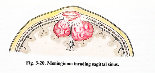
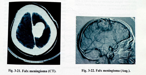
The meningiomas of the falx are those involving one or more of the structures superior sagittal sinus, inferior sagittal sinus or falx uni-or bilateral (Fig. 3-20; 3-21). Their location is subdivided according to which third of the superior sagittal sinus and falx (anterior, middle or posterior) they involve. They are usually supplied from bilateral arterial sources of dura and epicranial tissues, but in the anterior third enlarged supplying branches from the falx can be found. Venous drainage is much more complex. These tumours can often compromise the flow in the superior sagittal sinus. The occlusion can be partial or complete. The occlusion leads to venous collateralization. The enlarged collaterals can be at all levels, cortical, dural, diploic, epicranial. On their preservation very often depends the success of surgery. Some of the tumours attach only in areas close to the free edge of the falx, affecting only the inferior sagittal sinus. In these cases major consideration is given to the role of the inferior sagittal sinus in venous drainage and the relationship of the tumour with pericallosal arteries.
Investigations always include CT, MRI and angiography. They should reveal the exact attachment area of the tumour, its size and relationship to adjacent neural and vascular structures, involvement of sinuses and collateralization (Fig. 3-22). General information on structures and enhancement, indicating vascularity, is derived from the CT images. MRI is able to demonstrate involvement of sinuses and some other abnormal arterial and venous channels. Serial angiography gives the most precise vascular images as, being dynamic, it can also indicate flow and its direction.
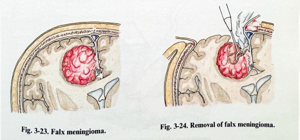
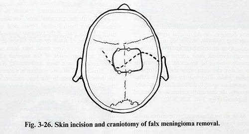
Surgical planning defines the goal of total removal of the lesion and the steps to be taken to this end. Complete feeder interruption and venous drainage preservation require sufficient access around the tumour and on both sides of the falx and the superior sagittal sinus. Craniotomy should provide this access. In a unilateral falx meningioma, craniotomy exposes an area across the midline only for access to the falx and the superior sagittal sinus, that is 1.5 - 2 cm away from the opposite sinus border (Fig 3-23;3-24). If the tumour is bilateral, then the craniotomy should provide space for tumour removal on both sides. Complete excision in superior sagittal sinus involvement entails risks.
Any significant patency of the sinus is an indication to preserve it, even with its reconstruction.
The completely occluded sinus permits excision including the involved area. Scalp incisions are extended on both sides, horseshoe or S type, considering the pattern of epicranial vascular supply.
Surgery is performed with the patient in a supine or lateral position, the first one being suitable only for tumours in the anterior third of the falx. Scalp incisions have no particularities, except excessive bleeding in case of significant epicranial vascular tumor supply. Craniotomies are often free flaps. Entry or burr holes are done on both sides of the superior sagittal sinus and the dura (sinus wall) is separated with dissectors. This avoids rupture of the sinus at bone cutting.
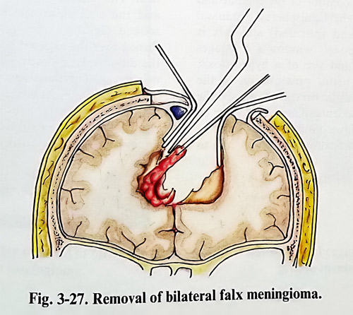
Rupture can also be provoked during bone flap lifting as a result of tumour bone infiltration at the sinus site, although potential danger also exists without infiltration at the places of emissary or diploic venous communications. After the flap is tailored with as much bilateral extent as needed, haemostasis is performed without sacrifice of venous channels. Oxidised cellulose packing must be the preferred method. The dura is opened with an incision distant from the midline and as close as possible to the tumour attach-ment, but not closer than 1.5 - 2 cm from the superior sagittal sinus edge. Incision of the dura continues towards the midline avoiding any dural venous drainer. The tumour is debulked in the usual way, leaving intact the part close to the falx. Debulking is done in the same way if bilateral extension exists (Figs. 3-25; 3-26; 3-27).
Debulking creates sufficient space on both sides of the falx. As preoperative information is available concerning the patency of the superior sagittal sinus, in cases of complete occlusion it is excised totally with all the affected dura. The lines of excision are a few millimetres anteriorly and posteriorly from the affected area. Care is taken not to affect any venous inflow from cortical or dural drainers just anteriorly or posteriorly to the resected part of the sinus. The remaining sinus ends are closed with sutures for perfect haemostasis.
All affected dura from the falx is also excised meticulously. When the superior sagittal sinus is partially or completely patent, but its walls involved by the tumour, the flow through it must be preserved. Excision is performed under temporary bypassing of the flow, and it is followed by sinus grafting at the end. Temporary bypassing is done by inserting a previously prepared siliconized bypass tube, corresponding by gauge to the sinus size, and fixing it with dural stitches through the falx around the sinus, close to the excision. Stitches are passed before opening the sinus. Final grafting is done with greater saphenous vein segments (keep in mind there can be valves) or artificial prostheses, similar to those used for artery replacement. Sinus bypassing and grafting carry risks of massive bleeding, air embolism and postoperative thrombosis. It is, therefore, highly advisable to undertake it only after having experience with it and strict evaluation for the need of it.
Meningiomas with deeper attachment to the falx requires opening of the dura uni- or bilateral, at 2-3 cm from the midline and parallel to it, turning the flaps medially. Spearing the bridging veins, the medial edge of the hemisphere is retracted (one non-important vein can occasionally be sacrificed). The most superficial affected part of the tumour is reached on one- or both sides.
We prefer to start the removal on the side of the larger part of the tumour in bilateral cases. If part of normal falx, close to the attachment can be incised, it is done and the tumour dura held on a stitch for easier manipulation. By careful retraction, part of the tumour surface is ex-posed, incised, biopsies taken and debulking is started. Debulking should be done with consideration given to the width and the depth of the tumour. With reducing tumour volume, excision from the falx close to the abnormal dura is continued in a stepwise fashion. When approaching the free edge, certain precaution is taken related to the inferior sagittal sinus, which is able to create problems of haemostasis only because of its depth. Its occlusion is practically always well tolerated. Even after the free edge of the falx is transsected on both sides of the tumour and the inferior sagittal sinus coagulated or clipped, part of the tumour may remain attached to the deeper part of the interhemi-spherical fissure. There the dissection must continue along the fissure reaching its depth, with complete debulking done, and following the pericallosal arteries under the arachnoid. The preservation of the distal anterior cerebral artery branches (pericallosal and callosomarginal) in the frontal and parietal areas and the posterior cerebral artery branches in the occipitome-dial areas is essential to avoid cortical deficit.
On completing the excision, the dura is repaired on the convexity to hermetic closure.
Bone and epicranial tissues are treated in the usual way for meningioma cases, by excising affected bone, plastic repair of the bone and epicranial closure, leaving an epidural drainage.

OLFACTORY GROOVE MENINGIOMAS
Consideration of this "classical" location for a meningioma, extensively studied by H.Cushing, brings together effectively all the principles of a meningioma's removal. Attachment is on the cribriform plate, affecting olfactory bulbi and tracts, in major lesions extending towards crista galli and planum sphenoidale.
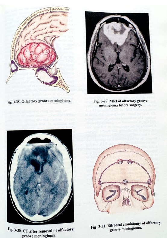
The tumour tends to occupy the inter-hemispherical fissure, indenting the frontal lobes from the base and medially, and being at least several centimetres in diameter. Since, even with substantial growth and significant frontal lobes compression and displacement, patients remain virtually asymptomatic. Often they present only with anosmia and mild personality changes that are easily neglected. Its blood supply is provided from the meningeal, ethmoidal and ophthalmic arteries. Part of the tumour can invade the ethmoidal paranasal cells. Tumour growth displaces the A2 arterial segments posteriorly and superiorly, and in big tumours they are close to and even embedded in the tumour surface. Although involved, these arterial segments can practically always be separated by dissection. This is not the case with the frontopolar branches, which often must be sacrificed. Olfactory nerves are severely distended and displaced. Posteriorly, the chiasmatic cistern is also compressed and displaced (Figs. 3-28; 3-29, 3-30).
As in all meningiomas, preoperative investigations begin with detailed contrast and non-contrast CT scans, performed also as direct coronal scans. The contour of the tumour and its relationship to the hemispheres, paranasal sinuses (ethmoid and sphenoid) and falx is delineated. The "bone" window CT images show the presence and size of a hyperostosis. MRI studies give better presentation of the position of arteries, optic nerves, chasm and the extent of perifocal oedema. Angiography can be reserved only for bigger tumours and it can be helpful for the identification of feeders, and to give additional information about major adjacent arteries and veins. Preoperative planning takes into consideration the size of the tumour, and its precise attachment before selecting the approach. Big and more anteriorly attached lesions are approached bifrontally, leaving the unilateral subfrontal or pterional way for small and posteriorly located tumours.
BIFRONTAL APPROACH
The bifrontal approach is performed in the supine position, with the upper part of the body elevated and the head slightly deflected. Skin incision follows the anterior hairline, forming a flap to be turned anteriorly towards the face, together with the epicranial layer. The bone flap extends over both frontal areas, close to the most anterior part of the superior temporal line with a height of 6 cm or more (larger in huge tumours). One or two small burr holes can be placed on both sides of the superior sagittal sinus area for safe bone undercutting (Fig. 3-31). Similar special care is taken in undercutting the bone flap close to the base midline, where the bone thickening as a crest inside requires special attention to preclude perforating the dura. As basal bone cutting lines are as close to the base as possible, the craniotomy widely opens the cavities of the frontal sinuses, and special care should be taken to isolate them from the aerated spaces below, and after that the dura to be opened. The mucosa of opened sinuses is removed and the sinuses are packed with antibiotic soaked Gelfoam. An periosteal flap can be turned over the exposed sinuses and sutured to the dura, isolating them during the next stages of surgery. The bone flap is a "free" one and immediately after its lifting, care is taken to deal with any superior sagittal sinus area bleeding. This is easily stopped with oxidised cellulose and moderate elevation of the head.
The dura is opened on both sides of the falx, 3-4 cm lateral from midline and 1- 1.5 cm from the basal craniotomy edge. Both incisions are enlarged to the midline, carefully approaching the falx and superior sagittal sinus. After safely reaching it, the medial surfaces of both frontal hemispheres are dissected from the falx and retracted with small spatulas, coagulating the bridging veins. The superior sagittal sinus and the falx are divided at their most basal point between two clips or ligatured, paying attention to the inner edge of falx where venous collectors can be found. The full mobility of the falx allows the retraction of the frontal lobes. They are lifted and slightly separated at the interhemispherical plane, until the anterior part of the tumour is seen. The arachnoid is dissected to determine the proper plane of separation of cortical vessels from the tumour.
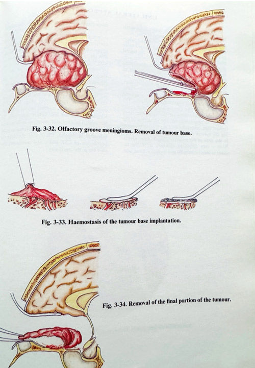
The classical advice of H.Cushing remains valid for tumour removal. After biopsies are taken, debulking and transsection of the base go together, removing bigger and bigger portions of the tumour, using ordinary suction, an ultrasonic aspirator, laser, or diathermy loop (Fig. 3-32). Arterial feeders often present difficulties in control. The traditional solution is a monopolar coagulation, connected to a dissector. However, if burning of the attachment in this way does not control the bleeding and bipolar coagulation is inefficient (it coagulates vessels inside soft tissues), the already exposed bony arterial channels are occluded with wax (Fig. 3-33). Leaving a thin layer of tumour over the attachment and hyperostosis can avoid the time and blood loss at this stage and postpone it for the moment when all the tumour mass is out, and space is much greater, the normal dural edges are seen and bleeding control can be faster. Debulking and detachment advance along the skull base in the cribriform palate area and the olfactory bulbs are rarely spared (may be only one of them at times). If both bulbs are sacrificed, both frontal lobes are retracted without limitations, but if one of them is preserved, the opposite lobe is predominantly retracted. Once the tumour has been debulked, the bleeding decreases, and the surgeon can follow the lateral border of the tumour to reach the lesser sphenoid wing and presumably the anterior clinoid area. The arachnoid is usually thick and provides protection of neural and vascular structures located behind it, but the translucent layer sometimes permits the identification of chiasm, optic nerves and supraclinoid carotids (Fig. 3-34).
Complete preservation of the cisternal arachnoid should be attempted. The posterior dome of the tumour, even in big tumours, is separated from A2 arterial segments, but the most upper part can involve (and sometimes encase) frontopolar arteries. They can be sacrificed if dissection proves to be impossible. After all parts of the tumour are removed, the attachment area is coagulated, the affected dura removed, and permanent haemostasis of feeders achieved after drilling the hyperostotic bone.
All tumour parts penetrating into ethmoid and/or sphenoid sinuses must be radically removed, as they are sources of recurrence. The resection of these portions must be completed at the same stage. The wide communication with paranasal sinuses is repaired with fascia - suturing it to a preserved part of the dura and gluing it to prevent CSF rhinorrhoea. Closure entails repairing the falx, simply tying the superior sagittal sinus ligatures one to an-other, and closing the dura along the incision.
Special care is taken to ensure water tight closure, as a CSF fistula can be a serious and more frequent complication in this type of meningioma removal. The bone flap is replaced and fixed, and epicranial layers are sutured in the usual way, leaving an epidural drain under mild suction.
UNILATERAL APPROACH
The unilateral approach, designed for smaller and more posterior tumours, is the one more often applied in planum sphenoidale and tuberculum sellae meningiomas or those located more unlaterally. The craniotomy exposes the lateral part of the frontal, pterional or anterior temporal regions after a skin incision from the midline at the anterior hairline to a point just anterior to the tragus. The flap can be left attached to the temporal muscle or be lifted as a "free" flap.
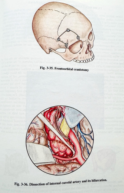
Frontoorbital craniotomy is very convenient for this purpose (Fig. 3-35). The dura is opened parallel to the basal edge of the craniotomy. The approach uses the convenience of the route along the lesser wing of the sphenoid, very familiar to neurosurgeons. The retraction of the frontal lobe, with release of the tension by opening the sylvian cistern if necessary, gives access to the area of the anterior clinoid. After identifying a very displaced olfactory tract, the tumour is seen anteriorly and medially. Even with more posteriorly attached tumours, the accessibility and safe separation of the chiasmatic cistern and the ipsilateral internal carotid are early and better, compared to the bifrontal approach. A tumour sample is taken for biopsy and the tumour is debulked in a similar way as with the bifrontal technique, but very often the olfactory tract must be sacrificed, as, due to retraction, the bulb tends to disrupt from the cribriform area. After debulking, the safe dissection of the residual part should be done following the posterior margin of the lesion, between the arachnoid and tumour (Fig. 3-36). The opposite optic nerve, internal carotid and olfactory tract are seen last, before completing the removal. The care and attention to hemostasis and radicalism of removal are essential as in the bifrontal technique. Closure of the dura for prevention of CSF leaks, and craniotomy closure are done in the usual way.
TUBERCULUM SELLAE MENINGIOMAS
These lesions arise from the area of the tuberculum sellae, sulcus chiasmatis and diaphragma sellae (Fig 3-37). The optic nerves are displaced laterally, and when the tumour is growing more posteriorly from the diaphragma, also upwards. The internal carotids are less displaced laterally. As the tumour continues growing, it exerts pressure on the infundibulum, ventral hypothalamus, and occupies the retro-sellar area and interpeduncullar fossa, although the area of attachment is relatively small. It often produces hyperostosis. The blood supply is very often provided by posterior ethmoidal arteries. Relations of the tumour to the anterior cerebral arteries are of the greatest importance.
Their displacement is often significant and small arterial branches can contribute to the supply of the tumour. Their course is sometimes difficult to follow, and these branches should be sacrificed after the surgeon is completely convinced that they are related only to the tumour. As with other meningiomas in this area, the optic canal can be invaded and the arteries (internal carotid and the anterior cerebral) encased. A total removal must always be attempted, but this can be impossible in some cases. The size of the tumour is also closely related to the outcome - the larger size is related to significant morbidity and mortality.
These meningiomas need thorough preoperative neuroophthalmological evaluation. Almost all symptomatic cases have visual affection - decreased visual acuity and/or limitation of visual fields. The tumour is usually detected by CT scan (Figs. 3-38; 3-39). It is best visualised on direct coronal scanning, which gives a better indication of the relationship to supra- and parasellar structures. MRI demonstrates the arteries well, especially if they are encased, and reveals the position of chasm and the perifocal oedema. Angiography is essential to demonstrate the collateralization through the anterior communicating complex between the internal carotid systems on both sides (Matas test) as temporary or permanent occlusion can be needed during intraoperative dissection.
Surgical planning depends on the size of the tumour and the position of the main mass regarding chasm and the optic nerves. When the main mass volume is located completely retro-chiasmatically, a view from the lesser sphenoidal wing, i.e. from the pterion, will be needed.
These lesions require a wide angle of approach as a rule and in some cases with big tumours and complex involvement of chasm, a broad subfrontal (even bifrontal) and pterional view will be required simultaneously. For small retro-chiasmatic lesions (more attached to diaphragm we recommend pterional craniotomy.When microsurgical work will be extensively needed between the nerves, over the chasm, on both sides of the nerves and both Al arterial segments, then bifrontal craniotomy is the option of choice, with an additional extension for a pterional view. The pterional view can be broader by opening the sylvian fissure and extending the craniotomy with an orbital part.

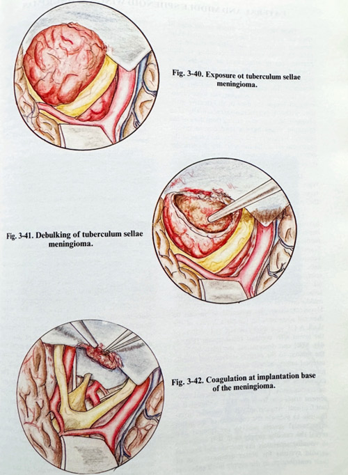
After performing the selected approach, the area is reached in a standard way, either along the lesser wing or the subfrontal route with the intention of sparing the olfactory tracts if possible. Two objectives have to be concidered at the beginning - detachment from the feeders and reduction of the volume. Both tasks are not so easy to perform in many cases. Part of the feeders can come from the anterior cerebral arteries, and interruption of feeders should be done at the beginning only at the attachment area. The main mass is approached between the optic nerves and under the chiasm and debulked if possible. The tumour parts are dissected from arteries, optic nerves and infundibulum, but it is better to follow first the anterior cerebral arteries and spare some fine branches, which enter the tumour and later exit supplying neural structures (“passing” arterties) (Fig. 3-40). Following the arteries do not invalidate the rule to preserve and follow arachnoid over chiasm, infundibulum and the rest of the basal brain surface, as this is the most important borderline of tumour separation. In huge tumours removal can compromise anterior cerebral arteries. Their damage has to be repaired with microsutures. This occurs more often in some cases of encasement and the possibility of removing the tumour completely depends on the existence of the arachnoid layer between the artery and the tumour. The arachnoid, although thicker, must be followed on the posterior surface, where the infundibulum and hypothalamic surface are separated, avoiding any microvascular sacrifice. Even if surgery must be prolonged to its acceptable maximum, complete removal must always be attempted, as second surgeries are as a rule much less efficient in removal due to the disrupted anatomy and adherence of tissues. There is no specific technique which differs from other similar approaches in completing and closing this surgical approach (Figs. 3-41; 3-42).
In big internally situated meningiomas it is essential to open the sylvian fissure widely to find the branches of the middle cerebral artery.
After enucleation of the tumour, these branches are followed proximally to the internal carotid artery to remove the tumour in a piecemeal fashion, never losing sight of the artery and its branches. Tumour invasion of the arterial wall and occlusion of the lumen is very rare. Dissection of the perforating vessels, the anterior choroidal artery and posterior communicating artery is time consuming, but it is the only way to diminish morbidity.
LATERAL AND MIDDLE SPHENOID WING MENINGIOMAS
These meningiomas attach to the edge of the lesser and part of the greater sphenoid wings between pterion and the anterior clinoid pro-cess. They are subdivided according to their precise area of attachment as this factor is crucial for assessing the technical difficulties at the time of removal and the outcome of surgical treatment. The majority of these tumours grow as an intracranial mass lesion, but some predominantly invade the dura on a broad base, without expanding intracranially ("en plaque"). Meningiomas arising from dura of the sphenoid wings have a tendency to invade adjacent structures and spaces. Invasions of the cavernous sinus and the orbit are of particular interest, as they increase the technical complexity of the required surgery.Despite some differences in classifying the types of location, there are several accepted types characterized by the common problems they present.
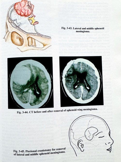
Lateral and middle sphenoid meningiomas have their attachment not involving important basal structures (Fig. 3-43). Their vascular supply is generally provided by enlarged branches of the middle meningeal artery and the venous drainage - by a variety of channels, the principal one commonly being the sphenoparietal sinus. Their growth has subfrontal, temporopolar and sylvian expansion. The tumour dome displaces the first segments of the middle cerebral artery, and rarely encases them. Hyperostosis on the sphenoid wings can be detected sometimes on the plain X-rays of the skull. A CT scan shows the size and position of the lesion after contrast enhancement (Fig. 3-44). MRI can demonstrate the relationship of the tumour to the cortical surface, and middle cerebral artery, as well as any medial displacement of arteries and the optic nerve, additional extensions to the orbit or involvement of the cavernous sinus. The suspicion of arterial encasement arises also from the MRI, although it is not frequent. Angiography is an essential investigation to indicate the condition of the major intracranial vessels next to the tumour and reveal the vascular supply to it. It is important to visualise separately the internal and external carotid systems for better demonstration of their contribution to the supply in all sphenoid meningiomas. Selective external carotid catheterisation can be used for preoperative embolisation of major feeders to reduce intraoperative blood loss.
Surgical planning focuses on a craniotomy, always including the pterional area and as extensive as frontal and temporal retraction may demand. The attachment place should be accessible both after subfrontal and temporobasal cortical retraction. The part of the sphenoid bone corresponding to the attachment place, usually infiltrated by the tumour and containing the feeders should be accessible through the basal part of the craniotomy (Fig. 3-45).
Skin incisions are usually curvilinear, frontotemporal, from the midline to the external projection of the temporal base towards the tragus or retroauricular. The craniotomy flap is lifted with special attention to its basal part, where it moght be close to the attachment, and bleeding can be serious. We recommend that access or burr holes be placed on both sides close, but not over the attachment at pterion and later that the affected bone be removed by nibbling or drilling. This removal is associated with bleeding, but the enlarged extracranial branches from the midportion of the maxillary artery can be coagulated. The extent of bone removal can reach the lateral part of the superior orbital fissure, spinous foramen and the course of the major superficial petrosal nerve, but removal to such an extent is not always required. This stage is important for the interruption of feeders. Bone removal can also be done after the opening of the dura, verifying the attachment place by direct observation. The dura is opened along the skull base, but outside the attachment area.
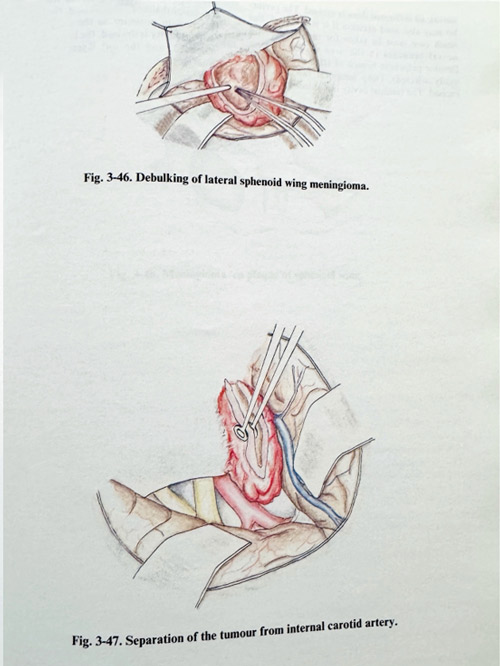
Additional dural incisions may be needed vertical to the first one, to ease the retraction of brain. The area of attachment is approached with minimal or no brain retraction. The base of the tumour is gradually transsected with bipolar coagulation and microscissors, firs debulking the adjacent part (Fig. 3-46). At the time of attachment transsection and debulking, much care should be taken with the most medial part of the involved dura, as it is close to important structures within the skull base and close to the anterior clinoid. It is recommended at the time of debulking and detaching the tumour, to continue dissecting the attachment in the sub-dural plane until such structures as the olfactory tract, optic nerve and supraclinoid internal carotid artery are seen under the arachnoid and by that the detachment can be completed safely.
Debulking is expanded sufficiently by one of the standard methods (cavitron aspirator, LASER, coagulation loop). Dissection of the main tumour mass as completely as possible has to be performed while preserving the arachnoid. The supraclinoid internal carotid is followed distally or the middle cerebral artery branches - proximally (usually in both directions, using first the better plane for dissection). Arterial feeding branches are coagulated and transsected as close as possible to the tumour (Fig. 3-47). After completing removal, the attachment dura is totally excised, haemostasis is completed and the dura is closed, ensuring the closure is water-tight. If enostosis remains despite the bone nibbling, it is drilled out before closure. The bone flap is secured in place with leaving an epidural drain, and the rest of the layers are closed in the usual way.
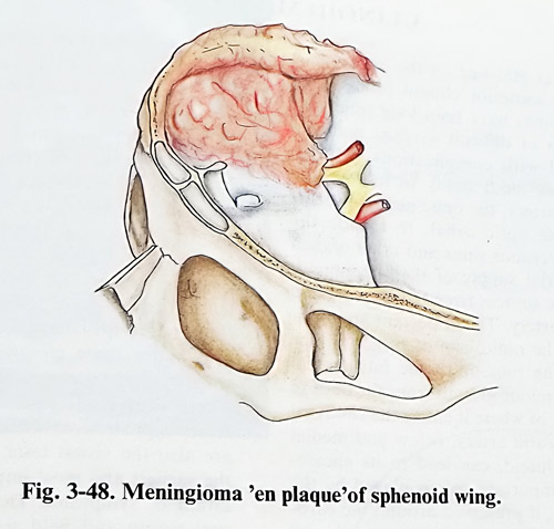
A very particular type of a meningioma, the so called " en plaque" type, which affects the sphenoid wings, presents some specific problems. This meningioma progressively invades the dura and the bone of the greater and lesser wing over many years. The dura is affected as a layer of just few millimetres thickness. The underlying bone haversian canals are invaded by meningotelial tumour cells, provoking reactive hyperostosis. It is the hyperostosis of the sphenoid which is responsible for the clinical symptoms. Without causing a mass effect, the lesion gradually involves the skull base anatomical structures passing through the apertures of the sphenoid. The most common symptom is from the involvement of the optic canal, followed by the nerves traversing the superior orbital fissure, the cavernous sinus, and the trigeminal branches in the foramen rotundum and foramen ovale. Presenting often in young and middle aged women, this lesion has a slow and insidious growth rate, detectable by the progressive exophthalmos and deformity in the temporal fossa. The essential investigations are the same as in the other menigiomas of this location.
Skull X-rays, CT, MRI and angiography selectively demonstrating the external and internal carotid systems provide the necessary information for surgery. Special attention is required for the visualisation of all involved bone and the position and condition of basal foramina. The orbital content is studied thoroughly, as it is commonly found to be invaded by the tumour.
The aim of surgery consists of removal of as much affected bone as possible, excision of affected dura and decompression of nerves along the foramina. Surgical planning is mainly aimed at determining essentially the extent of bone removal and dural excision. It should define how far the surgeon should go and which bony cavities and canals should be exposed and decompression performed. The plan refers particularly to the optic canal, the posterior orbital segments, the superior orbital fissure, foramen ovale and foramen rotundum. The amount of involved dura to be excised is planned according the pathologically enhancing areas on CT and MRI. After all these considerations, the needs of dural plastic repair, the type of material and the expected extent are determined. Cra-nioplasty is needed in cases of significant convexity or orbital wall removal (Fig. 3-48).
Surgery is conducted through a frontotemporal incision not anterior to the hairline, with the flap reflected to the base exposing the frontobasal and temporopolar areas, centred at the pterion. A fronto-temporo-pterional flap follows, overcoming the difficulty of turning it with the drill. Affected bone is definitely not included in the flap, and it is nibbled or drilled after flap-lifting. Surgery is performed in its initial part extradurally. Starting with the orbital roof, bone is drilled or nibbled, depending on what is more efficient in bone removal. We used to drill until the thickness was suitable for nibbling and we alternate these two manoeuvres. Once the orbital content is exposed, removal is extended to the greater and lesser wing until the superior orbital fissure is reached. The superior and inferior borders of the fissure are removed and the lesser wing drilling advanced to the anterior clinoid and the orbital canal. In a temporobasal direction bone removal reaches the greater and lesser superficial petrosal nerves, and foramina for the trigeminal 2nd and 3rd division. Any greater bone removal has to be weighted against the chances for radicalism and the threat of intra- and postoperative complications. Cavernous sinus area, petrosal apex or contralateral involvements are such conditions, when the continuation of removal has to be reconsidered seriously. After competition of the bone removal all infiltrated dura is excised. The periorbit may also need excision if it is involved, but much care must be taken for the arterial and neural branches in the area of Zinn's ring (levator palpebrae branch of Illrd nerve can be easily affected). Deep temporal fascia is also excised. The residual from bone removal cavity has a decompressive effect upon the orbital content and the preexist. ing examith meriTh to ure is repaired using pericranium posterior to the craniotomy. The residual cavity is drained, the bone flap replaced and fixed, and the soft layers sutured in the usual way.
CLINOID MENINGIOMAS
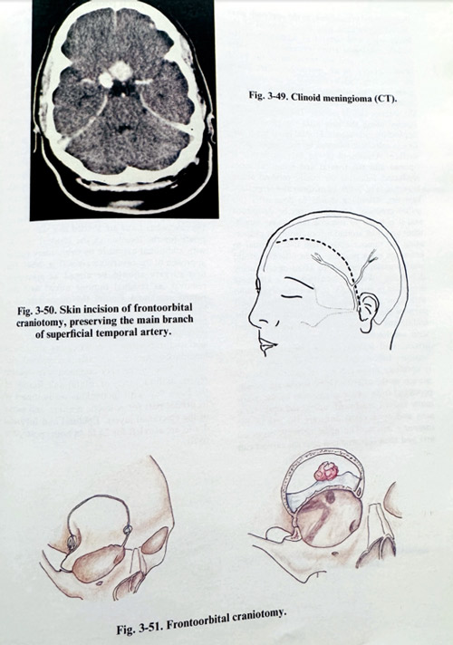
Meningiomas attached on the medial part of the sphenoid (anterior clinoid process), or clinoid meningiomas have been long time considered examples of difficult surgical removal, often associated with complications. Attachment in this area tends easily to involve the internal carotid artery, the optic nerve intracranially and in the optic canal, to invade the neighbouring cavernous sinus and other cranial nerves. The arterial supply of these meningiomas is of multiple sources from the external and internal carotid artery. The important and decisive feature for the radicalism of surgery is the relationship of the tumour to the intracranial carotid artery. Tumour growth from the dura in direct vicinity of where the internal carotid artery enters the subdural space, below and medial to the anterior clinoid, can lead to its encasеment. Here the the arachnoid plays a very important role, if it can be preserved around the carotid, dissection of the arterial wall from the surrounding tumour will be possible. It is the growth of the tumour that does or does not separate the arachnoid of the carotid cistern, and very often the preoperative studies are not able to establish it preoperatively. However, the details of these studies are decisive for the proper orientation during surgery and further on, for its success. These preoperative studies consist of the routine work-up of a meningioma patient: skull X-rays, CT scan of the skull and brain, including enhanced CT, MRI, and angiography. The CT images indicate the location of the tumour mass. They must be complemented with contrast enhanced, thin slice images and bone window CT studies (Fig.3-49). Additional valuable information is obtained from the coronal CT scanning. Coronal slices are better in demonstrating optic canal, intraorbital and intracavernous involvements. MRI gives initial information about the location of internal carotid arteries and the optic nerves, especially if encasement is suspected. MRI is also superior in depicting the peritumoral brain oedema, associated with the lesion. Angiographic features, with selective internal and external contrast injections, reveal the patency of the internal carotid and its branches as well as the tumour's arterial feeders.
As surgery can compromise carotid blood flow, some functional carotid tests, best known when intracavernous presurgical work-up is done, can help us to predict the outcome of temporary or permanent occlusion of the internal carotid during surgery. The first one, the balloon occlusion test, simulates by percutaneous intraarterial technique internal carotid artery occlusion and evaluates its consequences clinically and angiographically. The second is based on the transcranial doppler technique. With the transcranial doppler, the effect of carotid artery compression on the neck can be measured. It should demonstrate collateralisation (reversal of flow in the ipsilateral anterior cerebral artery) and under these circumstances the ipsilateral middle cerebral artery flow can be evaluated. These functional tests have however only a relative predictive value. Of basic importance are also the visual tests; visual affection is the earliest and most important clinical presentation of symptoms in these tumors. Detailed preoperative visual acuity and field studies are essential for preoperative evaluation. Endocrine tests can be a valuable aid for the complete assessment of the patient.
Surgical planning prepares the surgeon for the presumed technical difficulties to be faced during surgery. The craniotomy is usually a standard pterional or frontoorbital one, including adjacent frontal and temporal areas. The epicranial flap for this craniotomy should preserve the superficial temporal artery branches in case they are needed for an STA-MCA microanastomosis (Figs. 3-50; 3-51). The need for some arterial supply interruption has to be decided on in big tumours as detaching the lesion is risky if the attachment is broad, and its proximity to the optic nerve and internal carotid cannot be established visually; however more often this decision must be taken during surgery. Optic foramen and intraorbital involvement will require opening of the optic canal and orbitotomy. However, as details in microanatomy are not easily predictable, any decision must be taken after the lesion has been approached.
The bone flap is either free or attached to the temporal muscle. The flap should include the frontal, temporal and pterional areas, but significant widening of the approach is obtained by orbital wall removal and subperiosteal detachment of the zygomathic arch. We prefer the frontoorbital craniotomy, as the approach gives several advantages: easy direct removal of the lesser wing and control of feeders from the middle meningeal artery extradurally, a wider angle of view, and less retraction of the basal brain surfaces. In all approaches, after lifting the bone flap, follows removal of the lesser and great wing by nibbling and drilling. The removal can continue as much as possible towards the anterior clinoid process.
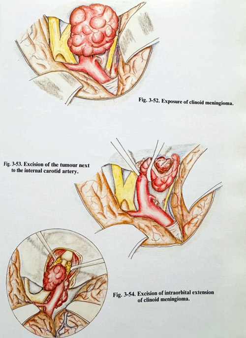
The dura is usually opened along the base after the effect of the applied osmotic agents is obtained. Frontal basal brain surface is retracted, followed by the temporopolar surface. Arachnoid in the Sylvian fissure is opened and the frontal and temporal lobes separated, following the middle cerebral artery branches (Fig. 3-52). In medium and large sized tumours, debulking should be done after this initial exposure. Debulking can be dangerous and it is better to visualise the tumour attachment area well around before cutting or evaporating the tumour mass. Here it is essential to know the exact position of the internal carotid and its major branches to avoid injury during debulking. We are accustomed to alternate dissection in retrograde to the artery's direction with slow debulking (Fig. 3-53). Often distal parts of the carotid, and practically always the middle and anterior cerebral arteries are vested with arachnoid, making the dissection of the arterial wall feasible. If the arterial dissection becomes difficult and risky, the attachment area is carefully inspected for an anterograde approach to the arteries. Optic nerves are usually protected from tumoural invasion by the chiasmatic cistern arachnoid, unless the optic foramen and optic canal dura are the site of the tumour's origin. The optic nerve is dissected first and then the proximal internal carotid can be exposed if this is possible. No attempts should be made to search for major arteries through the tumour, even with well-known artery location, as the invaded adventitia is disrupted easily together with the other arterial layers. If occasional rupture of the wall occurs, the artery should be clipped temporarily on both sides and the defect closed with 8/0 - 10/0 silk suture. If however a major rupture takes place, especially very proximal to the intracavernous portion, the technique of repair is more difficult and time consuming and is similar to that in cases of intracavernous internal carotid sacrifice - venous grafting of the artery or the EC-IC microanastomosis. Which technique to choose is indicated by the results of the balloon occlusion test.
All tumours prone to removal must be excised, the safest approach being with microscissors and bipolar coagu-lation. Affected dura is also excised at the end after being coagulated. Invasions of the orbit or the cavernous sinus are treated in a way appropriate for the location of the residual tumour: with additional exposure by orbitotomy and/or approach to the cavernous sinus (Fig. 3-54). The first surgery should be aimed at maximum removal, as residual tumour mixed with disrupted arachnoid around the major arteries is likely to lead to invasive growth later on. This makes second stage removals practically always incomplete and inefficient, because of this invasion and difficulties in demarcating anatomical landmarks. Once haemostasis is completed, closure is done in the very common way - plastic repairs of dura, rarely of orbital wall, fixation of the bone flap with meticulous readjustment of its orbital part for cosmetic reasons, and suture of the epicranial layers. Epidural and subgaleal drains are also left for 24 to 48 hours postoperatively.
SURGICAL MANAGEMENT OF SUPRATENTORIAL GLIOMAS
Last two decades have witnessed substantial progress in basic tumour research, which has been incomparably more successful than have been the real technical advances in glioma surgery. Despite this, no radical breakthrough in the treatment of these lesions has been achieved. The observed improvement in results obtained has rather derived more from a transfer of methods from other neurosurgical areas and adjuvant therapies; naturally the actual steps forward have been relatively modest.
Based on this type of limited progress, then, we can hardly predict, that surgery constitutes the solution of the glioma problem now or even that this will be the case in the near future.
Gliomas are a group of tumours with an infiltrative nature in general. Their histological types have recently been classified by the WHO, with the aim of indicating the degree of malignancy. Astrocytomas I and Il grades are low grade, while grades III and IV are considered high grade gliomas (glioblastomas). A specific type of nonmalignant glioma is the cystic pylocytic astrocytoma. Another common supratentorial glioma type is the oligodendroglioma; its malignant type is the oligodendroblastoma. It very often presents histologically as a mixed type of glioma. Malignant gliomas have a tendency to undergo cystic degeneration, with resultant intratumoural vascular proliferation, necrosis and hemorrhages.
Clinically, gliomas present with a great variety of focal symptoms and signs of neurological deficit, and epilepsy. When the effects of a mass lesion produced by the continued growth of the neoplasm are added to the affection of the primarily involved area, the clinical course shows deterioration due to compression of distant structures and increased intracranial pressure.
Surgery can be beneficial in two aspects of glioma treatment: to establish the nature of the lesion and to reduce its volume. Surgical radicalism is of dubious value in glioma surgery. The choice of method depends on the location of the lesion, the expected grade of malignancy and the general condition of the patient. The patient is investigated by CT and MRI which are aimed at determining the borders and intrinsic texture of the tumour, its vascularity, the possible differential diagnosis, the degree of perifocal oedema. Angiography is reserved only for very vascular tumours or for the needs of differential diagnosis.
The histopathological diagnosis has to be established in all symptomatic lesions or in those which are showing growth. The asymptomatic lesions not confirmed to be expanding can be followed up by imaging studies. Lesions with cortico-subcortical locations can be explored for biopsy by CT guided small craniotomy or burr hole techniques. Those located deeper in the centrum semiovale and in the basal ganglia are indicated for stereotactic biopsy (this technique is described in the corresponding chapter of the book). Multiple biopsies should always be taken, as the grade of malignancy varies among different tumour areas, and stereotactic sample taking yields maximal positive results in not all, but nearly 90% of the cases.
Removal of tumour tissue is necessary to obtain "internal" decompression of the still viable brain structures by reducing the amount of abnormal and nonfunctional tissues and evacuating tumoural cystic fluid. The indications for this are present when the patient's condition has deteriorated due to the mass effect and its at the very least temporary relief will benefit the patient with a sufficiently long period of low morbidity to justify it. The technique depends on the areas involved. Functionally important, "eloquent" cortical areas, the basal ganglia and some limbic structures must be spared. Therefore the technique of polar resection (also called "lobectomy") is reserved to frontal, temporal and occipital lobes and poles, taking into serious consideration the implications of cerebral dominance and the variability of cortical functional areas, and adjusting the extent of removal in accordance with this. All other gliomas with a deep location (affecting basal ganglia, thalamus and hypothalamus, and those crossing through the corpus callosum) are debulked very selectively to avoid functional damage.
Surgical planning should consider the extent of surgery. First to be decided are the place of biopsy taking and the amount of tissue to be removed. Then the craniotomy is planned to provide safe access and manipulation for the areas of interest for tissue removal.
Preoperative treatment is essential for the outcome of surgery. Medication includes two essential drugs: anticonvulsants and steroids.
Anticonvulsants have a place on both occasions: with and without previous seizures. Any preceding epilepsy with treatment already initiated requires evaluation for adequacy (using therapeutic serum levels), and the dose must be correspondingly adjusted. Perioperative prophylaxy is meant to increase the seizure threshold in the most vulnerable periods before and immediately after surgery. Diphenylhydantoin is administered in loading and maintenance dosages as the drug of choice, but phenobarbital, carbamazepine and some other anticonvulsants should be considered in some selected cases.
Steroid treatment is most often initiated immediately after the diagnosis has been established and possible surgery is expected. Dexamethasone is given, preferably a few days before surgery, under the protection of ranitidine or cimetidine, the high doses being reserved for those patient's in advanced stages of the disease - with increased intracranial pressure and a rapidly progressing neurological deficit. Surgical treatment is performed with special attention on control of intracranial pressure and seizure prophylaxis. Bolus doses of Dexamethasone and Diphenylhydantoin are given during the induction of anaesthesia if the patient has not been treated with them before. The patients who already have therapeutic levels of these drugs receive their regular doses then and at 6 hour intervals afterwards. An intracranial hypotensive effect is induced before opening the dura by the rapid infusion of 20% Mannitol and/or Furosemide. This will permit safe brain tissue manipulation. The patient is moderately hyperventilated during anaesthesia.
GLIOMA BIOPSY TAKING
As a separate procedure, the biopsy of a glioma is planned carefully on imaging data (CT and MRI). In those cases where stereotaxy will not be used, the intended point of approach through the skull should be visible (with a marker attached) on the imaging studies to establish the exact relationships between intended biopsy area and marker.
Biopsy taking through a large burr hole (or more than one) must be followed up by CT after the procedure. If a craniotomy is planned (partial removal to follow the biopsy), the intended margins of the craniotomy can be marked to provide orientation for the subcortical tumour borders (Figs. 3-55; 3-56).
Biopsy taking must yield tisuue for histopathological study which is representative of the structure of the tumour. Gliomas are non-uniform lesions inside. In more malignant tumours, areas of different grades of malignancy are found among cysts, necrotic tissue and haemorrhages. Taking several specimens will provide a more comprehensive histopathological image of the neoplastic lesion. The tissue should be vascularized and not necrotic, and different areas of the tumour are approached and sampled. If cysts are evacuated their walls are carefully inspected and biopsies from the wall are taken where it seems tumoral.
A biopsy should be taken at the beginning of tumour manipulation, and in case surgery has to be discontinued, a specimen will nonetheless have been provided. Tumour tissue is taken with specially designed forceps and its destruction - avoided until it is immersed in a fixation solution.
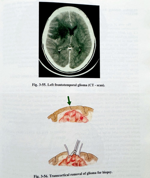
GLIOMA TISSUE RESECTION ("DEBULKING")
The area of proposed removal and the transcortical approach are prеplanned on the CT and MRI studies. The place of cortical incision is chosen according to the "eloquence" of the area to be penetrated and cerebral dominance. There are some preferable places for cortical incision indicated in the illustrations, using the depth of some main sulci (Fig. 3-57).
The incisions are at least 1.5 to 2 cm long. The abnormal tissue is usually seen at the expected depth. With some deeply located and smaller lesions splitting of the white matter can mislead the surgeon into passing beside his target. The recent development of intraoperative systems of localisation can significantly reduce the risks of such errors. If navigational systems are not available, the craniotomy can be done under stereotactic conditions, with account being taken of the displacements caused by dehydration and CSF evacuation. Cortical incisions are maintained with the minimum of retraction.
Initial biopsies are taken and the preliminary impressions of the tissue qualities on the “frozen” sections stains will determine the instrumentation to be used. High grade gliomas have a more variable, "mosaic" - like texture and the technique of their removal has to be changed on several occasions during the operation in order to deal effectively with them.
Cysts are punctured, fluid is collected for volume measurement and laboratory investigations. If a big cyst is present, and the intracranial pressure is uncontrollable, it can be punctured through a small coagulated area of dura and pia. Cysts should be evacuated at the beginning partially to avoid the serious shift that takes place afterwards (with the distorted anatomy that follows); the continuing leak from the puncture hole is used to guide the surgeon to the cystic cavity. A tumour tissue differing in appearance from the initial one is biopsied again. Soft and vascular tumour tissues, necrotie tissue (clearly non-bleeding yellowish tissue) and haemorrhages are usually suckable, and the opened vessels are controlled with bipolar coagulation. Harder tumour tissue is best removed with the ultrasonic aspirator, and we consider this technique to be preferable for the removal of gliomas in general (Fig. 3-58). The wall of the cavity produced is covered with oxidised cellulose and cottonoid. In the majority of gliomas, the lesion has no clear borders with adjacent brain. Some of the tumours form "pseudonodules displacing bundies of white matter; however this is not a real border, and following these margins will not lead to real radicalism. At the end the tumour removal should produce reduction of the mass effect, and after thorough haemostasis, the dura is closed in a watertight fashion. At closure, the craniotomy flap is repositioned and fixed, with the residual epidural space drained.
POLAR RESECTION (LOBECTOMY)
Polar resections and lobectomies aim at more extensive achievement of the same standard goals of internal decompression. By these methods the lesion is resected to its maximum, including some normally appearing brain areas towards the poles of the lobes, which have, however, been severely deafferentated by the lesion and very rarely lead to functional deficit after their removal (Fig. 3-59). Such amputations, although never guaranteed to be radical at the resection surface of the hemisphere, give the most substantial internal decompression over functional brain structures. However, every resection has its particular features of surgical anatomy and technique.
FRONTAL POLAR RESECTION (LOBECTOMY)
This technique is intended to remove the tumour, by including it in an amputated part of the frontal lobe. On the dominant side, the resection plane passes anteriorly to the frontal opercular part to avoid affection of speech. The extent of the resection is planned to incorporate the lesion as completely as possible, without affecting functionally important cortical areas.
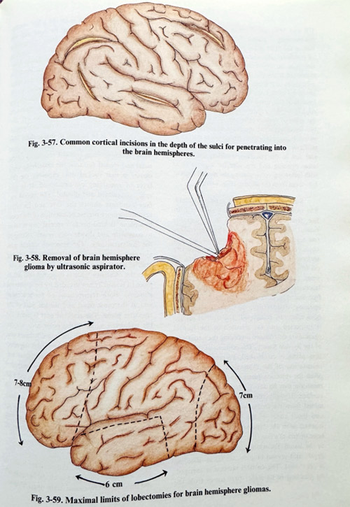
CT and MRI are the necessary investigations for planning in practically all cases. The resolution of the MRI image permits the identification of the main sulci and gyri. The plane of resection can be determined and some distances estimated from the frontal pole. These visible references are used: distances from the pole along the medial and frontobasal edges of hemispherical convexity, relationships to the free edge of the falx and how much it lies anterior to the genu of corpus callosum. MRI also allows the localization of the main branches of the anterior cerebral arteries (frontopolar arteries in particular). The bone flap is adjusted in position and size to the resection, avoiding the frontal sinus opening and the superior sagittal sinus with inflowing veins, placing the medial limit at 1.5 - 2 cm from the midline (Fig. 3-60).
Surgical techniques are rather standard. The incision is placed along the anterior hairline, from the zygomatic arch to the opposite frontal area, 3-4 cm across the midline (no scarring on the forehead will remain). The craniotomy flap covers an area of the convexity surface of the frontal lobe. Its borders are 1.5 cm from the midline and just out of the frontal sinus, and just superior to the orbit. The flap is fractured in the temporal area or is lifted as a free one. The dura should not be tense before opening; dehydration measures should have been efficiently completed. The cortical surface is inspected and the position of the frontal operculum determined according the initial part of the Sylvian fissure. The position of the resection plane is established, keeping permanent orientation of the midsagittal plane as very often the resection plane can be tilted and the resection can be too posterior on the medial surface. Usually the measurements are estimated from the frontal pole along the sagittal and basal edges of the lobe. The points are marked and are later united as a line (Fig. 3-61). In an area close to the lesion, the white matter is incised to the depth, and several biopsies are taken. Bleeding is controlled. The cortical incision is enlarged.
As bleeding in its major part comes from the cortical supply, once it is in the white matter, the transection of tissue can be done with the forceps and suction tip. Penetrating into tumour tissue is heralded usually by an increase in bleeding (Pig. 3-62) The penetration plane should however be maintained. If resection "en block" is difficult, subpial tissue removal provides space for manipulation. Retraction is applied to the side to be resected, avoiding pressure on the opposite side to the remaining hemisphere. The edges of the lobes are also transected. Special attention is paid to the bridging veins, in a way to be able to divide them after coagulation away, with a stump on the superior sagittal sinus. When dividing pia and arachnoid on the basal and medial surfaces, the underlying white matter is first aspirated out, exposing the thin layer of remaining pia-arachnoid. Then it is easily coagulated and divided. The medial surface exposure shows the falx and its edge, permitting more precise orientation of the resection plane. Behind the falx special care is taken to coagulate and divide only vessels related to the ipsilateral lobe. The resection should pass anterior to the corpus callosum and the big arteries anterior to the genu. On the basal surface, the olfactory tract is dissected and left in place (it is usually not invaded by hemispherical gliomas). Both transections of pia-arachnoid join at the most basal and medial part of the resection plane. The resected part is lifted and the remaining veins in the polar area are dissected and divided. If on the non-dominant side, the plane is located more posteriorly and the resection opens the frontal horn of the lateral ventricle. The pericallosal and callosomarginal arteries are dissected carefully close to the midline and the genu of the corpus callosum. In cases of larger lesions, infiltrated areas are seen on the remaining resection surface. If they do not penetrate deeper into the basal ganglia, such remaining parts can be additionally resected (Fig. 3-63).
After haemostasis, the residual cavity is filled with saline, the dura is closed hermetically and the craniotomy closed in the usual fashion.
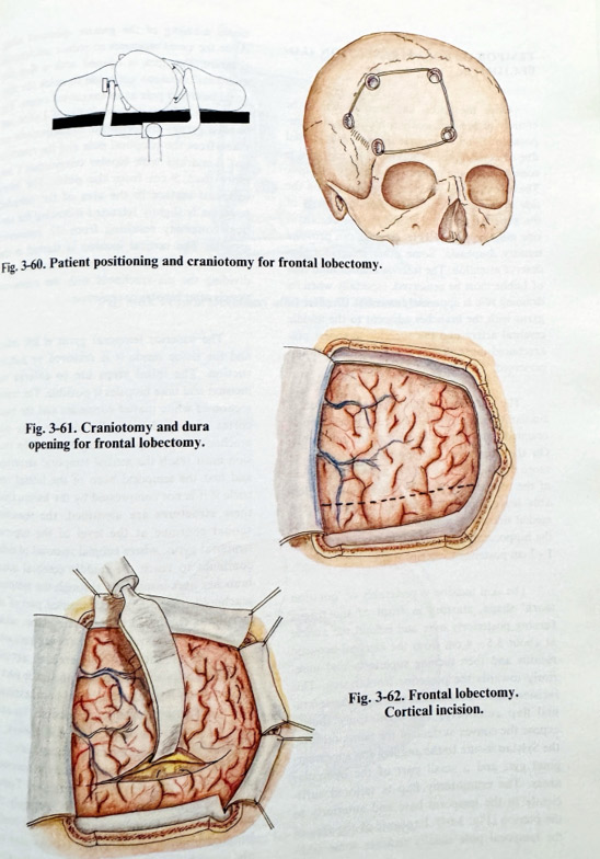
TEMPORAL POLAR RESECTION (LOВЕСТОMY)
Once the lesion has been shown to be entirely or predominantly confined to one temporal lobe and the neural function is impaired due to compression, temporal lobe resection is considered the preferable surgical technique.
The extent of this resection also depends on the side of cerebral dominance. Posterior parts of the superior temporal gyrus on the dominant side must be spared as its damage can provoke sensory dysphasia. Some other structures also deserve attention. The inferior anastomotic vein of Labbe must be preserved, especially when its draining role is apparently essential. The Hershl gyrus with the branches adjacent to the middle cerebral artery and the superior temporal pia-arachnoid must also be carefully dissected and preserved.
The planning of this type of resection is similar to the frontal. The resection plane is oriented to the most posterior acceptable position.
On the dominant side, posterior resection of usually more than 4.5 cm can lead to a dysphasic deficit of the receptive type. The plane should aim leaving intact the vein of Labbe. On the medial side of the temporal lobe the resection of the hippocampus is preferably not greater than 1 - 2 cm posterior to its tip.
The skin incision is preferably of 'question mark' shape, starting in front of the tragus turning posteriorly over and behind the auricle at about 3.5 - 4 cm from the external acoustic meatus and then turning superiorly and anteriorly towards the posterior frontal area. This incision also makes possible the turning of an epicranial flap anteriorly. The craniotomy should expose the convex surface of the temporal lobe, the Sylvian fissure to the angular and supramarginal gyri and a small part of the opercular areas. The craniotomy flap is tailored sufficiently to the temporal base and anteriorly to the pterion (Fig. 3-64). Exposure of the area of the temporal pole usually requires some additional nibbling of the greater sphenoid wing
After the usual measures to reduce intracranial pressure, the dura is opened with a flap and additional incisions are made to reach the temporal base and pole at its maximum extent. The Sylvian fissure, vein of Labbe and angular gyrus are identified. Measurements are taken from the temporal pole and the resection line is marked ( not more than 5 cm from the pole). The basal temporal surface in the area of the intended resection is slightly retracted following the cortical concavity resulting from the eminentia arcuata. The vertical incision is started in the area of the middle and inferior temporal gyri, dividing the pia-arachnoid with the contained vessels after bipolar coagulation.
The superior temporal gyrus is left intact and the tissue inside it is removed by subpial suction. The initial steps are to enlarge the incision and take biopsies if possible. The transsection of white matter continues and the basal cortex is aspirated subpially, dividing the pia-arachnoid after bipolar coagulation. The incision must reach the medial temporal structures first opening the temporal horn of the lateral ventricle if it is not compressed by the lesion. Once these structures are identified, the resection should continue at the level of the superior temporal gyrus, where subpial removal of tissue continues to reach the middle cerebral artery branches over insula seen through the temporal arachnoid (Fig. 3-65). The superior part of the incision is continued at the same plane, taking care not to penetrate insula (never removing tissue under the middle cerebral arterial branches); it must join up with the other part of the resection inside the temporal horn, exposing the hippocampus as fully as possible. Its anterior 2.5 cm can usually, but not always, be resected without significant memory deficit. Its tumoral infiltration will determine the necessary extent of the sacrifice of this structure. Technically the hippocampus is divided, but only the part inside the ventricle and not the underlying parahippocampal gyrus. Its vascular supply, provided medially, is interrupted afterwards from temporo-basal at the division of the pia-arachnoid. At this stage the tentorial edge and arachnoid can be seen there, and it should not be disrupted. The additional removal of tissue superiorly can be carried out cautiously; there is danger that the striatum can be penetrated in the same manner.
Always when the tumour masks the anatomy, the debulking must be limited to the margins of safety already mentioned. Venous collectors at the temporal base and the pole, and the temporal arterial branches are coagulated. The dura is closed hermetically. The bone flap is fixed in place and the epicranial layers repaired.
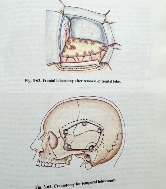
OCCIPITAL POLAR RESECTION (LOВЕСТОМY)
The planning of this resection can consider removing up to 7 cm in average from the pole of the occipital lobe. A significant part of the calcarine fissure is sacrificed and the occipital horn of the lateral ventricle is opened. The cingulate gyrus and cingulum should be spared.
On the convex surface, the resection line should pass posterior to the supramarginal and angular gyri. On the dominant side, however, the extent is much less, at times as little as 3.5 - 4 cm from the pole. Main arterial feeders are provided from the posterior cerebral artery on the medial occipital surface and the distal middle cerebral branches, very often the angular artery. Involvement of the splenium has a particular importance for the deficit, as producing hemianopia and splenial comissural damage leads to complex high cortical functional deficit (hemianopia with visual hemiagnosia).
The incision starts at the midoccipital area at about 2 cm below the external occipital protuberance, and continues superiorly to the vertex, to turn laterally toward the squamose suture in its posterior part. The flap is turned to the base, preserving the occipital artery and the greater occipital nerve. The craniotomy (more often as a free flap) exposes the dura along the transverse and posterior part of superior sagittal sinuses at about 1.5 cm from their edges and anteriorly to the supramarginal and angular areas. The length of the sagittal border of the craniotomy is at least 7-8 cm (Fig. 3-66). The dura is opened with a T-shaped incision, the long part pointing to the torcular, in such a way that flaps are turned over both sinuses - transverse and superior sagittal. As in all other lobectomies, intracranial hypotension is obtained before dural opening. Cortical incision is initiated on the convexity, dividing the feeders from the distal middle cerebral artery. After the pia-arachnoid is incised and biopsies taken from the depth, the cortical incision is enlarged and deepened to reach the occipital horn. The bridging veins from the part to be resected are coagulated and transsected. The medial wall of the occipital horn is identified and the eminence on it, called the calcar avis. It is incised vertically. By suction and bipolar coagulation the medial pia-arachnoid is exposed and the underlying branches of the posterior cerebral artery coagulated and cut. The remaining part of the lobe is then less vascularized and the resection is completed. The cavity is filled with saline after meticulous haemostasis, the dura is sutured hermetically, the flap is repositioned and fixed, and epicranial layers are closed in the usual way (Fig. 3-67).
REOPERATION FOR GLIOMAS
These operations are intended to prolong life and reduce morbidity, although in such recurrent cases that is not easy to be decided with certainty. Technically they are aimed at the removal of tumour volume and in that way to reduce damage to functionally active brain and reduce the intracranial pressure. Necrotic and cystic portions are very often taken into consideration (for cysts, inserting reservoirs to perform multiple percutaneous evacuations), but enlargement of tumor tissue resections already performed can be done too. The effect of surgery in the majority of cases is uncertain, and policies often vary significantly between surgeons and departments. However, if it is to be attempted, the same skin incision and craniotomy must be used to their maximal extent. The dura is opened at a previously unaffected place (to avoid the scar which firmly attaches to the brain tissue). The tumour can be found easily and the intended removal is done in the usual way. The technique is the same when the tissue for removal is radiation necrosis. Its resection can produce an internal decompressive effect with the same benefit as obtained from removing tumour parts. The closure of the craniotomy is in the same way as in all other cases.