
MENINGIOMAS: GENERAL RULES OF REMOVAL
Meningiomas are benign tumours arising from the arachnoid layer which expand as mass lesions with an extracerebral intracranial location, infiltrating dura and bone. The majority are benign, they grow slowly, do not infiltrate adjacent brain tissue, and are well demarcated, making possible their total removal. Parts of these lesions have an atypical hystological pattern with more pronounced characteristics of malignancy. Although it is not always the case, those meningiomas with a certain degree of malignancy tend to have a greater incidence of recurrence, infiltration of adjacent cortex and other tissues, and this lowers the rate of success of surgical treatment. In some major clinical statistics they are in the range of 13 to 18% of all primary intracranial tumour cases.
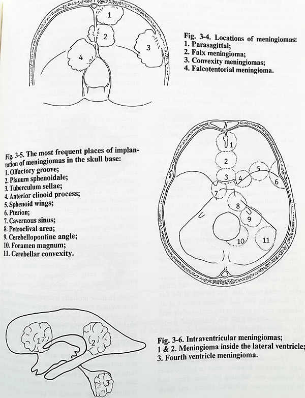
Meningiomas tend to grow in predilected locations (Figs. 3-4: 3-5; 3-6). As these sites determine the choice of surgical technique, location has proved to be the best factor for surgical classification. Meningiomas can be over the convexity or skull base, supratentorial or infratentorial, and intraventricular. The tumour is described according its place of attachment (initial growth) at the dura.
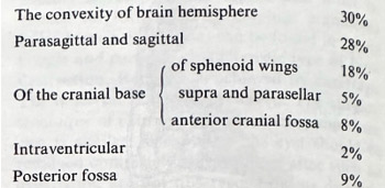
Convexity meningiomas are truly “convex”, parasagittal and falx. In their most posterior location as parasagittal they also involve also the confluens - torcular meningiomas, or falx and tentorium - falcotentorial meningiomas. Occasionally they can be attached only to the free edge of the tentorium - tentorial notch meningiomas. Skull base locations involve the midline of the anterior fossa - olfactory groove, tuberculum and diaphragma sellae, sphenoid wings - from pterion to the clinoid, the cavernous sinus, superior petrous surface and petroclival area, posterior petrous surface, cerebellar convexity and foramen magnum. After this classification, a rough approximation of the distribution in some major clinical statistics shows:
Meningiomas are usually slowly growing and vascular tumours. Planning surgery for a meningioma requires proper preoperative studies eliciting some specific information. Imaging studies (CT and MRI) should reveal the exact relationship of the tumour to the neural structures (especially brain stem structures and cranial nerves), the major vascular structures (arteries, important veins and venous sinuses) and to important skull base structures, if related. Angiography is needed for the establishment of the vascular supply of the tumour and the identification of the major tributaries participating in it. All investigations should clearly define the place of dural attachment of the meningioma and the bony changes that coexist (infiltration of bone and enostosis).
The basic steps of meningioma removal include: 1. Epicranial and bone flaps. 2. Dural opening. 3. Approach to the attachment of the tumour and interruption of arterial feeders. 4. Debulking of the tumour. 5. Dissection of the tumour from adjacent structures. 6. Removal of infiltrated structures and enostosis. 7. Repair of defects at attachment. 8. Closure of craniotomy.
These rules can be approximately the same and valid in all meningioma cases, except those which are intraventricular. Surgery is done under general anaesthesia, with intracranial hypotension by osmotic agents and diuretics, and with well preplanned blood loss estimation, (providing the necessary number of blood units plus one or two).
Epicranial and bone flaps have to comply with some important requirements. The most essential are that the exposed area of craniotomy has to give broad and comfortable access to the attachment place with sufficient space of manipulation around it. In the case of a meningioma on the convexity, the task is easier - between the attachment and border of the craniotomy there should be enough distance to allow easy work upon the dura (it is usually 1-2 cm at least). When the tumour is attached to a dural fold (falx, tentorium), or on the skull base, the craniotomy edge should widely expose the accessible border of the structure of attachment in such a way, that after retraction of the hemisphere there will be access from both sides of the attachment (Fig. 3-7). Although these rules are followed strictly, exceptions are possible, for instance, in a case where wider access has to be gained to important structures around the attachment, especially venous sinuses.
It should always be kept in mind that craniotomies in meningioma cases can lead to substantial blood loss because of pathologically hypervascular bone and dura. Some practical tips can be used. For instance, burr or entry hole bleeding is controlled easily by wax, bipolar coagulation and packing, but bone cutting lines are not. That suggests that the surgeon should do in advance as many holes as are needed, but slightly away from the hypervascular bone. The moment of major uncontrolled bleeding is between the bone cutting and the lifting of the flap, so the anaesthesia team should be alert for that. This also requires shortening the time between bone cutting and lifting of the flap, as after elevation there is much better access to the bleeders. The most vascular bone cutting is left last. The turning of the flap is followed by fast and efficient control of all bleeding, first by covering the entire surface with cottonoids and surgicel, wet gauzes, and gradually uncovering small areas of dura for controlling bleeders with coagulation.
Epicranial flaps are planned in a standard way. Attention, however, must be paid to cases of meningiomas penetrating the bone. These lead to hypervascular epicranial tissues, and therefore to flaps with altered vascular pattern and that increases blood loss during surgery.
Dural opening is adjusted to the place of attachment. It is done after application of dehydrating agents for intracranial hypotension. The rule is to open the dura at the border between affected and unaffected dura, starting with the interruption of major arterial feeders of the tumour. Affected and unaffected dura is held on traction sutures. In cases where the attachment is not seen after the craniotomy, the dura is opened according to the plan of access intracranially.
Approach to the attachment place and interruption of arterial feeders will provide the proper conditions for a removal with less bleeding and injury to the neural structures. In the case of a convexital meningioma the goal is achieved with the craniotomy. More important and time consuming is the step in meningiomas with attachment locations deep and far from the most convenient craniotomy site. Approaches have always been very much dependent on this step. The attachment site is optimally approached both intradurally and extradurally.
However, the extradural approach is not always possible. In tumours attached to falx and tentorium it cannot be performed. The extradural approach, if planned, has to follow craniotomy and haemostasis. Now skull base attachment places are approached with techniques additional to those of specific skull base approach. Skull base exposure aims early, extracranial control of the arterial supply to the lesion and prevention of damage of important structures within the skull base during detachment of the tumour. At the end of surgery it permits radical removal of the lesion. In more medially located tumours which have invaded the skull base, that is a very difficult task and requires high neurosurgical skills. The intradural part of the approach (routinely done under magnification) consists of brain retraction until the attachment place is seen and further separation of the lesion with bipolar coagulation and micro-instrumentation follows. Important structures in relation to the attachment are dissected and separated. Removal of a small part of the tumour may be needed to provide space for manipulation and access. Care should be taken at this stage not to separate the dura from the bone, as the bleeding provoked is from bony vascular channels, which cannot be coagulated and have to be waxed (not an easy task in the depth of soft abnormal tissue). Intradural separation is much easier, if it has been anticipated, by extradural control of arterial supply. At present, with the advent of endovascular techniques (interventional neuroradiology), a session of feeder obliteration can take place a day before surgery.
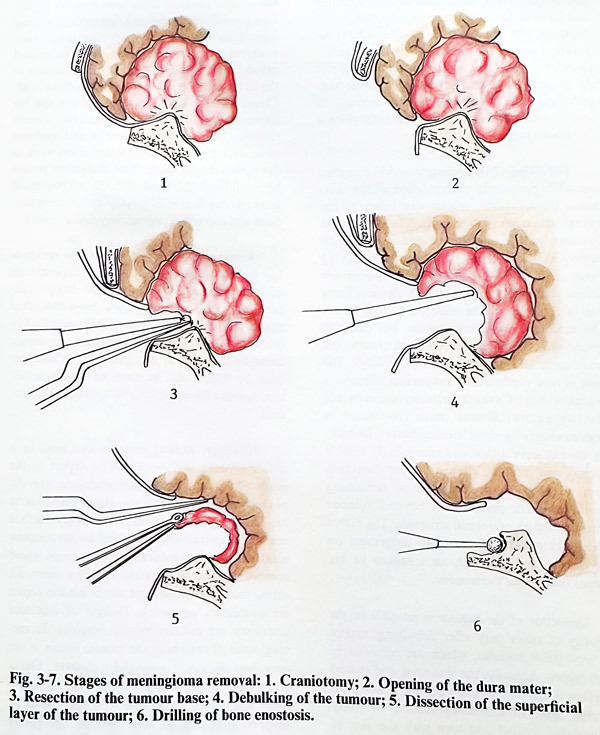
Debulking of the tumour should be carried out while preventing manipulation of the mass lesion intracranially, minimising injury to the neural structures in its vicinity. The already less vascular tumour must be reduced in volume by removing its internal part without applying force and provoking movements. There are several techniques to do this, the classical being use of the loop of the monopolar coagulator.
The cutting loop removes slices, but can give spread of currents (dangerous when close to the brain stem and cranial nerves especially). The laser is now preferred. It evaporates tissue in a touchless way. Cavitational destruction (ultrasonic aspiration) is more frequently used, but is not effective against fibrous tissue (contained in large amounts by some meningiomas). Debulking should leave a thin and increasingly flexible layer of the tumour, allowing easier dissection from neural structures. However, debulking should not risk penetration beyond tumour limits and injury of normal structure. The extent of debulking requires thorough judgement on how far to go, especially in a region of big arteries overlaying the tumour.
Dissection of the tumour is possible in the majority of cases only after debulking, except for very small nodules, which are removed in small pieces. Actually, the dissected and spared structure is the surrounding normal tissue, and the thin layer of remaining tumour is freely manipulated, pulled and torn with the aim of protecting the important neural and vascular structures. There are several important points on the dissection of meningiomas which should be emphasised. The first of these is the location of the tumour tissue regarding arachnoid layers.
The completely benign tumour which is noninvasive of the adjacent tissues should be covered by a single arachnoid layer. This should be the natural plane of dissection. Another important point is to identify and follow from normal anatomical sites all main important intracranial anatomical structures (especially arteries and nerves) with attention when they are involved on the surface of the tumour (there will be significant distortion).
Classical examples are the optic nerve and the internal carotid artery at the anterior clinoid process. The aim is to preserve and mobilise some structures and this can also allow work in the subarachnoid space. This dissection should be considered the most difficult microsurgical work, as the degree of postoperative complications and morbidity depend very much upon it.
Removal of infiltrated tissues and enostosis has a direct relationship to the recurrence rate of meningiomas. To achieve the lowest rate (about 5%), all infiltrated dura, bone and other tissues should be meticulously resected. The infiltrated dura is excised and any bony enostosis, commonly found at the place of attachment must be drilled out. In cases of deeply seated meningioma the most suitable tool for that is the micro-drill.
However, radical excision can lead to a defect of dura and bone, whose repair at the place of attachment is important to prevent postoperative complications. Basal dura has to be patched if there is no intact bony surface remaining instead and there will be no communication with the epidural space. In cases where communication is possible with an open aerated sinus, muscle packing and dural plastic repair are absolutely necessary. Defects of falx and tentorium are not necessarily repaired. In me-ningiomas of convexity the plastic repair is the only way to get a watertight closure. Dural layers can be split and the upper one turned to cover the defect. Otherwise it is fascia or allograft that can be used. We do not prefer allografts, unless the options to use other materials are completely limited.
If a bone defect remains because of removal of infiltrated bony tissue, the plastic repair can be done at the same session or at a later stage. The decision depends on the expectation and control of perifocal and postoperative brain oedema.
The closure of craniotomy is done in the routine way.