LATERAL AND MIDDLE SPHENOID WING MENINGIOMAS
These meningiomas attach to the edge of the lesser and part of the greater sphenoid wings between pterion and the anterior clinoid pro-cess. They are subdivided according to their precise area of attachment as this factor is crucial for assessing the technical difficulties at the time of removal and the outcome of surgical treatment. The majority of these tumours grow as an intracranial mass lesion, but some predominantly invade the dura on a broad base, without expanding intracranially ("en plaque"). Meningiomas arising from dura of the sphenoid wings have a tendency to invade adjacent structures and spaces. Invasions of the cavernous sinus and the orbit are of particular interest, as they increase the technical complexity of the required surgery.Despite some differences in classifying the types of location, there are several accepted types characterized by the common problems they present.
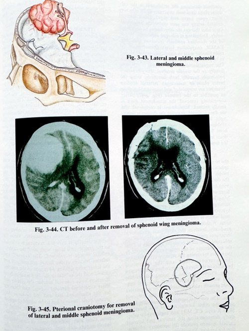
Lateral and middle sphenoid meningiomas have their attachment not involving important basal structures (Fig. 3-43). Their vascular supply is generally provided by enlarged branches of the middle meningeal artery and the venous drainage - by a variety of channels, the principal one commonly being the sphenoparietal sinus. Their growth has subfrontal, temporopolar and sylvian expansion. The tumour dome displaces the first segments of the middle cerebral artery, and rarely encases them. Hyperostosis on the sphenoid wings can be detected sometimes on the plain X-rays of the skull. A CT scan shows the size and position of the lesion after contrast enhancement (Fig. 3-44). MRI can demonstrate the relationship of the tumour to the cortical surface, and middle cerebral artery, as well as any medial displacement of arteries and the optic nerve, additional extensions to the orbit or involvement of the cavernous sinus. The suspicion of arterial encasement arises also from the MRI, although it is not frequent. Angiography is an essential investigation to indicate the condition of the major intracranial vessels next to the tumour and reveal the vascular supply to it. It is important to visualise separately the internal and external carotid systems for better demonstration of their contribution to the supply in all sphenoid meningiomas. Selective external carotid catheterisation can be used for preoperative embolisation of major feeders to reduce intraoperative blood loss.
Surgical planning focuses on a craniotomy, always including the pterional area and as extensive as frontal and temporal retraction may demand. The attachment place should be accessible both after subfrontal and temporobasal cortical retraction. The part of the sphenoid bone corresponding to the attachment place, usually infiltrated by the tumour and containing the feeders should be accessible through the basal part of the craniotomy (Fig. 3-45).
Skin incisions are usually curvilinear, frontotemporal, from the midline to the external projection of the temporal base towards the tragus or retroauricular. The craniotomy flap is lifted with special attention to its basal part, where it moght be close to the attachment, and bleeding can be serious. We recommend that access or burr holes be placed on both sides close, but not over the attachment at pterion and later that the affected bone be removed by nibbling or drilling. This removal is associated with bleeding, but the enlarged extracranial branches from the midportion of the maxillary artery can be coagulated. The extent of bone removal can reach the lateral part of the superior orbital fissure, spinous foramen and the course of the major superficial petrosal nerve, but removal to such an extent is not always required. This stage is important for the interruption of feeders. Bone removal can also be done after the opening of the dura, verifying the attachment place by direct observation. The dura is opened along the skull base, but outside the attachment area.
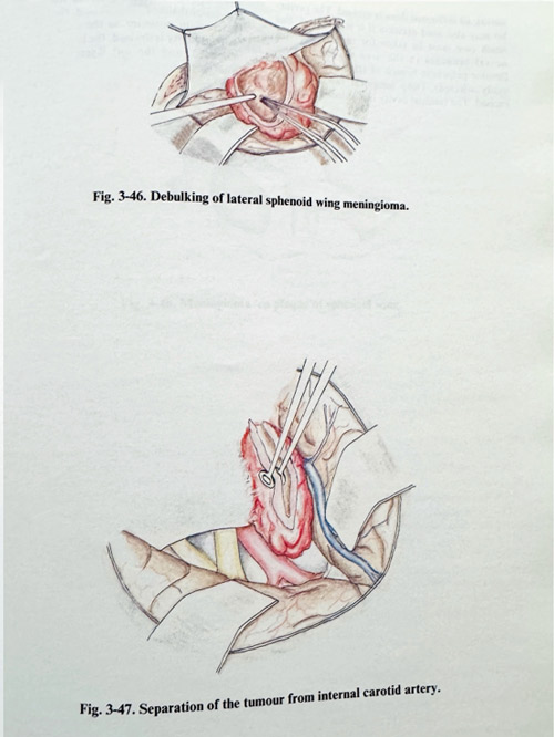
Additional dural incisions may be needed vertical to the first one, to ease the retraction of brain. The area of attachment is approached with minimal or no brain retraction. The base of the tumour is gradually transsected with bipolar coagulation and microscissors, firs debulking the adjacent part (Fig. 3-46). At the time of attachment transsection and debulking, much care should be taken with the most medial part of the involved dura, as it is close to important structures within the skull base and close to the anterior clinoid. It is recommended at the time of debulking and detaching the tumour, to continue dissecting the attachment in the sub-dural plane until such structures as the olfactory tract, optic nerve and supraclinoid internal carotid artery are seen under the arachnoid and by that the detachment can be completed safely.
Debulking is expanded sufficiently by one of the standard methods (cavitron aspirator, LASER, coagulation loop). Dissection of the main tumour mass as completely as possible has to be performed while preserving the arachnoid. The supraclinoid internal carotid is followed distally or the middle cerebral artery branches - proximally (usually in both directions, using first the better plane for dissection). Arterial feeding branches are coagulated and transsected as close as possible to the tumour (Fig. 3-47). After completing removal, the attachment dura is totally excised, haemostasis is completed and the dura is closed, ensuring the closure is water-tight. If enostosis remains despite the bone nibbling, it is drilled out before closure. The bone flap is secured in place with leaving an epidural drain, and the rest of the layers are closed in the usual way.
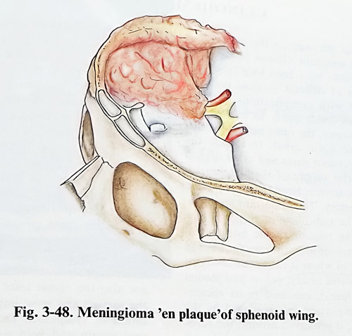
A very particular type of a meningioma, the so called " en plaque" type, which affects the sphenoid wings, presents some specific problems. This meningioma progressively invades the dura and the bone of the greater and lesser wing over many years. The dura is affected as a layer of just few millimetres thickness. The underlying bone haversian canals are invaded by meningotelial tumour cells, provoking reactive hyperostosis. It is the hyperostosis of the sphenoid which is responsible for the clinical symptoms. Without causing a mass effect, the lesion gradually involves the skull base anatomical structures passing through the apertures of the sphenoid. The most common symptom is from the involvement of the optic canal, followed by the nerves traversing the superior orbital fissure, the cavernous sinus, and the trigeminal branches in the foramen rotundum and foramen ovale. Presenting often in young and middle aged women, this lesion has a slow and insidious growth rate, detectable by the progressive exophthalmos and deformity in the temporal fossa. The essential investigations are the same as in the other menigiomas of this location.
Skull X-rays, CT, MRI and angiography selectively demonstrating the external and internal carotid systems provide the necessary information for surgery. Special attention is required for the visualisation of all involved bone and the position and condition of basal foramina. The orbital content is studied thoroughly, as it is commonly found to be invaded by the tumour.
The aim of surgery consists of removal of as much affected bone as possible, excision of affected dura and decompression of nerves along the foramina. Surgical planning is mainly aimed at determining essentially the extent of bone removal and dural excision. It should define how far the surgeon should go and which bony cavities and canals should be exposed and decompression performed. The plan refers particularly to the optic canal, the posterior orbital segments, the superior orbital fissure, foramen ovale and foramen rotundum. The amount of involved dura to be excised is planned according the pathologically enhancing areas on CT and MRI. After all these considerations, the needs of dural plastic repair, the type of material and the expected extent are determined. Cra-nioplasty is needed in cases of significant convexity or orbital wall removal (Fig. 3-48).
Surgery is conducted through a frontotemporal incision not anterior to the hairline, with the flap reflected to the base exposing the frontobasal and temporopolar areas, centred at the pterion. A fronto-temporo-pterional flap follows, overcoming the difficulty of turning it with the drill. Affected bone is definitely not included in the flap, and it is nibbled or drilled after flap-lifting. Surgery is performed in its initial part extradurally. Starting with the orbital roof, bone is drilled or nibbled, depending on what is more efficient in bone removal. We used to drill until the thickness was suitable for nibbling and we alternate these two manoeuvres. Once the orbital content is exposed, removal is extended to the greater and lesser wing until the superior orbital fissure is reached. The superior and inferior borders of the fissure are removed and the lesser wing drilling advanced to the anterior clinoid and the orbital canal. In a temporobasal direction bone removal reaches the greater and lesser superficial petrosal nerves, and foramina for the trigeminal 2nd and 3rd division. Any greater bone removal has to be weighted against the chances for radicalism and the threat of intra- and postoperative complications. Cavernous sinus area, petrosal apex or contralateral involvements are such conditions, when the continuation of removal has to be reconsidered seriously. After competition of the bone removal all infiltrated dura is excised. The periorbit may also need excision if it is involved, but much care must be taken for the arterial and neural branches in the area of Zinn's ring (levator palpebrae branch of Illrd nerve can be easily affected). Deep temporal fascia is also excised. The residual from bone removal cavity has a decompressive effect upon the orbital content and the preexist. ing examith meriTh to ure is repaired using pericranium posterior to the craniotomy. The residual cavity is drained, the bone flap replaced and fixed, and the soft layers sutured in the usual way.