FALX MENINGIOMAS
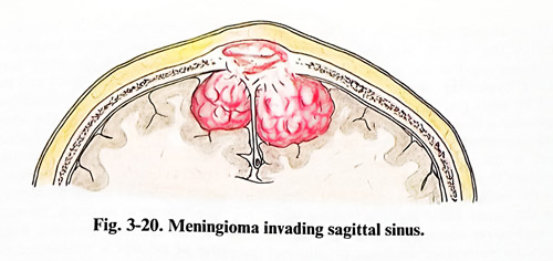
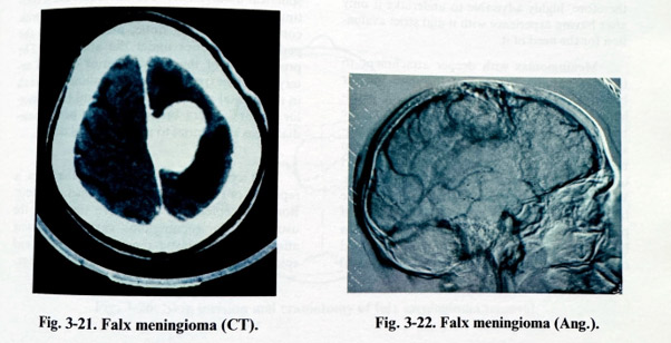
The meningiomas of the falx are those involving one or more of the structures superior sagittal sinus, inferior sagittal sinus or falx uni-or bilateral (Fig. 3-20; 3-21). Their location is subdivided according to which third of the superior sagittal sinus and falx (anterior, middle or posterior) they involve. They are usually supplied from bilateral arterial sources of dura and epicranial tissues, but in the anterior third enlarged supplying branches from the falx can be found. Venous drainage is much more complex. These tumours can often compromise the flow in the superior sagittal sinus. The occlusion can be partial or complete. The occlusion leads to venous collateralization. The enlarged collaterals can be at all levels, cortical, dural, diploic, epicranial. On their preservation very often depends the success of surgery. Some of the tumours attach only in areas close to the free edge of the falx, affecting only the inferior sagittal sinus. In these cases major consideration is given to the role of the inferior sagittal sinus in venous drainage and the relationship of the tumour with pericallosal arteries.
Investigations always include CT, MRI and angiography. They should reveal the exact attachment area of the tumour, its size and relationship to adjacent neural and vascular structures, involvement of sinuses and collateralization (Fig. 3-22). General information on structures and enhancement, indicating vascularity, is derived from the CT images. MRI is able to demonstrate involvement of sinuses and some other abnormal arterial and venous channels. Serial angiography gives the most precise vascular images as, being dynamic, it can also indicate flow and its direction.
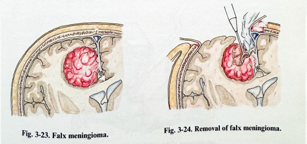
Surgical planning defines the goal of total removal of the lesion and the steps to be taken to this end. Complete feeder interruption and venous drainage preservation require sufficient access around the tumour and on both sides of the falx and the superior sagittal sinus. Craniotomy should provide this access. In a unilateral falx meningioma, craniotomy exposes an area across the midline only for access to the falx and the superior sagittal sinus, that is 1.5 - 2 cm away from the opposite sinus border (Fig 3-23;3-24). If the tumour is bilateral, then the craniotomy should provide space for tumour removal on both sides. Complete excision in superior sagittal sinus involvement entails risks.
Any significant patency of the sinus is an indication to preserve it, even with its reconstruction.
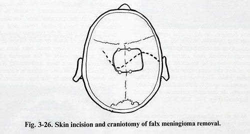
The completely occluded sinus permits excision including the involved area. Scalp incisions are extended on both sides, horseshoe or S type, considering the pattern of epicranial vascular supply.
Surgery is performed with the patient in a supine or lateral position, the first one being suitable only for tumours in the anterior third of the falx. Scalp incisions have no particularities, except excessive bleeding in case of significant epicranial vascular tumor supply. Craniotomies are often free flaps. Entry or burr holes are done on both sides of the superior sagittal sinus and the dura (sinus wall) is separated with dissectors. This avoids rupture of the sinus at bone cutting.
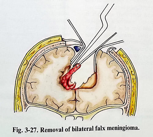
Rupture can also be provoked during bone flap lifting as a result of tumour bone infiltration at the sinus site, although potential danger also exists without infiltration at the places of emissary or diploic venous communications. After the flap is tailored with as much bilateral extent as needed, haemostasis is performed without sacrifice of venous channels. Oxidised cellulose packing must be the preferred method. The dura is opened with an incision distant from the midline and as close as possible to the tumour attach-ment, but not closer than 1.5 - 2 cm from the superior sagittal sinus edge. Incision of the dura continues towards the midline avoiding any dural venous drainer. The tumour is debulked in the usual way, leaving intact the part close to the falx. Debulking is done in the same way if bilateral extension exists (Figs. 3-25; 3-26; 3-27).
Debulking creates sufficient space on both sides of the falx. As preoperative information is available concerning the patency of the superior sagittal sinus, in cases of complete occlusion it is excised totally with all the affected dura. The lines of excision are a few millimetres anteriorly and posteriorly from the affected area. Care is taken not to affect any venous inflow from cortical or dural drainers just anteriorly or posteriorly to the resected part of the sinus. The remaining sinus ends are closed with sutures for perfect haemostasis.
All affected dura from the falx is also excised meticulously. When the superior sagittal sinus is partially or completely patent, but its walls involved by the tumour, the flow through it must be preserved. Excision is performed under temporary bypassing of the flow, and it is followed by sinus grafting at the end. Temporary bypassing is done by inserting a previously prepared siliconized bypass tube, corresponding by gauge to the sinus size, and fixing it with dural stitches through the falx around the sinus, close to the excision. Stitches are passed before opening the sinus. Final grafting is done with greater saphenous vein segments (keep in mind there can be valves) or artificial prostheses, similar to those used for artery replacement. Sinus bypassing and grafting carry risks of massive bleeding, air embolism and postoperative thrombosis. It is, therefore, highly advisable to undertake it only after having experience with it and strict evaluation for the need of it.
Meningiomas with deeper attachment to the falx requires opening of the dura uni- or bilateral, at 2-3 cm from the midline and parallel to it, turning the flaps medially. Spearing the bridging veins, the medial edge of the hemisphere is retracted (one non-important vein can occasionally be sacrificed). The most superficial affected part of the tumour is reached on one- or both sides.
We prefer to start the removal on the side of the larger part of the tumour in bilateral cases. If part of normal falx, close to the attachment can be incised, it is done and the tumour dura held on a stitch for easier manipulation. By careful retraction, part of the tumour surface is ex-posed, incised, biopsies taken and debulking is started. Debulking should be done with consideration given to the width and the depth of the tumour. With reducing tumour volume, excision from the falx close to the abnormal dura is continued in a stepwise fashion. When approaching the free edge, certain precaution is taken related to the inferior sagittal sinus, which is able to create problems of haemostasis only because of its depth. Its occlusion is practically always well tolerated. Even after the free edge of the falx is transsected on both sides of the tumour and the inferior sagittal sinus coagulated or clipped, part of the tumour may remain attached to the deeper part of the interhemi-spherical fissure. There the dissection must continue along the fissure reaching its depth, with complete debulking done, and following the pericallosal arteries under the arachnoid. The preservation of the distal anterior cerebral artery branches (pericallosal and callosomarginal) in the frontal and parietal areas and the posterior cerebral artery branches in the occipitome-dial areas is essential to avoid cortical deficit.
On completing the excision, the dura is repaired on the convexity to hermetic closure.
Bone and epicranial tissues are treated in the usual way for meningioma cases, by excising affected bone, plastic repair of the bone and epicranial closure, leaving an epidural drainage.