TUBERCULUM SELLAE MENINGIOMAS
These lesions arise from the area of the tuberculum sellae, sulcus chiasmatis and diaphragma sellae (Fig 3-37). The optic nerves are displaced laterally, and when the tumour is growing more posteriorly from the diaphragma, also upwards. The internal carotids are less displaced laterally. As the tumour continues growing, it exerts pressure on the infundibulum, ventral hypothalamus, and occupies the retro-sellar area and interpeduncullar fossa, although the area of attachment is relatively small. It often produces hyperostosis. The blood supply is very often provided by posterior ethmoidal arteries. Relations of the tumour to the anterior cerebral arteries are of the greatest importance.
Their displacement is often significant and small arterial branches can contribute to the supply of the tumour. Their course is sometimes difficult to follow, and these branches should be sacrificed after the surgeon is completely convinced that they are related only to the tumour. As with other meningiomas in this area, the optic canal can be invaded and the arteries (internal carotid and the anterior cerebral) encased. A total removal must always be attempted, but this can be impossible in some cases. The size of the tumour is also closely related to the outcome - the larger size is related to significant morbidity and mortality.
These meningiomas need thorough preoperative neuroophthalmological evaluation. Almost all symptomatic cases have visual affection - decreased visual acuity and/or limitation of visual fields. The tumour is usually detected by CT scan (Figs. 3-38; 3-39). It is best visualised on direct coronal scanning, which gives a better indication of the relationship to supra- and parasellar structures. MRI demonstrates the arteries well, especially if they are encased, and reveals the position of chasm and the perifocal oedema. Angiography is essential to demonstrate the collateralization through the anterior communicating complex between the internal carotid systems on both sides (Matas test) as temporary or permanent occlusion can be needed during intraoperative dissection.
Surgical planning depends on the size of the tumour and the position of the main mass regarding chasm and the optic nerves. When the main mass volume is located completely retro-chiasmatically, a view from the lesser sphenoidal wing, i.e. from the pterion, will be needed.
These lesions require a wide angle of approach as a rule and in some cases with big tumours and complex involvement of chasm, a broad subfrontal (even bifrontal) and pterional view will be required simultaneously. For small retro-chiasmatic lesions (more attached to diaphragm we recommend pterional craniotomy.When microsurgical work will be extensively needed between the nerves, over the chasm, on both sides of the nerves and both Al arterial segments, then bifrontal craniotomy is the option of choice, with an additional extension for a pterional view. The pterional view can be broader by opening the sylvian fissure and extending the craniotomy with an orbital part.

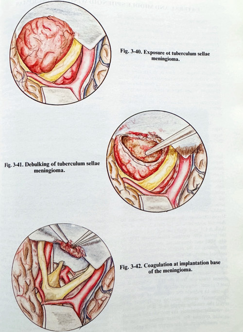
After performing the selected approach, the area is reached in a standard way, either along the lesser wing or the subfrontal route with the intention of sparing the olfactory tracts if possible. Two objectives have to be concidered at the beginning - detachment from the feeders and reduction of the volume. Both tasks are not so easy to perform in many cases. Part of the feeders can come from the anterior cerebral arteries, and interruption of feeders should be done at the beginning only at the attachment area. The main mass is approached between the optic nerves and under the chiasm and debulked if possible. The tumour parts are dissected from arteries, optic nerves and infundibulum, but it is better to follow first the anterior cerebral arteries and spare some fine branches, which enter the tumour and later exit supplying neural structures (“passing” arterties) (Fig. 3-40). Following the arteries do not invalidate the rule to preserve and follow arachnoid over chiasm, infundibulum and the rest of the basal brain surface, as this is the most important borderline of tumour separation. In huge tumours removal can compromise anterior cerebral arteries. Their damage has to be repaired with microsutures. This occurs more often in some cases of encasement and the possibility of removing the tumour completely depends on the existence of the arachnoid layer between the artery and the tumour. The arachnoid, although thicker, must be followed on the posterior surface, where the infundibulum and hypothalamic surface are separated, avoiding any microvascular sacrifice. Even if surgery must be prolonged to its acceptable maximum, complete removal must always be attempted, as second surgeries are as a rule much less efficient in removal due to the disrupted anatomy and adherence of tissues. There is no specific technique which differs from other similar approaches in completing and closing this surgical approach (Figs. 3-41; 3-42).
In big internally situated meningiomas it is essential to open the sylvian fissure widely to find the branches of the middle cerebral artery.
After enucleation of the tumour, these branches are followed proximally to the internal carotid artery to remove the tumour in a piecemeal fashion, never losing sight of the artery and its branches. Tumour invasion of the arterial wall and occlusion of the lumen is very rare. Dissection of the perforating vessels, the anterior choroidal artery and posterior communicating artery is time consuming, but it is the only way to diminish morbidity.