SURGICAL MANAGEMENT OF SUPRATENTORIAL GLIOMAS
Last two decades have witnessed substantial progress in basic tumour research, which has been incomparably more successful than have been the real technical advances in glioma surgery. Despite this, no radical breakthrough in the treatment of these lesions has been achieved. The observed improvement in results obtained has rather derived more from a transfer of methods from other neurosurgical areas and adjuvant therapies; naturally the actual steps forward have been relatively modest.
Based on this type of limited progress, then, we can hardly predict, that surgery constitutes the solution of the glioma problem now or even that this will be the case in the near future.
Gliomas are a group of tumours with an infiltrative nature in general. Their histological types have recently been classified by the WHO, with the aim of indicating the degree of malignancy. Astrocytomas I and Il grades are low grade, while grades III and IV are considered high grade gliomas (glioblastomas). A specific type of nonmalignant glioma is the cystic pylocytic astrocytoma. Another common supratentorial glioma type is the oligodendroglioma; its malignant type is the oligodendroblastoma. It very often presents histologically as a mixed type of glioma. Malignant gliomas have a tendency to undergo cystic degeneration, with resultant intratumoural vascular proliferation, necrosis and hemorrhages.
Clinically, gliomas present with a great variety of focal symptoms and signs of neurological deficit, and epilepsy. When the effects of a mass lesion produced by the continued growth of the neoplasm are added to the affection of the primarily involved area, the clinical course shows deterioration due to compression of distant structures and increased intracranial pressure.
Surgery can be beneficial in two aspects of glioma treatment: to establish the nature of the lesion and to reduce its volume. Surgical radicalism is of dubious value in glioma surgery. The choice of method depends on the location of the lesion, the expected grade of malignancy and the general condition of the patient. The patient is investigated by CT and MRI which are aimed at determining the borders and intrinsic texture of the tumour, its vascularity, the possible differential diagnosis, the degree of perifocal oedema. Angiography is reserved only for very vascular tumours or for the needs of differential diagnosis.
The histopathological diagnosis has to be established in all symptomatic lesions or in those which are showing growth. The asymptomatic lesions not confirmed to be expanding can be followed up by imaging studies. Lesions with cortico-subcortical locations can be explored for biopsy by CT guided small craniotomy or burr hole techniques. Those located deeper in the centrum semiovale and in the basal ganglia are indicated for stereotactic biopsy (this technique is described in the corresponding chapter of the book). Multiple biopsies should always be taken, as the grade of malignancy varies among different tumour areas, and stereotactic sample taking yields maximal positive results in not all, but nearly 90% of the cases.
Removal of tumour tissue is necessary to obtain "internal" decompression of the still viable brain structures by reducing the amount of abnormal and nonfunctional tissues and evacuating tumoural cystic fluid. The indications for this are present when the patient's condition has deteriorated due to the mass effect and its at the very least temporary relief will benefit the patient with a sufficiently long period of low morbidity to justify it. The technique depends on the areas involved. Functionally important, "eloquent" cortical areas, the basal ganglia and some limbic structures must be spared. Therefore the technique of polar resection (also called "lobectomy") is reserved to frontal, temporal and occipital lobes and poles, taking into serious consideration the implications of cerebral dominance and the variability of cortical functional areas, and adjusting the extent of removal in accordance with this. All other gliomas with a deep location (affecting basal ganglia, thalamus and hypothalamus, and those crossing through the corpus callosum) are debulked very selectively to avoid functional damage.
Surgical planning should consider the extent of surgery. First to be decided are the place of biopsy taking and the amount of tissue to be removed. Then the craniotomy is planned to provide safe access and manipulation for the areas of interest for tissue removal.
Preoperative treatment is essential for the outcome of surgery. Medication includes two essential drugs: anticonvulsants and steroids.
Anticonvulsants have a place on both occasions: with and without previous seizures. Any preceding epilepsy with treatment already initiated requires evaluation for adequacy (using therapeutic serum levels), and the dose must be correspondingly adjusted. Perioperative prophylaxy is meant to increase the seizure threshold in the most vulnerable periods before and immediately after surgery. Diphenylhydantoin is administered in loading and maintenance dosages as the drug of choice, but phenobarbital, carbamazepine and some other anticonvulsants should be considered in some selected cases.
Steroid treatment is most often initiated immediately after the diagnosis has been established and possible surgery is expected. Dexamethasone is given, preferably a few days before surgery, under the protection of ranitidine or cimetidine, the high doses being reserved for those patient's in advanced stages of the disease - with increased intracranial pressure and a rapidly progressing neurological deficit. Surgical treatment is performed with special attention on control of intracranial pressure and seizure prophylaxis. Bolus doses of Dexamethasone and Diphenylhydantoin are given during the induction of anaesthesia if the patient has not been treated with them before. The patients who already have therapeutic levels of these drugs receive their regular doses then and at 6 hour intervals afterwards. An intracranial hypotensive effect is induced before opening the dura by the rapid infusion of 20% Mannitol and/or Furosemide. This will permit safe brain tissue manipulation. The patient is moderately hyperventilated during anaesthesia.
GLIOMA BIOPSY TAKING
As a separate procedure, the biopsy of a glioma is planned carefully on imaging data (CT and MRI). In those cases where stereotaxy will not be used, the intended point of approach through the skull should be visible (with a marker attached) on the imaging studies to establish the exact relationships between intended biopsy area and marker.
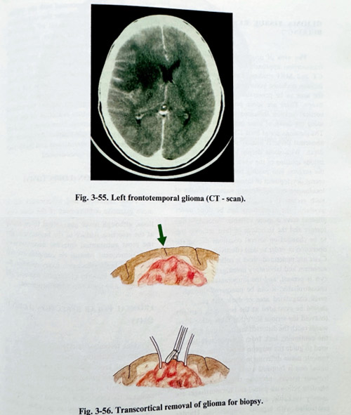
Biopsy taking through a large burr hole (or more than one) must be followed up by CT after the procedure. If a craniotomy is planned (partial removal to follow the biopsy), the intended margins of the craniotomy can be marked to provide orientation for the subcortical tumour borders (Figs. 3-55; 3-56).
Biopsy taking must yield tisuue for histopathological study which is representative of the structure of the tumour. Gliomas are non-uniform lesions inside. In more malignant tumours, areas of different grades of malignancy are found among cysts, necrotic tissue and haemorrhages. Taking several specimens will provide a more comprehensive histopathological image of the neoplastic lesion. The tissue should be vascularized and not necrotic, and different areas of the tumour are approached and sampled. If cysts are evacuated their walls are carefully inspected and biopsies from the wall are taken where it seems tumoral.
A biopsy should be taken at the beginning of tumour manipulation, and in case surgery has to be discontinued, a specimen will nonetheless have been provided. Tumour tissue is taken with specially designed forceps and its destruction - avoided until it is immersed in a fixation solution.
GLIOMA TISSUE RESECTION ("DEBULKING")
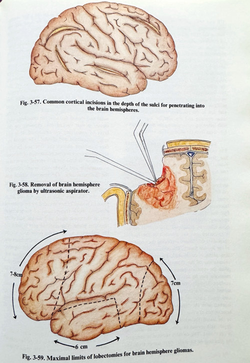
The area of proposed removal and the transcortical approach are prеplanned on the CT and MRI studies. The place of cortical incision is chosen according to the "eloquence" of the area to be penetrated and cerebral dominance. There are some preferable places for cortical incision indicated in the illustrations, using the depth of some main sulci (Fig. 3-57).
The incisions are at least 1.5 to 2 cm long. The abnormal tissue is usually seen at the expected depth. With some deeply located and smaller lesions splitting of the white matter can mislead the surgeon into passing beside his target. The recent development of intraoperative systems of localisation can significantly reduce the risks of such errors. If navigational systems are not available, the craniotomy can be done under stereotactic conditions, with account being taken of the displacements caused by dehydration and CSF evacuation. Cortical incisions are maintained with the minimum of retraction.
Initial biopsies are taken and the preliminary impressions of the tissue qualities on the “frozen” sections stains will determine the instrumentation to be used. High grade gliomas have a more variable, "mosaic" - like texture and the technique of their removal has to be changed on several occasions during the operation in order to deal effectively with them.
Cysts are punctured, fluid is collected for volume measurement and laboratory investigations. If a big cyst is present, and the intracranial pressure is uncontrollable, it can be punctured through a small coagulated area of dura and pia. Cysts should be evacuated at the beginning partially to avoid the serious shift that takes place afterwards (with the distorted anatomy that follows); the continuing leak from the puncture hole is used to guide the surgeon to the cystic cavity. A tumour tissue differing in appearance from the initial one is biopsied again. Soft and vascular tumour tissues, necrotie tissue (clearly non-bleeding yellowish tissue) and haemorrhages are usually suckable, and the opened vessels are controlled with bipolar coagulation. Harder tumour tissue is best removed with the ultrasonic aspirator, and we consider this technique to be preferable for the removal of gliomas in general (Fig. 3-58). The wall of the cavity produced is covered with oxidised cellulose and cottonoid. In the majority of gliomas, the lesion has no clear borders with adjacent brain. Some of the tumours form "pseudonodules displacing bundies of white matter; however this is not a real border, and following these margins will not lead to real radicalism. At the end the tumour removal should produce reduction of the mass effect, and after thorough haemostasis, the dura is closed in a watertight fashion. At closure, the craniotomy flap is repositioned and fixed, with the residual epidural space drained.
POLAR RESECTION (LOBECTOMY)
Polar resections and lobectomies aim at more extensive achievement of the same standard goals of internal decompression. By these methods the lesion is resected to its maximum, including some normally appearing brain areas towards the poles of the lobes, which have, however, been severely deafferentated by the lesion and very rarely lead to functional deficit after their removal (Fig. 3-59). Such amputations, although never guaranteed to be radical at the resection surface of the hemisphere, give the most substantial internal decompression over functional brain structures. However, every resection has its particular features of surgical anatomy and technique.
FRONTAL POLAR RESECTION (LOBECTOMY)
This technique is intended to remove the tumour, by including it in an amputated part of the frontal lobe. On the dominant side, the resection plane passes anteriorly to the frontal opercular part to avoid affection of speech. The extent of the resection is planned to incorporate the lesion as completely as possible, without affecting functionally important cortical areas.
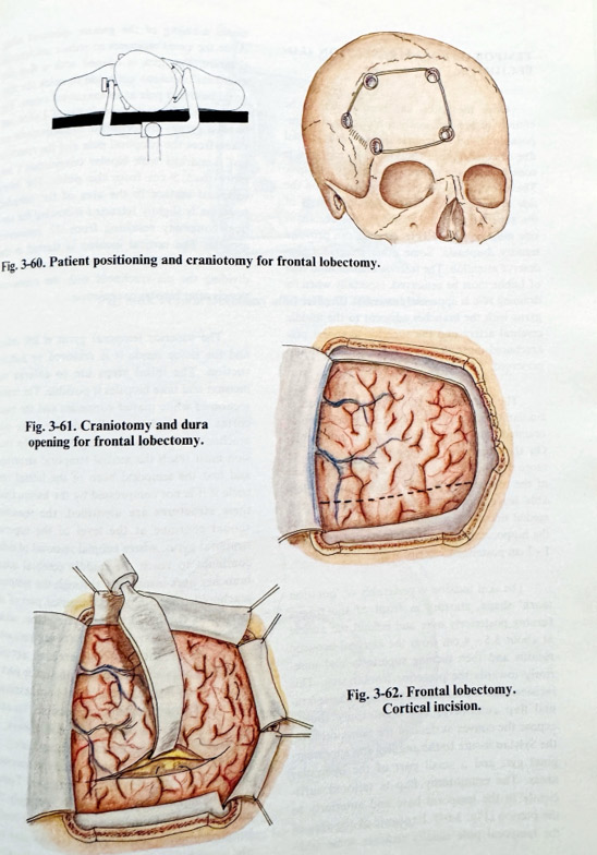
CT and MRI are the necessary investigations for planning in practically all cases. The resolution of the MRI image permits the identification of the main sulci and gyri. The plane of resection can be determined and some distances estimated from the frontal pole. These visible references are used: distances from the pole along the medial and frontobasal edges of hemispherical convexity, relationships to the free edge of the falx and how much it lies anterior to the genu of corpus callosum. MRI also allows the localization of the main branches of the anterior cerebral arteries (frontopolar arteries in particular). The bone flap is adjusted in position and size to the resection, avoiding the frontal sinus opening and the superior sagittal sinus with inflowing veins, placing the medial limit at 1.5 - 2 cm from the midline (Fig. 3-60).
Surgical techniques are rather standard. The incision is placed along the anterior hairline, from the zygomatic arch to the opposite frontal area, 3-4 cm across the midline (no scarring on the forehead will remain). The craniotomy flap covers an area of the convexity surface of the frontal lobe. Its borders are 1.5 cm from the midline and just out of the frontal sinus, and just superior to the orbit. The flap is fractured in the temporal area or is lifted as a free one. The dura should not be tense before opening; dehydration measures should have been efficiently completed. The cortical surface is inspected and the position of the frontal operculum determined according the initial part of the Sylvian fissure. The position of the resection plane is established, keeping permanent orientation of the midsagittal plane as very often the resection plane can be tilted and the resection can be too posterior on the medial surface. Usually the measurements are estimated from the frontal pole along the sagittal and basal edges of the lobe. The points are marked and are later united as a line (Fig. 3-61). In an area close to the lesion, the white matter is incised to the depth, and several biopsies are taken. Bleeding is controlled. The cortical incision is enlarged.
As bleeding in its major part comes from the cortical supply, once it is in the white matter, the transection of tissue can be done with the forceps and suction tip. Penetrating into tumour tissue is heralded usually by an increase in bleeding (Pig. 3-62) The penetration plane should however be maintained. If resection "en block" is difficult, subpial tissue removal provides space for manipulation. Retraction is applied to the side to be resected, avoiding pressure on the opposite side to the remaining hemisphere. The edges of the lobes are also transected. Special attention is paid to the bridging veins, in a way to be able to divide them after coagulation away, with a stump on the superior sagittal sinus. When dividing pia and arachnoid on the basal and medial surfaces, the underlying white matter is first aspirated out, exposing the thin layer of remaining pia-arachnoid. Then it is easily coagulated and divided. The medial surface exposure shows the falx and its edge, permitting more precise orientation of the resection plane. Behind the falx special care is taken to coagulate and divide only vessels related to the ipsilateral lobe. The resection should pass anterior to the corpus callosum and the big arteries anterior to the genu. On the basal surface, the olfactory tract is dissected and left in place (it is usually not invaded by hemispherical gliomas). Both transections of pia-arachnoid join at the most basal and medial part of the resection plane. The resected part is lifted and the remaining veins in the polar area are dissected and divided. If on the non-dominant side, the plane is located more posteriorly and the resection opens the frontal horn of the lateral ventricle. The pericallosal and callosomarginal arteries are dissected carefully close to the midline and the genu of the corpus callosum. In cases of larger lesions, infiltrated areas are seen on the remaining resection surface. If they do not penetrate deeper into the basal ganglia, such remaining parts can be additionally resected (Fig. 3-63).
After haemostasis, the residual cavity is filled with saline, the dura is closed hermetically and the craniotomy closed in the usual fashion.
TEMPORAL POLAR RESECTION (LOВЕСТОMY)
Once the lesion has been shown to be entirely or predominantly confined to one temporal lobe and the neural function is impaired due to compression, temporal lobe resection is considered the preferable surgical technique.
The extent of this resection also depends on the side of cerebral dominance. Posterior parts of the superior temporal gyrus on the dominant side must be spared as its damage can provoke sensory dysphasia. Some other structures also deserve attention. The inferior anastomotic vein of Labbe must be preserved, especially when its draining role is apparently essential. The Hershl gyrus with the branches adjacent to the middle cerebral artery and the superior temporal pia-arachnoid must also be carefully dissected and preserved.
The planning of this type of resection is similar to the frontal. The resection plane is oriented to the most posterior acceptable position.
On the dominant side, posterior resection of usually more than 4.5 cm can lead to a dysphasic deficit of the receptive type. The plane should aim leaving intact the vein of Labbe. On the medial side of the temporal lobe the resection of the hippocampus is preferably not greater than 1 - 2 cm posterior to its tip.
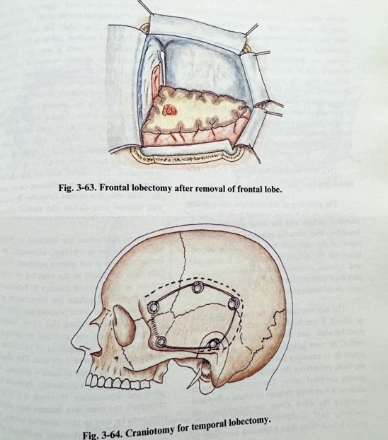
The skin incision is preferably of 'question mark' shape, starting in front of the tragus turning posteriorly over and behind the auricle at about 3.5 - 4 cm from the external acoustic meatus and then turning superiorly and anteriorly towards the posterior frontal area. This incision also makes possible the turning of an epicranial flap anteriorly. The craniotomy should expose the convex surface of the temporal lobe, the Sylvian fissure to the angular and supramarginal gyri and a small part of the opercular areas. The craniotomy flap is tailored sufficiently to the temporal base and anteriorly to the pterion (Fig. 3-64). Exposure of the area of the temporal pole usually requires some additional nibbling of the greater sphenoid wing
After the usual measures to reduce intracranial pressure, the dura is opened with a flap and additional incisions are made to reach the temporal base and pole at its maximum extent. The Sylvian fissure, vein of Labbe and angular gyrus are identified. Measurements are taken from the temporal pole and the resection line is marked ( not more than 5 cm from the pole). The basal temporal surface in the area of the intended resection is slightly retracted following the cortical concavity resulting from the eminentia arcuata. The vertical incision is started in the area of the middle and inferior temporal gyri, dividing the pia-arachnoid with the contained vessels after bipolar coagulation.
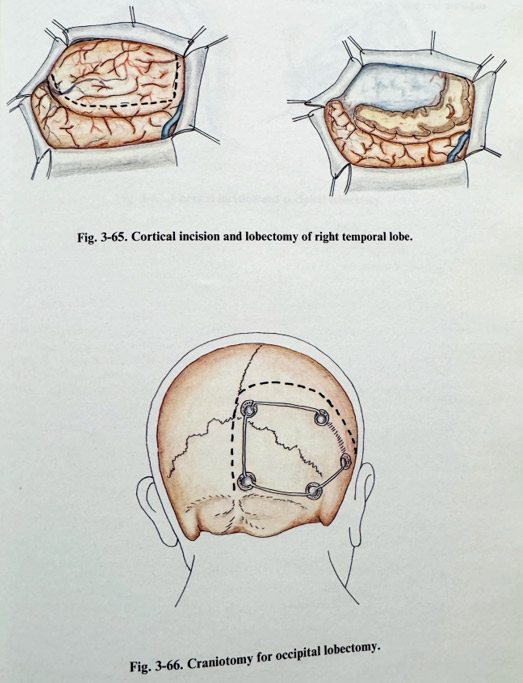
The superior temporal gyrus is left intact and the tissue inside it is removed by subpial suction. The initial steps are to enlarge the incision and take biopsies if possible. The transsection of white matter continues and the basal cortex is aspirated subpially, dividing the pia-arachnoid after bipolar coagulation. The incision must reach the medial temporal structures first opening the temporal horn of the lateral ventricle if it is not compressed by the lesion. Once these structures are identified, the resection should continue at the level of the superior temporal gyrus, where subpial removal of tissue continues to reach the middle cerebral artery branches over insula seen through the temporal arachnoid (Fig. 3-65). The superior part of the incision is continued at the same plane, taking care not to penetrate insula (never removing tissue under the middle cerebral arterial branches); it must join up with the other part of the resection inside the temporal horn, exposing the hippocampus as fully as possible. Its anterior 2.5 cm can usually, but not always, be resected without significant memory deficit. Its tumoral infiltration will determine the necessary extent of the sacrifice of this structure. Technically the hippocampus is divided, but only the part inside the ventricle and not the underlying parahippocampal gyrus. Its vascular supply, provided medially, is interrupted afterwards from temporo-basal at the division of the pia-arachnoid. At this stage the tentorial edge and arachnoid can be seen there, and it should not be disrupted. The additional removal of tissue superiorly can be carried out cautiously; there is danger that the striatum can be penetrated in the same manner.
Always when the tumour masks the anatomy, the debulking must be limited to the margins of safety already mentioned. Venous collectors at the temporal base and the pole, and the temporal arterial branches are coagulated. The dura is closed hermetically. The bone flap is fixed in place and the epicranial layers repaired.
OCCIPITAL POLAR RESECTION (LOВЕСТОМY)
The planning of this resection can consider removing up to 7 cm in average from the pole of the occipital lobe. A significant part of the calcarine fissure is sacrificed and the occipital horn of the lateral ventricle is opened. The cingulate gyrus and cingulum should be spared.
On the convex surface, the resection line should pass posterior to the supramarginal and angular gyri. On the dominant side, however, the extent is much less, at times as little as 3.5 - 4 cm from the pole. Main arterial feeders are provided from the posterior cerebral artery on the medial occipital surface and the distal middle cerebral branches, very often the angular artery. Involvement of the splenium has a particular importance for the deficit, as producing hemianopia and splenial comissural damage leads to complex high cortical functional deficit (hemianopia with visual hemiagnosia).
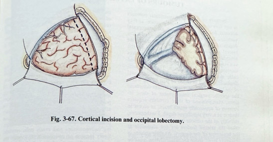
The incision starts at the midoccipital area at about 2 cm below the external occipital protuberance, and continues superiorly to the vertex, to turn laterally toward the squamose suture in its posterior part. The flap is turned to the base, preserving the occipital artery and the greater occipital nerve. The craniotomy (more often as a free flap) exposes the dura along the transverse and posterior part of superior sagittal sinuses at about 1.5 cm from their edges and anteriorly to the supramarginal and angular areas. The length of the sagittal border of the craniotomy is at least 7-8 cm (Fig. 3-66). The dura is opened with a T-shaped incision, the long part pointing to the torcular, in such a way that flaps are turned over both sinuses - transverse and superior sagittal. As in all other lobectomies, intracranial hypotension is obtained before dural opening. Cortical incision is initiated on the convexity, dividing the feeders from the distal middle cerebral artery. After the pia-arachnoid is incised and biopsies taken from the depth, the cortical incision is enlarged and deepened to reach the occipital horn. The bridging veins from the part to be resected are coagulated and transsected. The medial wall of the occipital horn is identified and the eminence on it, called the calcar avis. It is incised vertically. By suction and bipolar coagulation the medial pia-arachnoid is exposed and the underlying branches of the posterior cerebral artery coagulated and cut. The remaining part of the lobe is then less vascularized and the resection is completed. The cavity is filled with saline after meticulous haemostasis, the dura is sutured hermetically, the flap is repositioned and fixed, and epicranial layers are closed in the usual way (Fig. 3-67).
REOPERATION FOR GLIOMAS
These operations are intended to prolong life and reduce morbidity, although in such recurrent cases that is not easy to be decided with certainty. Technically they are aimed at the removal of tumour volume and in that way to reduce damage to functionally active brain and reduce the intracranial pressure. Necrotic and cystic portions are very often taken into consideration (for cysts, inserting reservoirs to perform multiple percutaneous evacuations), but enlargement of tumor tissue resections already performed can be done too. The effect of surgery in the majority of cases is uncertain, and policies often vary significantly between surgeons and departments. However, if it is to be attempted, the same skin incision and craniotomy must be used to their maximal extent. The dura is opened at a previously unaffected place (to avoid the scar which firmly attaches to the brain tissue). The tumour can be found easily and the intended removal is done in the usual way. The technique is the same when the tissue for removal is radiation necrosis. Its resection can produce an internal decompressive effect with the same benefit as obtained from removing tumour parts. The closure of the craniotomy is in the same way as in all other cases.